. 2024 Sep 28;12(1):181.
doi: 10.1186/s40168-024-01924-8.

Akkermansia muciniphila improve cognitive dysfunction by regulating BDNF and serotonin pathway in gut-liver-brain axis

Affiliations

Akkermansia muciniphila improve cognitive dysfunction by regulating BDNF and serotonin pathway in gut-liver-brain axis

Eun Ji Kang et al. Microbiome. .

Abstract

Backrground: Akkermansia muciniphila, a next-generation probiotic, is known as a cornerstone regulating the gut-organ axis in various diseases, but the underlying mechanism remains poorly understood. Here, we revealed the neuronal and antifibrotic effects of A. muciniphila on the gut-liver-brain axis in liver injury.

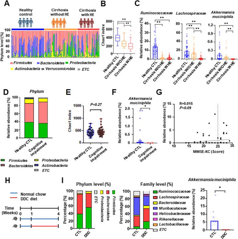
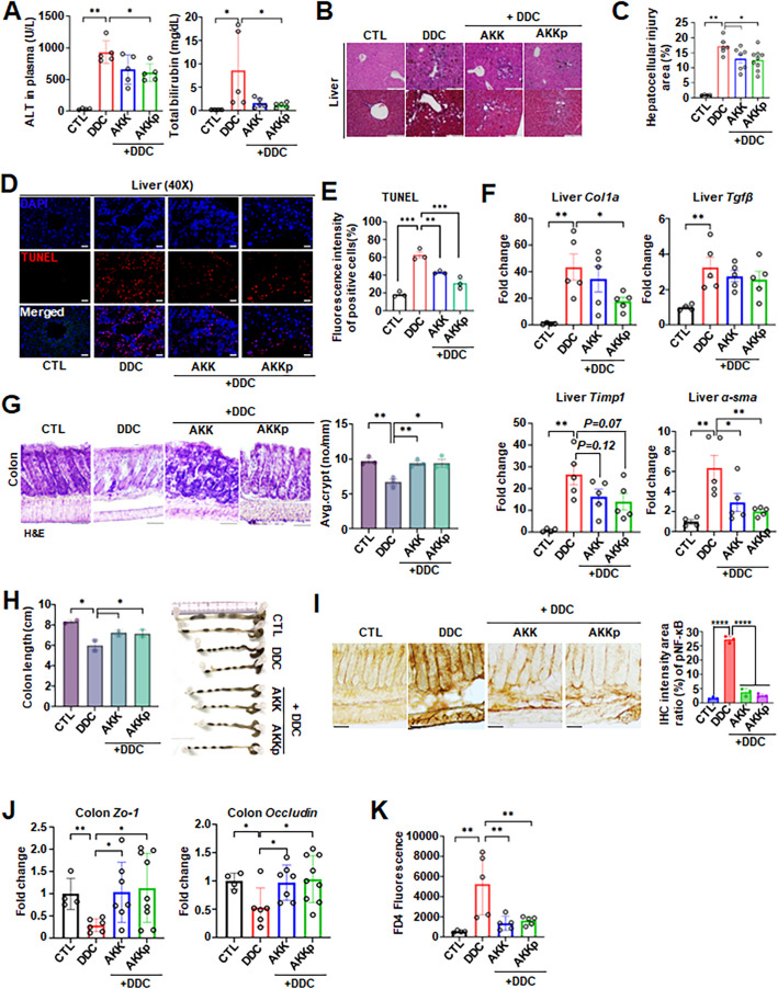
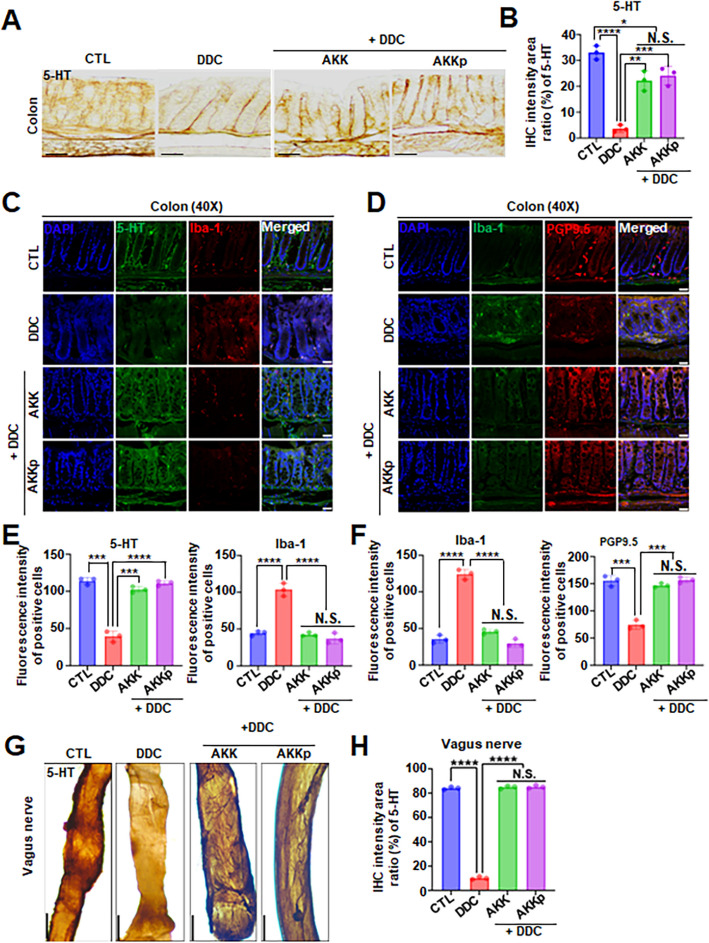
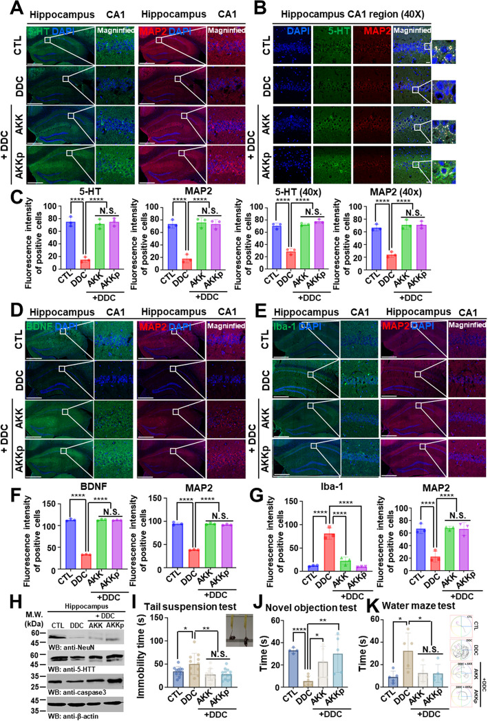
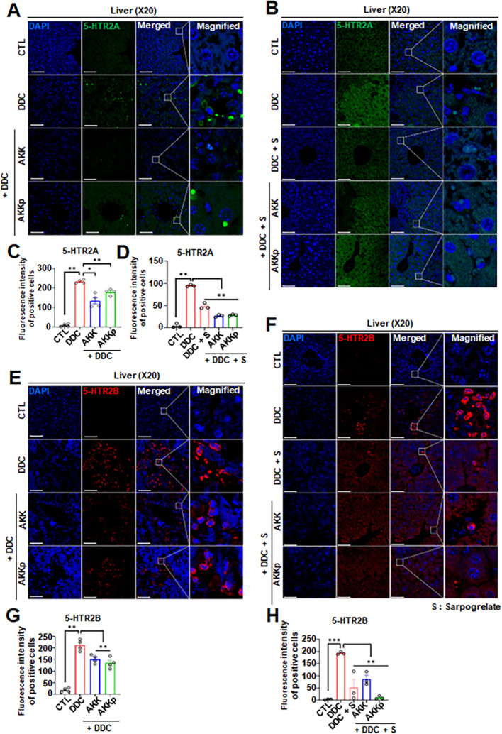
Results: To investigate neurologic dysfunction and characteristic gut microbiotas, we performed a cirrhosis cohort (154 patients with or without hepatic encephalopathy) and a community cognition cohort (80 participants in one region for three years) and validated the existence of cognitive impairment in a 3,5-diethoxycarbonyl-1,4-dihydrocollidine-induced hepatic injury mouse model. The effects of the candidate strain on cognition were evaluated in animal models of liver injury. The expression of brain-derived neurotrophic factor (BDNF) and serotonin receptors was accessed in patients with fibrosis (100 patients) according to the fibrosis grade and hepatic venous pressure gradient. The proportion of A. muciniphila decreased in populations with hepatic encephalopathy and cognitive dysfunction. Tissue staining techniques confirmed gut-liver-brain damage in liver injury, with drastic expression of BDNF and serotonin in the gut and brain. The administration of A. muciniphila significantly reduced tissue damage and improved cognitive dysfunction and the expression of BDNF and serotonin. Isolated vagus nerve staining showed a recovery of serotonin expression without affecting the dopamine pathway. Conversely, in liver tissue, the inhibition of injury through the suppression of serotonin receptor (5-hydroxytryptamine 2A and 2B) expression was confirmed. The severity of liver injury was correlated with the abundance of serotonin, BDNF, and A. muciniphila.

Conclusions: A. muciniphila, a next-generation probiotic, is a therapeutic candidate for alleviating the symptoms of liver fibrosis and cognitive impairment.

Keywords: Akkermansia muciniphila; BDNF; Cognitive impairment; Gut-organ axis; Liver injury; Serotonin.


Conflict of interest statement

The authors declare no competing interests.

Figures

Fig. 1
Fig. 1
Cirrhotic patients with hepatic encephalopathy and cognitive impairment patients exhibited a decreased fecal bacterial abundance of Akkemansia muciniphila, and which was also identified in animal models. a Taxa summary of bacterial phyla level obtained by 16S rDNA sequencing of fecal samples. b Microbial alpha diversity Chao1 in the context of disease progression classifications. **P < 0.01; Wilcoxon rank-sum test. c Relative abundances of species with significantly different representations in human groups. Data represent the means ± SEM; **P < 0.01; one-way ANOVA. d Relative abundance of bacterial phyla level obtained by 16S rDNA sequencing of fecal samples. e Microbial alpha diversity in patients with cognitive impairment. f Relative abundances of A. muciniphila in healthy control and cognitive impairment group. Data represent the means ± SEM; *P < 0.05, unpaired t-test. g Pearson's correlation coefficients, p values, and linear relationships of A. muciniphila relative abundance (%) and MMSE-KC score. h Schematic of intervention with DDC diet (red) during the 3 weeks of 4 weeks DDC-induced liver injury animal model. i Relative abundance of phylum, family and A. muciniphila in control mice and DDC diet mice. AKK, Akkermansia muciniphila; AKKp, pasteurized Akkermansia muciniphila. Data represent the means ± SEM; n ≥ 3; *P < 0.05; unpaired t-test
Fig. 2
Fig. 2
Oral administration of A. muciniphila alleviates liver injury and protects intestinal barrier in a mouse model. a Plasma ALT and total bilirubin levels. Data represent the means ± SEM; n ≥ 4; **P < 0.01, *P < 0.05; one-way ANOVA. b, c Histological assessment of liver injury with representative images of H&E and Masson's trichrome stained liver sections. Data represent the means ± SEM; n ≥ 3; **P < 0.01, *P < 0.05; one-way ANOVA. Scale bar, 200 μm. d, e TUNEL staining in the liver from mouse. Data represent the means ± SEM; n ≥ 3; **P < 0.01 one-way ANOVA. Scale bar, 50 μm. f Hepatic mRNA expression of liver genes (Col1a, Tgfβ, Timp1, and α-sma). Data represent the means ± SEM; n ≥ 4; **P < 0.01, * P < 0.05; one-way ANOVA. g Histological assessment of colon crypt with representative images of H&E stained colon sections. Data represent the means ± SEM; n ≥ 3; **P < 0.01, *P < 0.05; one-way ANOVA. Scale bar, 200 μm. h GI tract dysfunction characterization measured by the colon length. Data represent the means ± SEM; n = 3; *P < 0.05; one-way ANOVA. i Immunohistochemical staining of phosphorylated NFκB in the mouse colon. Data represent the means ± SEM; n ≥ 3; ****P < 0.0001 one-way ANOVA. Scale bar, 200 μm. j Colon mRNA expression of tight junction-related genes (Zo-1, Occludin). Data represent the means ± SEM; n ≥ 4; **P < 0.01, * P < 0.05; one-way ANOVA. k Plasma FD4 (fluorescein isothiocyanate dextran 4) level of the in vivo gut permeability assay. Data represent the means ± SEM; n ≥ 4; **P < 0.01; one-way ANOVA
Fig. 3
Fig. 3
Serotonin deficiency and activation of neuroinflammatory mechanisms in the gut and brain axis of a liver disease animal model and neuronal cell deaths alleviated by A. muciniphila administration. a, b Immunohistochemistry staining of 5-HT in the colon of mice. Quantitative analysis of 5-HT-positive cells. Data represent the means ± SEM; representative data of 3 samples; ****P < 0.0001; N.S. = not significant, one-way ANOVA. Scale bar, 100 μm. c Immunofluorescent staining of anti-5-HT/anti-Iba-1 in the colon of mice and the fluorescent signals were quantified (d) Immunofluorescent staining of anti-Iba-1/anti-PGP9.5 in the colon of mice and the fluorescent signals were quantified (e) Scale bar, 10 μm. Data represent the means ± SEM; representative data of 3 samples; ****P < 0.0001; N.S. = not significant, one-way ANOVA. f Scale bar, 10 μm. Data represent the means ± SEM; representative data of 3 samples; ****P < 0.0001; N.S. = not significant, one-way ANOVA. g, h Therapeutic effect of AKK or pasteurized AKK (AKKp) treated groups in the vagus nerve of mice were analyzed by immunohistochemistry staining. Scale bar, 20 μm. Data represent the means ± SEM; representative data of 3 samples; ****P < 0.0001; N.S. = not significant, one-way ANOVA
Fig. 4
Fig. 4
Both BDNF and 5-HT are reduced in DDC only treated group hippocampus, while increased in A. muciniphila treated group. a, b, c Co-immunofluorescence staining analysis of anti-5-HT/anti-MAP2 in the hippocampus CA1 region of mice and the fluorescent signals (a, b) were quantified (c). Scale bar, 100 μm. Data represent the means ± SEM; representative data of 3 samples; ****P < 0.0001; N.S. = not significant, one-way ANOVA. d, e, f, g The immunofluorescence co-staining of anti-BDNF/anti-MAP2 (d, f) and anti-Iba-1/anti-MAP2 (e, g) in the hippocampus of the above animals. Scale bar, 100 μm. Data represent the means ± SEM; representative data of 3 samples; ****P < 0.0001; N.S. = not significant, one-way ANOVA. h Western blot analysis of prefrontal cortex and hippocampus lysates from the animals treated with DDC only or both treated DDC + AKK group or DDC + AKKp group. i Immobility time graph of tail suspension test. Data represent the means ± SEM; n ≥ 11; *P < 0.05, **P < 0.01; N.S. = not significant, unpaired t-test. j Cognitive impairment tests of novel objection test and water maze test (k) were performed. Data represent the means ± SEM; n ≥ 4; *P < 0.05, **P < 0.01; N.S. = not significant, unpaired t-test
Fig. 5
Fig. 5
5-HT2A/2B receptor expression are increased in DDC only treated group liver tissue, while reduced in A. muciniphila treated group. a, b, c, d immunofluorescence staining analysis of anti-5-HTR2A in the liver tissue of mice and the fluorescent signals (a, b) were quantified (c, d). Scale bar, 100 μm. Data represent the means ± SEM; n ≥ 3; **P < 0.01, *P < 0.05; one-way ANOVA. e, f, g, h immunofluorescence staining analysis of anti-5-HTR2B in the liver tissue of the above mice. The fluorescent signals (e, f) were quantified (g, h). Scale bar, 100 μm. Data represent the means ± SEM; n ≥ 3; ***P < 0.005, **P < 0.01; one-way ANOVA
Fig. 6
Fig. 6
BDNF/serotonin is associated with the progression of liver cirrhosis. a mRNA expression of 5-HT2A receptor and 5-HT2B receptor, and BDNF in human liver biopsy. Western blot analysis of 5-HT2A receptor and 5-HTR2B receptor in human liver biopsy. Data represent the means ± SEM; n = 10; **P < 0.01, *P < 0.05. one-way ANOVA. b Concentrations of serotonin and BDNF measured in portal blood of patients with liver cirrhosis. Data represent the means ± SEM; n ≥ 12; **P < 0.01, *P < 0.05. one-way ANOVA. c Pearson's correlation coefficients, P values, and linear relationships of serotonin and BDNF concentration, A. muciniphila relative abundance, and HVPG. d Pearson's correlation coefficients, p values, and linear relationships of serotonin and BDNF concentration, A. muciniphila relative abundance, and Child–Pugh score

References

    1. Butterworth RF. Hepatic Encephalopathy in Cirrhosis: Pathology and Pathophysiology. Drugs. 2019;79(Suppl 1):17–21. - PMC - PubMed
    1. Shawcross DL, Thabut D, Amodio P. Ammonia - an enduring foe - What evaluating whole body ammonia metabolism can teach us about cirrhosis and therapies treating hepatic encephalopathy. J Hepatol. 2023;79(2):266–8. - PubMed
    1. Korean Association for the Study of the L. KASL clinical practice guidelines for liver cirrhosis: Varices, hepatic encephalopathy, and related complications. Clin Mol Hepatol. 2020;26(2):83–127. - PMC - PubMed
    1. Haeussinger D, Dhiman RK, Felipo V, Görg B, Jalan R, Kircheis G, et al. Hepatic encephalopathy Nature Reviews Disease Primers. 2022;8(1):43. - PubMed
    1. Tonon M, Piano S. Acute on chronic liver failure in cirrhosis. Clin Mol Hepatol. 2022;28(2):273–5. - PMC - PubMed

Supplementary concepts