. 2024 Sep 28;57(1):69.
doi: 10.1186/s40659-024-00536-8.

LYP regulates SLP76 and other adaptor proteins in T cells

Affiliations

LYP regulates SLP76 and other adaptor proteins in T cells

Virginia Ruiz-Martín et al. Biol Res. .

Abstract

Background: The LYP tyrosine phosphatase presents a SNP (1858C > T) that increases the risk of developing autoimmune diseases such as type I diabetes and arthritis. It remains unclear how this SNP affects LYP function and promotes the development of these diseases. The scarce information about LYP substrates is in part responsible for the poor understanding of LYP function.

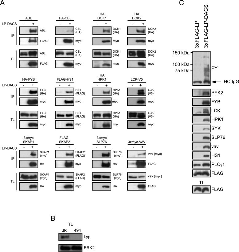
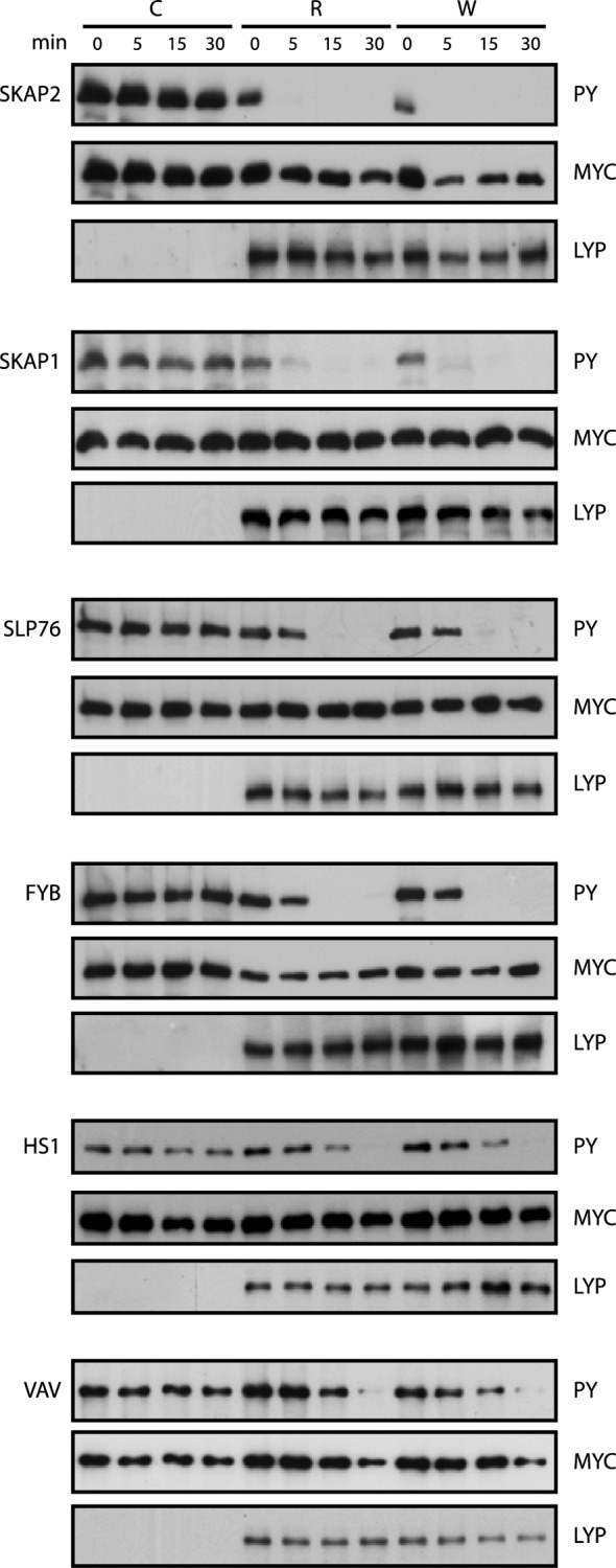
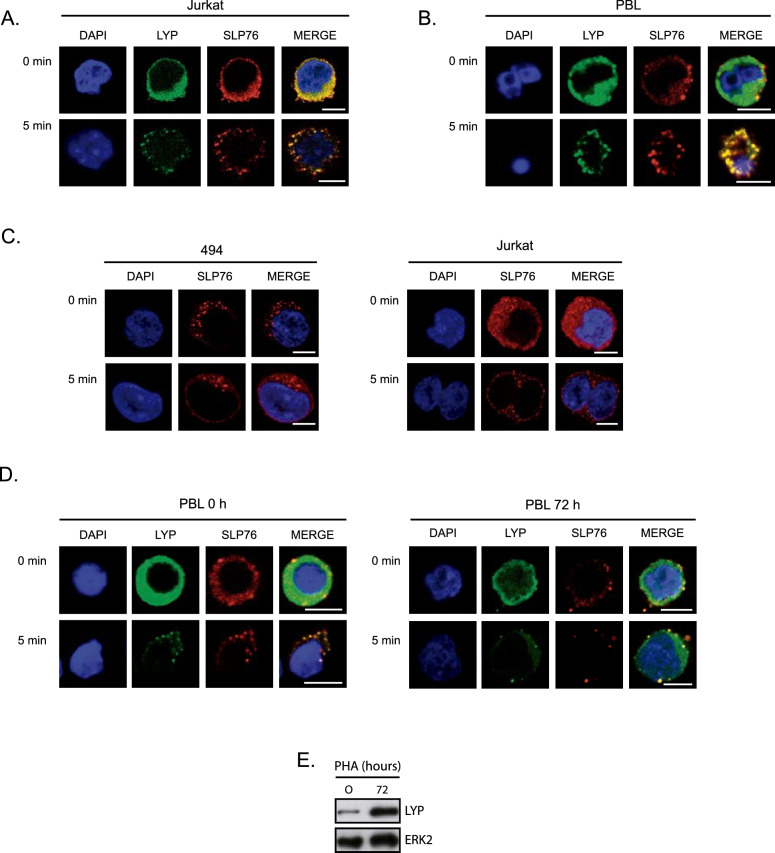
Results: In this study, we identify in T lymphocytes several adaptor proteins as potential substrates targeted by LYP, including FYB, SLP-76, HS-1, Vav, SKAP1 and SKAP2. We also show that LYP co-localizes with SLP76 in microclusters, upon TCR engagement.

Conclusions: These data indicate that LYP may modulate T cell activation by dephosphorylating several adaptor proteins, such as FYB, SLP-76, HS-1, Vav, SKAP1 and SKAP2 upon TCR engagement.

Keywords: FYB; LYP (lymphoid phosphatase); Protein tyrosine phosphatase (PTP); SKAP2; SLP76; T-cell; T-cell receptor (TCR).


Conflict of interest statement

The authors declare that they have no competing interests.

Figures

Fig. 1
Fig. 1
Interaction of the substrate trapping mutant of LYP phosphatase domain, LP-DACS, with several potential substrates. A HEK293 cells were transiently transfected with 3xmyc, 3xHA or 3xFLAG-LP-DACS along with plasmids that express potential substrates, as indicated on the top of each panel. After PV treatment for 5 min, cells were lysed and LP-DACS was immunoprecipitated with specific antibodies against myc, HA or FLAG epitopes bound to sepharose beads. Proteins were transferred to nitrocellulose membranes after SDS-PAGE, and the presence of potential substrates in the precipitates was detected by Western Blot. B Detection of LYP and ERK2, as loading controls, in Jurkat cells and the LYP-deficient Jurkat-cell line (494). C Jurkat cells deficient in LYP (494) were transiently transfected with plasmids that express 3xFLAG-LP, the active phosphatase domain of LYP, and 3xFLAG-LP-DACS. After PV treatment for 5 min, the phosphatase domain of LYP was immunoprecipitated with FLAG antibody bound to Sepharose beads, and proteins present in the precipitates were identified by Western Blot. HC-IgG heavy chain IgG, IP immunoprecipitates, TL total lysates
Fig. 2
Fig. 2
In vitro dephosphorylation assays with LYP-R620 and LYP-W620. Several proteins were tested for dephosphorylation by either LYP-R or LYP-W at the time points indicated on the top of the panel. Phosphatase assays were carried out with a full-length version of LYP and its putative substrates as indicated in Material and Methods. Dephosphorylation was detected by Western blot with p-Tyr (4G10) antibody
Fig. 3
Fig. 3
Identification of the Tyr targeted by LYP in several potential substrates. A HEK293 cells were transiently transfected with 3xHA-LP-DACS along with expression plasmids of SKAP2 wild type and mutated to Phe in the Tyr indicated. After PV treatment for 5 min, cells were lysed, and LP-DACS was immunoprecipitated with HA Ab bound to Sepharose beads. Proteins were separated by SDS-PAGE and transferred to nitrocellulose membranes. The presence of SKAP2 in the precipitates was detected by WB, as indicated. B Graph representing the mean data obtained by densitometry in at least three experiments (n = 4 for single Y to F mutant and n = 3 for 3YF) like the one shown in (A). Densitometry values of the myc blot on the top, expressed as percentage of the WT are indicated below. C As before, SKAP1 wild type and the mutants in Tyr232 and Tyr271 were assayed for binding to LP-DACS. D Interaction of HS1 YF mutants with 3xHA-LP-DACS, as before. E Interaction of Vav YF, as indicated, with 3xHA-LP-DACS. F Graph representing the mean data obtained by densitometry in three experiments like the one shown in (E). Densitometry values of the myc blot on the top, expressed as percentage of the WT are indicated below. G Interaction of FYB wild type or mutated in five of the six tyrosines tested, as indicated, with 3xHA-LP-DACS. H As before, interaction of SLP76 YF full length mutants with 3xHA-LP-DACS. NS nonsignificant, *P < 0.05,**P < 0.01 and ***P < 0.001 for comparison of proteins mutated in Tyr with the wild type protein
Fig. 4
Fig. 4
Regulation of TCR signaling by LYP downstream of ZAP70. A Activation of a luciferase reporter gene driven by the IL-2 minimal promoter in Jurkat cells transfected with Vav and stimulated with CD3 plus CD28 antibodies for 6 h, as indicated. The insert shows the IB of the Vav and LYP proteins expressed. *P < 0.05 and **P < 0.01 for comparison of cells transfected with different plasmids and cells transfected with empty vector (pEF). B Activation of a luciferase reporter gene driven by the IL-2 minimal promoter in Jurkat cells transfected with rac-QL, a dominant active mutant of rac, and LYP and and stimulated with CD3 plus CD28 antibodies for 6 h. The insert shows the IB of LYP. *P < 0.05 and **P < 0.01 for comparison of cells transfected with different plasmids and cells transfected with empty vector (pEF) C Activation of a luciferase reporter gene driven by the IL-2 minimal promoter in wild type and 494 Jurkat cells transfected with LYP and SLP76 as indicated. The cells were left untreated or stimulated with CD3/CD28 antibodies for for 6 h. The insert shows the IB of the SLP76 and LYP proteins expressed. *P < 0.05 **P < 0.01 and ***P < 0.001 for comparison of cells transfected with different plasmids and cells transfected with empty vector (pEF). D Wild type and 494 JK cells were stimulated with CD3/CD28 antibodies during the indicated periods of time. Phosphorylation of Y128 of SLP76 in each condition was measured in cell lysates by IB. Similarly, phosphorylation of LCK in Y394 and Y493 are shown. E, Wild type Jurkat cells and cells deficient in LYP (JK 494) transfected with LYP were stimulated with CD3/CD28 antibodies for 15 min and the phosphorylation of Y128 of SLP76, and Y191 of LAT were detected in each condition by IB
Fig. 5
Fig. 5
LYP co-localizes with SLP76 in T cell microclusters. Jurkat cells (A) or PBL (B) were plated on coverslips covered with antibody for CD3 for the indicated periods of time. Then, cells were fixed and stained with specific antibodies for SLP76 and LYP, as indicated. Images were taken with a confocal microscope and representative images are shown. C, Wild type and 494 Jurkat cell lines were plated on coverslips covered with CD3 antibody for the indicated periods of time. Then, cells were fixed and stained with specific antibodies for SLP76. D, PBL cells were left untreated or stimulated with PHA for 72 h to induce the expression of LYP. As before, they were plated on stimulatory coverslips and processed to detect LYP and SLP76 by immunofluorescence with a confocal microscope. E, LYP expression in PLB treated with PHA. Scale bar represents 5 μm

Similar articles

References

    1. Vang T, Miletic A V, Arimura Y, Tautz L, Rickert RC, Mustelin T. Protein tyrosine phosphatases in autoimmunity. 2008; 26:29–55. 10.1146/annurev.immunol.26.021607.090418 - PubMed
    1. Cloutier JF, Veillette A. Cooperative inhibition of T-cell antigen receptor signaling by a complex between a kinase and a phosphatase. J Exp Med. 1999;189(1):111–21. 10.1084/jem.189.1.111 - PMC - PubMed
    1. Badour K, Zhang J, Shi F, Leng Y, Collins M, Siminovitch KA. Fyn and PTP-PEST-mediated regulation of Wiskott-Aldrich Syndrome Protein (WASp) tyrosine phosphorylation is required for coupling T cell antigen receptor engagement to WASp effector function and T cell activation. J Exp Med. 2004;199(1):99–111. 10.1084/jem.20030976 - PMC - PubMed
    1. Marcos T, Ruiz-Martín V, De La Puerta ML, Trinidad AG, Rodríguez MDC, De La Fuente MA, et al. Proline-serine-threonine phosphatase interacting protein 1 inhibition of T-cell receptor signaling depends on its SH3 domain. FEBS J. 2014;281(17):3844–54. 10.1111/febs.12912 - PubMed
    1. Manso JA, Marcos T, Ruiz-Martín V, Casas J, Alcón P, Sánchez Crespo M, et al. PSTPIP1-LYP phosphatase interaction: structural basis and implications for autoinflammatory disorders. Cell Mol Life Sci. 2022;79(2):131. 10.1007/s00018-022-04173-w. - PMC - PubMed

MeSH terms

Substances

LinkOut - more resources