Review
. 2024 Nov;300(11):107834.
doi: 10.1016/j.jbc.2024.107834. Epub 2024 Sep 27.

Structural basis for the participation of the SARS-CoV-2 nucleocapsid protein in the template switch mechanism and genomic RNA reorganization

Affiliations
Review

Structural basis for the participation of the SARS-CoV-2 nucleocapsid protein in the template switch mechanism and genomic RNA reorganization

Peter R Bezerra et al. J Biol Chem. 2024 Nov.

Abstract

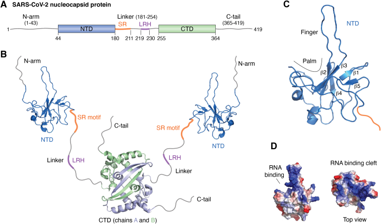
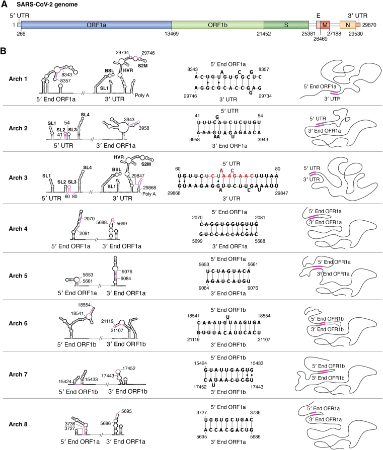
The COVID-19 pandemic has resulted in a significant toll of deaths worldwide, exceeding seven million individuals, prompting intensive research efforts aimed at elucidating the molecular mechanisms underlying the pathogenesis of SARS-CoV-2 infection. Despite the rapid development of effective vaccines and therapeutic interventions, COVID-19 remains a threat to humans due to the emergence of novel variants and largely unknown long-term consequences. Among the viral proteins, the nucleocapsid protein (N) stands out as the most conserved and abundant, playing the primary role in nucleocapsid assembly and genome packaging. The N protein is promiscuous for the recognition of RNA, yet it can perform specific functions. Here, we discuss the structural basis of specificity, which is directly linked to its regulatory role. Notably, the RNA chaperone activity of N is central to its multiple roles throughout the viral life cycle. This activity encompasses double-stranded RNA (dsRNA) annealing and melting and facilitates template switching, enabling discontinuous transcription. N also promotes the formation of membrane-less compartments through liquid-liquid phase separation, thereby facilitating the congregation of the replication and transcription complex. Considering the information available regarding the catalytic activities and binding signatures of the N protein-RNA interaction, this review focuses on the regulatory role of the SARS-CoV-2 N protein. We emphasize the participation of the N protein in discontinuous transcription, template switching, and RNA chaperone activity, including double-stranded RNA melting and annealing activities.

Keywords: RNA chaperone; RNA-binding; SARS-CoV-2; coronavirus; nucleocapsid protein.


Conflict of interest statement

Conflict of interest The authors declare that they have no conflicts of interest with the contents of this article.

Figures

Figure 1
Figure 1
SARS-CoV-2 nucleocapsid protein structural organization.A, schematic representation of the primary sequence distinguishing all structural regions. The rectangles represent the globular domains, the N-terminal (NTD, 44--180) and C-terminal (CTD, 255--364) domains. The gray lines represent the three intrinsically disordered regions: the N-arm (1--43), linker (181--254), and C-tail (365--419). The orange line shows the serine-arginine-rich motif (SR motif) (181--211) and the purple line shows the leucine-rich helix (LRH) (219--230) within the linker IDR (181--254). B, representation of the structural elements of the N protein. The structure of the globular domains was obtained from the following PDB identifiers: NTD, 6YI3, CTD, and 7VBF. The NTD (blue) is the regulatory RNA-binding domain, whereas the CTD (light blue, chain A, and light green, chain B) is the dimerization domain, which also binds RNA, mainly in the nucleocapsid assembly. The gray lines represent the IDRs, the orange lines represent the SR motif, and the purple lines represent LRH. The orientation of the IDRs and NTD relative to the CTD domain was freely sketched to reflect the dynamic behavior due to the IDRs. C, NMR structure of the NTD highlighting the binding cleft formed by the finger and palm (6YI3). The finger is composed of a β2 to β3 loop, and the palm is composed of the surface of the twisted β-sheet (β1–β5). Note that the SR region (orange) is immediately adjacent to the C-terminus of the NTD. D, the electrostatic surface potential of the NTD showing the positively charged RNA binding cleft (blue) located right between the palm and the finger. The negatively charged potential is shown in red, and the neutral potential is shown in white. The structures were generated with PyMOL version 2.5.4 Schrödinger, LLC (112).
Figure 2
Figure 2
Template switching, discontinuous transcription, and transcription regulatory sequences of SARS-CoV-2.A, representation of the genomic RNA (gRNA) of SARS-CoV-2, highlighting the open reading frames (ORFs) for the nonstructural protein and structural and accessory proteins. The structural and accessory regions contain TRS-B upstream of the ORF (black rectangles). The polyproteins pp1a and pp1ab are represented in orange. The blue lines represent the sgmRNAs for each ORF of the structural and accessory proteins. We show the TRS-B sequences for each ORF. The question mark (?) at TRS-ORF6 is due to the high distance between the TRS-B (27041) and AUG (27202), and for TRS-ORF10 is because the sgmRNA has never been detected, despite the presence of TRS-B. B, alignment of the TRSs of SARS-CoV-2, showing the TRS start position and the start codon (AUG) position in the gRNA. C, schematic mechanism of the canonical TRS-dependent template switch: first, the nascent negative-sense chain (red) is encoded by the RNA-dependent RNA polymerase, represented as a green ellipse. In addition to negative-sense synthesis, genome rearrangement prompts the genome to adopt a conformation suitable for polymerase jumping, favoring the template switch. Once cTRS-B is synthesized, the negative-sense strand detaches through the melting of dsTRS-B. The template switch occurs through the RNA polymerase jump, promoting base pairing (annealing) between TRS-L and cTRS-B. The cyclization of the gRNA placing the cTRS-B close to the TRS-L is essential. After cTRS-B/TRS-L annealing, the polymerase continues to encode until the 5′ end of the gRNA. The resulting nascent chain is the sgRNA (red), which is used as a template for the RNA polymerase to synthesize the sgmRNA (light blue).
Figure 3
Figure 3
RNA recognition by the NTD.A, schematic representation of the palm of the NTD (β1–β5) showing the interacting MHV RNA strand (TRS, AUCUAAACUU) in orange. The binding polarity was determined by paramagnetic resonance relaxation enhancement (PRE) (50). The TRS-L binding orientation is defined by the proximity to the nitroxide spin label at U2 and U9 (asterisk). Residues H107, R125, Y127, and Y190 stand out as important for the interaction. The homologous residues in the SARS-CoV-2 NTD are shown in parentheses: A90, R107, Y109, and Y127. B, ribbon representation of one of the poses of the crystal structure (PDB ID: 7XWZ) (52) of the complex between the SARS-CoV-2 NTD and a dsRNA (5′-GUCAGUG-3’ (red) and complementary strand (blue)). The residues participating in the interaction are in orange. The finger region is not observed in the structure because of its flexibility. We drew the finger in cyan to represent its dynamics, possibly transiently embracing the dsRNA. C, SARS-CoV-2 N-NTD structure (PDB ID: 6YI3) (blue), highlighting the highly conserved residues among betacoronaviruses. Y109 is changed to a phenylalanine in the bat coronavirus HKU4 (asterisk). D, structure of the SARS-CoV-2 NTD (PDB ID: 6YI3) showing the residues with the largest chemical shift perturbation (CSP) according to Dinesh et al. (47).
Figure 4
Figure 4
Models of the double-stranded RNA (dsRNA) melting mechanism promoted by the N protein.A, the sandwich model was proposed by Grossoehme and colleagues (55).The polymerase synthesizes the negative-sense nascent chain (red). The dimeric form of the N protein scans the duplex RNA and recognizes TRS-B/cTRS-B (dsTRS), making a sandwich: one N-terminal domain (NTD) subunit binds to TRS-B, and the other binds to cTRS-B. dsTRS-B is destabilized, leading to the dissociation (melting) of the dsTRS. Each strand (TRS-B, cTRS-B) becomes associated with each NTD subunit. The melting of dsTRS promoted by N facilitates template switching, as described in Figure 2. Note that for the annealing of cTRS-B with TRS-L, the unwinding of SL3, which contains the TRS, is necessary. B, in the second model, only one NTD recognizes and melts the dsTRS. The possibility of two subunits exerting independent activities increases the efficiency of the mechanism. C, NTD motions filtered from principal component analysis of 25 runs of MD simulation of the SARS-CoV-2 NTD binding a duplex TRS-L and cTRS-L (dsTRS-L) (99). The colored gradient represents the direction of the motions from blue to red. PC1 converges to a tweezer-like motion, mainly through the finger region. PC2 shows a twist-like motion of the palm, which involves the long β4/β5 loop, β1/β2 loop, and finger. As a reference, the figure shows that Y109 is located at the center of the binding site.
Figure 5
Figure 5
SARS-CoV-2 genome architectures experimentally reported by Ziv et al. (95).A, representation of the SARS-CoV-2 gRNA used as a guide. The size of each region is scaled to the size of the genome. B, Ziv architectures described in a previous paper (95). The first column shows the name of each architecture, numbered from one to eight. The second column shows the secondary structure of each interacting region calculated by RNAfold WebServer (113). The complementary regions involved in the interactions are colored in magenta. The third column shows the RNA sequences and their positions at the gRNA. Noncanonical base pairs are represented as black dots. The sequence in red for Arch 3 is TRS-L. The fourth column is a free drawing of the interaction to help visualize the effect of each gRNA cyclization.

Similar articles

References

    1. Sola I., Almazán F., Zúñiga S., Enjuanes L. Continuous and discontinuous RNA synthesis in coronaviruses. Annu. Rev. Virol. 2015;2:265–288. - PMC - PubMed
    1. Robson F., Khan K.S., Le T.K., Paris C., Demirbag S., Barfuss P., et al. Coronavirus RNA proofreading: molecular basis and therapeutic targeting. Mol. Cell. 2020;79:710–727. - PMC - PubMed
    1. Yuan Y., Jiao B., Qu L., Yang D., Liu R. The development of COVID-19 treatment. Front. Immunol. 2023;14 - PMC - PubMed
    1. Cai Z., Bai H., Ren D., Xue B., Liu Y., Gong T., et al. Integrin αvβ1 facilitates ACE2-mediated entry of SARS-CoV-2. Virus Res. 2024;339 - PMC - PubMed
    1. Almazan F., Galan C., Enjuanes L. The nucleoprotein is required for efficient coronavirus genome replication. J. Virol. 2004;78:12683–12688. - PMC - PubMed

Publication types

MeSH terms

Substances

LinkOut - more resources