

Insulin-like growth factor II mRNA binding protein 3 is highly expressed in primary diffuse large B-cell lymphoma of the CNS
- PMID: 39343609
- PMCID: PMC11528255
- DOI: 10.3960/jslrt.24025
Insulin-like growth factor II mRNA binding protein 3 is highly expressed in primary diffuse large B-cell lymphoma of the CNS
Abstract
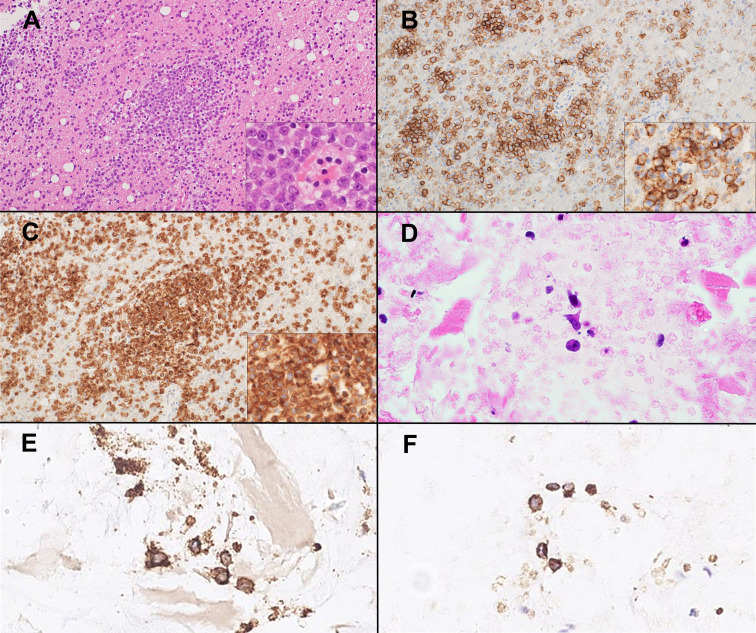
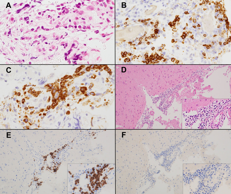
Primary diffuse large B-cell lymphoma of the central nervous system (CNS-DLBCL) can be difficult to diagnose because of the limited amount of biopsy tissue. Here, we analyzed the utility of insulin-like growth factor II mRNA binding protein 3 (IMP3) immunohistochemistry (IHC) as an adjunctive diagnostic tool for CNS-DLBCL. IHC was performed on 57 biopsy samples (55 brain biopsy samples and two vitreous cell blocks) from 54 patients with CNS-DLBCL, including three biopsy samples initially diagnosed as negative or indeterminate for CNS-DLBCL. Additionally, IMP3 IHC was performed on 68 DLBCLs other than CNS-DLBCL and 12 inflammatory brain diseases. Cytoplasmic IMP3 expression was noted in ≥50% of tumor cells in 100% (57/57) of CNS-DLBCLs and 88.2% (60/68) of non-CNS-DLBCLs. In contrast, no IMP3-positive CD20-positive B cells were observed in the inflammatory brain disease (P < 0.0001). In conclusion, IMP3 is highly expressed in CNS-DLBCL. However, it is also expressed in other types of DLBCLs, making it less specific. Most CNS-DLBCL cases can be diagnosed without performing IHC for IMP3 expression, but it may be a useful adjunctive tool to differentiate from reactive lesions when tumor cells are few or deformed.
Keywords: immunohistochemistry; insulin-like growth factor II mRNA binding protein 3; primary diffuse large B-cell lymphoma of the central nervous system.
Conflict of interest statement
CONFLICT OF INTEREST
None declared
Figures
References
-
- Deckert M, Ferry JA, Paulus W, et al. Primary diffuse large B-cell lymphoma of the CNS. In: WHO Classification of Tumours Editorial Board (ed): Central Nervous System Tumours. WHO Classification of Tumours Series, 5th ed, Volume 6. Lyon, International Agency for Research on Cancer. 2021; pp. 351-355.
-
- Air EL, Leach JL, Warnick RE, McPherson CM. Comparing the risks of frameless stereotactic biopsy in eloquent and noneloquent regions of the brain: a retrospective review of 284 cases. J Neurosurg. 2009; 111: 820-824. - PubMed
-
- Ranheim EA. Pearls and pitfalls in the diagnostic workup of small lymph node biopsies. Mod Pathol. 2019; 32: 38-43. - PubMed
-
- King RL, Pasha T, Roullet MR, Zhang PJ, Bagg A. IMP-3 is differentially expressed in normal and neoplastic lymphoid tissue. Hum Pathol. 2009; 40: 1699-1705. - PubMed