

Activation of fibroblasts by plasma cells via PDGF/PDGFR signaling in IgG4-related sialadenitis
- PMID: 39343610
- PMCID: PMC11528260
- DOI: 10.3960/jslrt.24040
Activation of fibroblasts by plasma cells via PDGF/PDGFR signaling in IgG4-related sialadenitis
Abstract
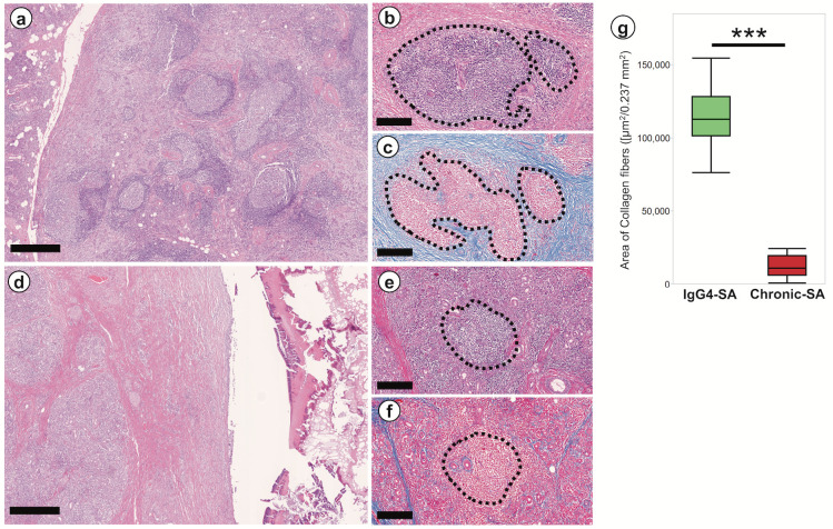
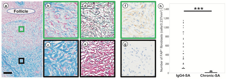
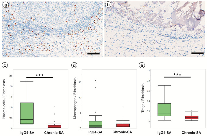
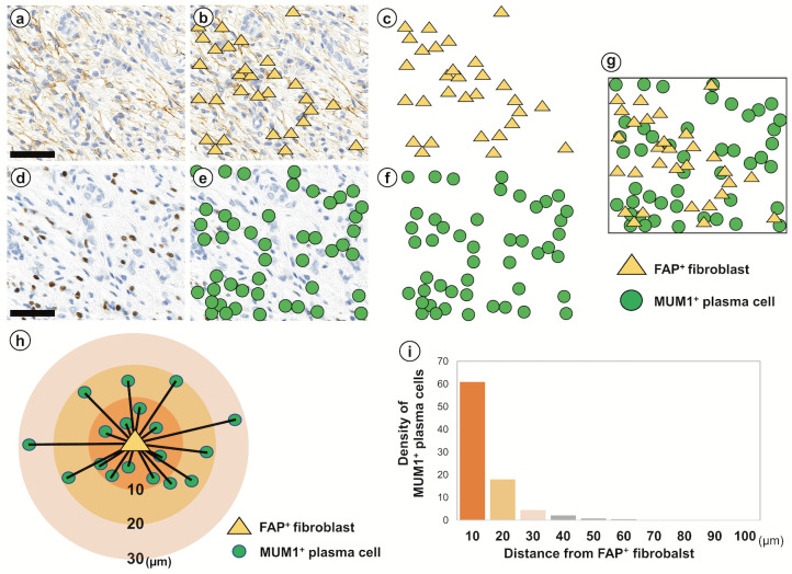
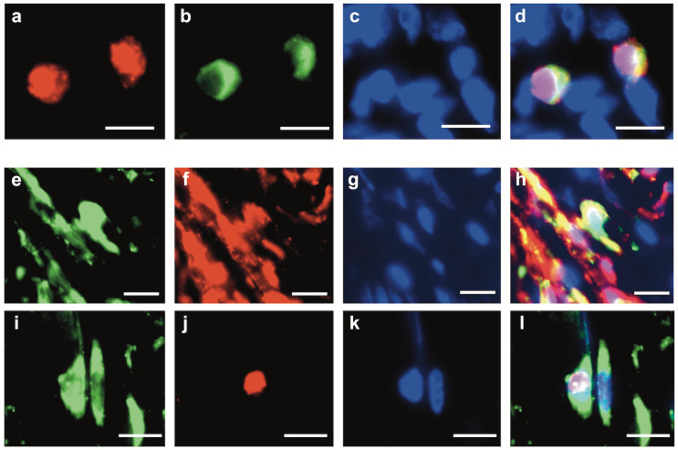
IgG4-related sialadenitis (IgG4-SA) is one of the IgG4-related disease. The histological features of IgG4-SA include dense lymphoplasmacytic infiltrates and fibrosis. This study aimed to reveal the involvement of plasma cells in the development of fibrosis and the mechanism underlying fibrosis in IgG4-SA. Hematoxylin-eosin staining, Azan staining, silver staining, and immunohistochemistry (IHC) were performed on IgG4-SA and chronic sialadenitis specimens, and theses samples were analyzed by image analysis software. Histological spatial analysis was used to analyze the localization of IHC-positive cells and the distances between these cells. In the IgG4-SA group, many secondary lymphoid follicles with germinal centers were found, and many collagen fibers developed around these germinal centers. Collagen fibers composed mainly of type I collagen was abundant at sites away from secondary lymphoid follicles, and reticular fibers composed of type III collagen was abundant near secondary lymphoid follicles. Many FAP+ fibroblasts and MUM1+ plasma cells were localized near secondary lymphoid follicles. Histological spatial analysis demonstrated that 90.4% of MUM1+ plasma cells accumulated within 20 µm of FAP+ fibroblasts. Multiple immunofluorescence assays revealed that MUM1+ plasma cells expressed platelet-derived growth factor (PDGF) β, and FAP+ fibroblasts expressed PDGF receptor (PDGFR) β and pSTAT3 in IgG4-SA. We have shown that fibrosis is localized around secondary lymphoid follicles and that fibroblasts are activated by plasma cells via PDGF/PDGFR signaling in IgG4-SA.
Keywords: IgG4-related sialadenitis; PDGF-PDGFR signaling; fibroblast; histological spatial analysis; plasma cell.
Conflict of interest statement
CONFLICT OF INTEREST
The authors have no competing interests to declare that are relevant to the content of this article.
Figures
References
-
- Satou A, Notohara K, Zen Y, et al. Clinicopathological differential diagnosis of IgG4-related disease: A historical overview and a proposal of the criteria for excluding mimickers of IgG4-related disease. Pathol Int. 2020; 70: 391-402. - PubMed
-
- Deshpande V, Zen Y, Chan JK, et al. Consensus statement on the pathology of IgG4-related disease. Mod Pathol. 2012; 25: 1181-1192. - PubMed
-
- Katz G, Stone JH. Clinical Perspectives on IgG4-Related Disease and Its Classification. Annu Rev Med. 2022; 73: 545-562. - PubMed
-
- Kamisawa T, Zen Y, Pillai S, Stone JH. IgG4-related disease. Lancet. 2015; 385: 1460-1471. - PubMed
MeSH terms
Substances
LinkOut - more resources
Full Text Sources
Miscellaneous