. 2024 Sep 30;56(1):57.
doi: 10.1007/s00726-024-03419-0.

HECTD2 as a target for veratric acid in the regulation of ferroptosis in renal cell carcinoma

Affiliations

HECTD2 as a target for veratric acid in the regulation of ferroptosis in renal cell carcinoma

Dong Lv et al. Amino Acids. .

Abstract

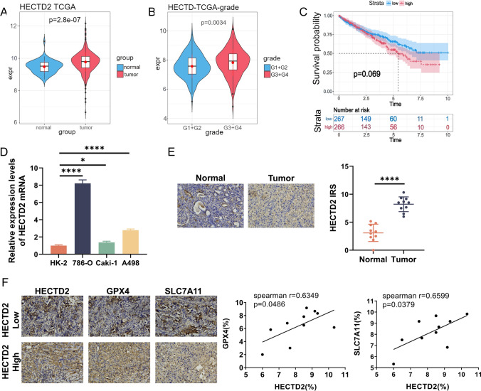
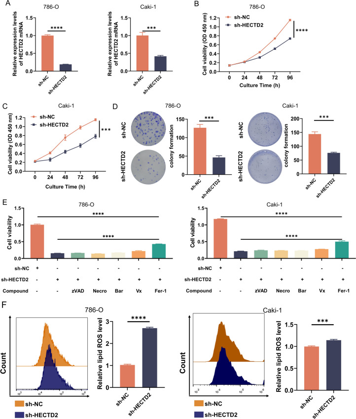
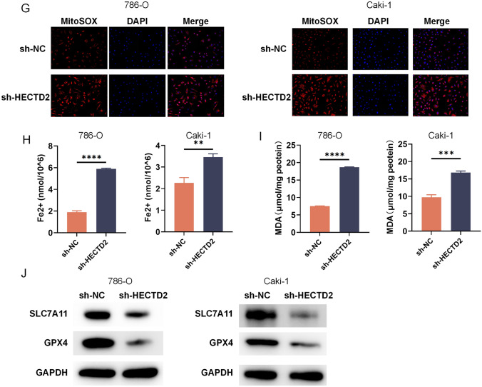
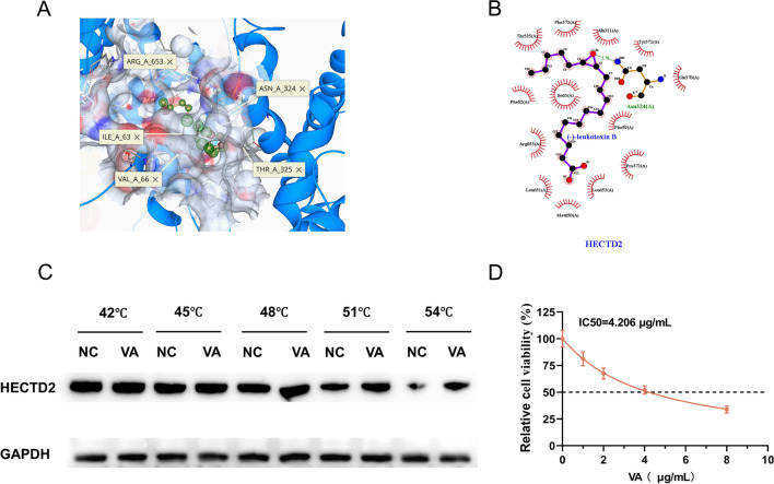
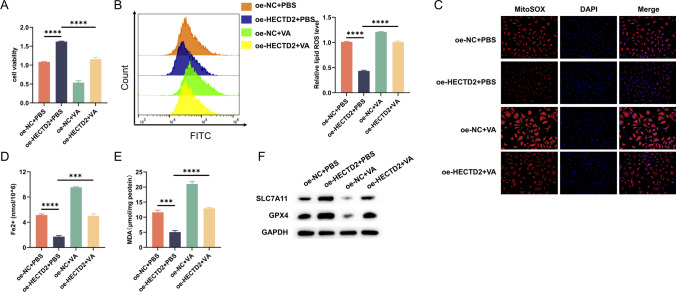
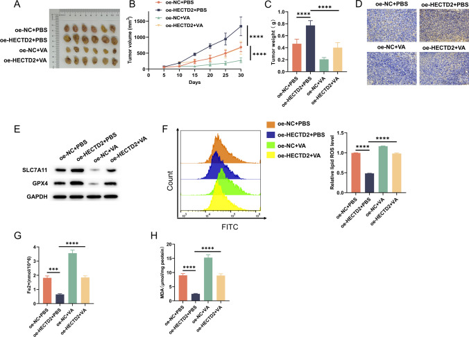
Function of HECTD2 in renal cell carcinoma malignant progression is undefined. Molecular mechanism behind anti-cancer effects of veratric acid (VA) from traditional Chinese medicine (TCM) is underexplored. The Cancer Genome Atlas was leveraged to study HECTD2 expression in renal cell carcinoma and its relationship with histological grading. Kaplan-Meier survival analysis was performed. HECTD2 expression was detected in cancer cells and tissues via qRT-PCR and immunohistochemistry. GPX4 and SLC7A11 expression in tumor samples with high or low HECTD2 expression was examined by immunohistochemistry, cell viability by CCK8, cell proliferation by colony formation assay, lipid ROS and mitochondrial superoxide levels by flow cytometry, Fe2+ and MDA content by assay kits, and GPX4 and SLC7A11 proteins by western blot. SeeSAR software screened TCM small molecule compounds with highest affinity to HECTD2, confirmed with cellular thermal shift assay. VA IC50 was measured by CCK8. Xenograft model was developed and treated with VA. Tumor size and weight were monitored, with immunohistochemistry to detect HECTD2 expression in tumors and assess ferroptosis-related markers. HECTD2 was overexpressed in tumor tissues and cells, which positively correlated with histological grading. HECTD2 depletion inhibited cell vitality and proliferation, raised intracellular lipid ROS, mitochondrial superoxide, Fe2+, and MDA. HECTD2 was a target with highest VA affinity. In vitro and vivo experiments concurred that VA treatment hindered malignancy of renal cell carcinoma and enhanced its susceptibility to ferroptosis. HECTD2 supports ferroptosis resistance in renal cell carcinoma, but VA, through its targeting of HECTD2, initiates ferroptosis, showcasing its anti-cancer efficacy.

Keywords: Ferroptosis; HECTD2; Renal cell carcinoma; Traditional Chinese medicine; Veratric acid.


Conflict of interest statement

The authors have no conflicts of interest to declare.

Figures

Fig. 1
Fig. 1
HECTD2 expression profiles in renal cell carcinoma. A Comparative analysis of HECTD2 mRNA levels in renal cell carcinoma-normal tissue pairs as per TCGA data. B Comparative mRNA expression analysis of HECTD2 across various stages of renal cell carcinoma as per TCGA data. C Survival analysis via Kaplan–Meier estimators depicting the impact of HECTD2 expression levels on the OS of renal cancer patients as per TCGA data. OS overall survival. D qRT-PCR detection of HECTD2 mRNA in the normal renal cell line HK-2 and the renal cell carcinoma cell lines 786-O, Caki-1, and A498. E Visual representation and IRS of HECTD2 protein levels in 10 renal cell carcinoma-normal tissue pairs detected by IHC. F IHC analysis (representative images) of GPX4 and SLC7A11 protein expression in renal cancer samples with high and low HECTD2 expression, as classified by IRS. *Represents P < 0.05. ****Represents P < 0.0001
Fig. 2
Fig. 2
Effects of HECTD2 silencing on renal cell carcinoma cell function and ferroptosis. 786-O cells and Caki-1 cells were transfected with sh-NC or sh-HECTD2. A qRT-PCR validation of transfection efficacy. B, C Impacts of HECTD2 on cell viability assessed by CCK8. D Impacts of HECTD2 on cell proliferation confirmed by colony formation assay. E Specific inhibitors targeting distinct cell death modalities were applied to HECTD2-depleted 786-O cells, encompassing z-VAD-FMK (z-VAD) for apoptosis, necrostatin 1 (Necro) for necroptosis, bafilomycin A1 (Bar) for autophagy, VX-765 (Vx) for pyroptosis, and ferrostatin-1 (Fer) for ferroptosis, with cell viability measured by CCK8. F Intracellular lipid ROS levels detected by the C11-BODIPY probe across the groups. G Mitochondrial superoxide levels detected by the MitoSOX probe across the groups. H Intracellular Fe2+ levels detected by a Fe2+ assay kit for each group. I MDA levels within cells detected by a MDA quantification kit for each group. J WB detection of SLC7A11 and GPX4 protein levels in each group. **Represents P < 0.01, ***Represents P < 0.001, ****Represents P < 0.0001
Fig. 2
Fig. 2
Effects of HECTD2 silencing on renal cell carcinoma cell function and ferroptosis. 786-O cells and Caki-1 cells were transfected with sh-NC or sh-HECTD2. A qRT-PCR validation of transfection efficacy. B, C Impacts of HECTD2 on cell viability assessed by CCK8. D Impacts of HECTD2 on cell proliferation confirmed by colony formation assay. E Specific inhibitors targeting distinct cell death modalities were applied to HECTD2-depleted 786-O cells, encompassing z-VAD-FMK (z-VAD) for apoptosis, necrostatin 1 (Necro) for necroptosis, bafilomycin A1 (Bar) for autophagy, VX-765 (Vx) for pyroptosis, and ferrostatin-1 (Fer) for ferroptosis, with cell viability measured by CCK8. F Intracellular lipid ROS levels detected by the C11-BODIPY probe across the groups. G Mitochondrial superoxide levels detected by the MitoSOX probe across the groups. H Intracellular Fe2+ levels detected by a Fe2+ assay kit for each group. I MDA levels within cells detected by a MDA quantification kit for each group. J WB detection of SLC7A11 and GPX4 protein levels in each group. **Represents P < 0.01, ***Represents P < 0.001, ****Represents P < 0.0001
Fig. 3
Fig. 3
VA targets HECTD2. A 3D diagram of the interaction between VA and surrounding residues of HECTD2. B 2D diagram of the interaction between VA and surrounding residues of HECTD2. C Cellular thermal shift assay to validate HECTD2 as a direct binding target of VA. D Renal cell carcinoma cells treated with a gradient concentration of VA (0, 1, 2, 4, 8 μg/mL) to pinpoint the IC50
Fig. 4
Fig. 4
VA targets HECTD2 to induce ferroptosis in renal cell carcinoma cells. 787-O cells transfected with SH-NC or SH-HECTD2 and treated with PBS or VA respectively. A Cell viability detected via CCK8. B Determination of intracellular lipid ROS levels in each group using the C11-BODIPY probe. C Detection of mitochondrial superoxide levels in each group using the MitoSOX probe. D Measurement of intracellular Fe2+ levels in each group using a Fe2+ assay kit. E Quantification of MDA content within each group using a MDA assay kit. F WB analysis of SLC7A11 and GPX4 protein levels in each group. ***Represents P < 0.001, ****Represents P < 0.0001
Fig. 5
Fig. 5
Animal models confirming the inhibition of renal cell carcinoma growth by VA targeting HECTD2. A Mouse xenograft models constructed using 786-O cells with stable HECTD2 overexpression, followed by subcutaneous injection of PBA or VA. B, C Comparative study of the tumor volume and weight in the various groups of mice. D IHC analysis of HECTD2 protein levels in tumor tissues. E WB analysis of SLC7A11 and GPX4 protein expression in tumor tissues. F Assessment of lipid ROS in the tumor tissues with the C11-BODIPY fluorescent probe. G Determination of Fe2+ levels in the tumor tissues using a Fe2+ assay kit. H Quantification of MDA levels in the tumor tissues using an MDA assay kit. ***Represents P < 0.001, ****Represents P < 0.0001

Similar articles

References

    1. Abotaleb M, Liskova A, Kubatka P, Büsselberg D (2020) Therapeutic potential of plant phenolic acids in the treatment of cancer. Biomolecules 10:221 - PMC - PubMed
    1. Bahadoram S, Davoodi M, Hassanzadeh S, Bahadoram M, Barahman M, Mafakher L (2022) Renal cell carcinoma: an overview of the epidemiology, diagnosis, and treatment. G Ital Nefrol. 39:2022 - PubMed
    1. Bernassola F, Chillemi G, Melino G (2019) HECT-Type E3 ubiquitin ligases in cancer. Trends Biochem Sci 44:1057–1075 - PubMed
    1. Chen YW, Wang L, Panian J, Dhanji S, Derweesh I, Rose B et al (2023) Treatment landscape of renal cell carcinoma. Curr Treat Options Oncol 24:1889–1916 - PMC - PubMed
    1. Cockram PE, Kist M, Prakash S, Chen SH, Wertz IE, Vucic D (2021) Ubiquitination in the regulation of inflammatory cell death and cancer. Cell Death Differ 28:591–605 - PMC - PubMed

MeSH terms

Substances

LinkOut - more resources