

Evaluating palatal mucosal thickness in orthodontic miniscrew sites using cone-beam computed tomography
- PMID: 39343869
- PMCID: PMC11439259
- DOI: 10.1186/s12903-024-04935-x
Evaluating palatal mucosal thickness in orthodontic miniscrew sites using cone-beam computed tomography
Abstract
Purpose: To comprehensively analyze the palatal thickness of soft tissues and determine optimal regions for the placement of palatal orthodontic miniscrews.
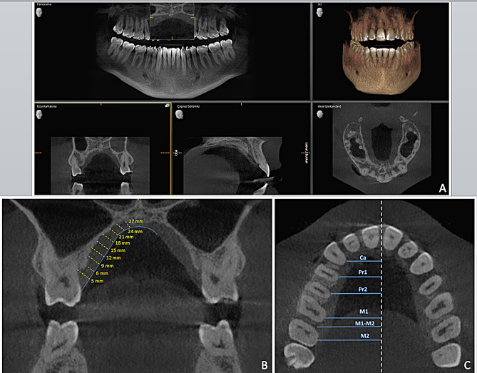
Methods: Cone-beam computed tomography (CBCT) images on the coronal plane were obtained from 60 patients (30 female, 30 male; age range 19-45; mean age 32 ± 11), with 3-mm intervals in the regions of the canine (Ca), first premolar (Pr1), second premolar (Pr2), midpoint between the first and second molars (M1-M2), first molar (M1), second molar (M2) and midpalate.
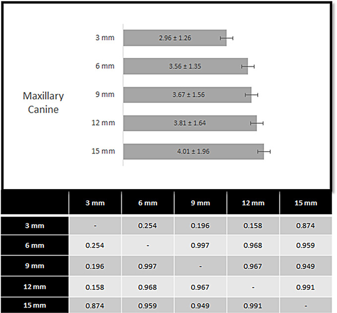
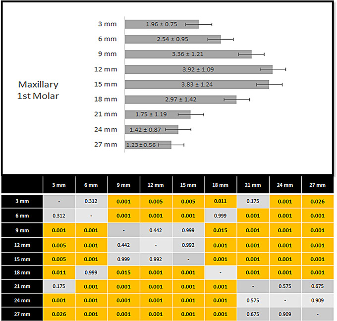
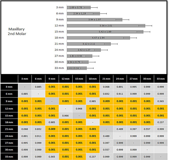
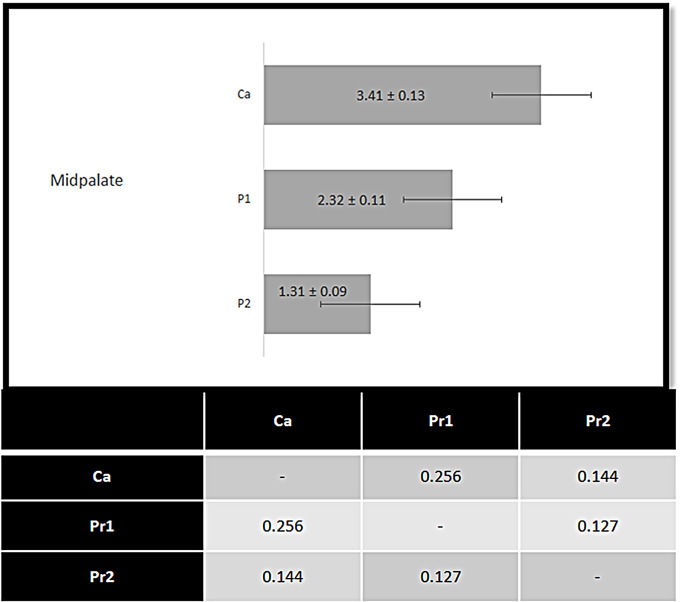
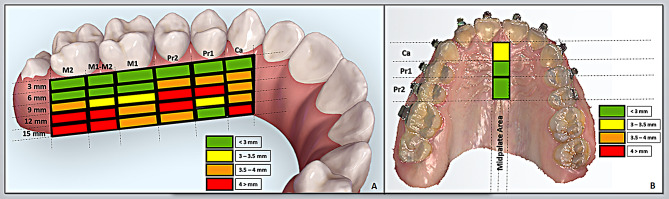
Results: The mucosal thickness measurements between the teeth showed significantly greater thickness in the Ca region at the 3 mm point, in the Pr1 region at the 6 mm point, and in the Pr2 region at the 9 and 12 mm points. At the 9 mm point, the Pr1 region demonstrated greater thickness than the M1-M2 whereas the Pr2 region was thicker than the M1 and M1-M2 regions. At the 12 and 15 mm points, the thickness increased from anterior to posterior: the Pr1 region was thinner than the Pr2, M1, and M2 regions and the Pr2 region was thinner than the M2 region. A schematization was devised for the optimal areas recommended for miniscrews in the palatal region.
Conclusion: The mucosal thickness in the palatal region increases from anterior to posterior except the midpalatal region. Based on the results, in terms of soft tissue, the most suitable place for miniscrew placement is 6 mm from the gingival margin of the teeth and in the median portion of the palate. The findings may guide clinicians in choosing the optimal sites for palatal mini-implants.
Keywords: CBCT; Mini-implants; Miniscrews; Orthodontics; Palatal thickness.
© 2024. The Author(s).
Conflict of interest statement
The authors declare no competing interests.
Figures


References
-
- Maino BG, Mura P, Gianelly A. A retrievable palatal implant for absolute anchorage in orthodontics. World J Orthod. 2004;3:125–34.
-
- Angle EH. Treatment of Malocclusion of Teeth. 7th ed. Philadelphia, PA: SS White Dental Manufacturing Comp; 1907.
-
- Diedrich PA. Critical consideration of various orthodontic anchorage systems. J Orofac Orthop. 1993;54:156–71. - PubMed
-
- Ülgen M. Anomaliler, sefalometri, etioloji, büyüme ve gelişim, tanı. Yeditepe Üniversitesi Yayınları; 2000.
MeSH terms
LinkOut - more resources
Full Text Sources
