. 2024 Sep 30;43(1):268.
doi: 10.1186/s13046-024-03181-x.

UBASH3B-mediated MRPL12 Y60 dephosphorylation inhibits LUAD development by driving mitochondrial metabolism reprogramming

Affiliations

UBASH3B-mediated MRPL12 Y60 dephosphorylation inhibits LUAD development by driving mitochondrial metabolism reprogramming

Xingzhao Ji et al. J Exp Clin Cancer Res. .

Abstract

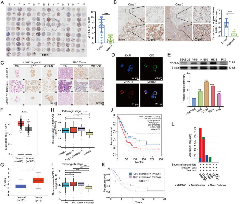
Background: Metabolic reprogramming plays a pivotal role in tumorigenesis and development of lung adenocarcinoma (LUAD). However, the precise mechanisms and potential targets for metabolic reprogramming in LUAD remain elusive. Our prior investigations revealed that the mitochondrial ribosomal protein MRPL12, identified as a novel mitochondrial transcriptional regulatory gene, exerts a critical influence on mitochondrial metabolism. Despite this, the role and regulatory mechanisms underlying MRPL12's transcriptional activity in cancers remain unexplored.

Methods: Human LUAD tissues, Tp53fl/fl;KrasG12D-driven LUAD mouse models, LUAD patient-derived organoids (PDO), and LUAD cell lines were used to explored the expression and function of MRPL12. The posttranslational modification of MRPL12 was analyzed by mass spectrometry, and the oncogenic role of key phosphorylation sites of MRPL12 in LUAD development was verified in vivo and in vitro.

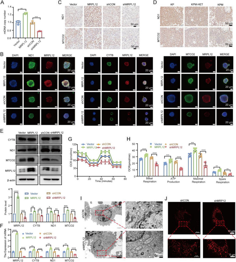
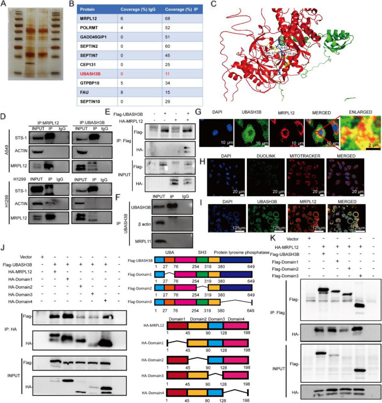
Results: MRPL12 was upregulated in human LUAD tissues, Tp53fl/fl;KrasG12D-driven LUAD tissues in mice, LUAD PDO, and LUAD cell lines, correlating with poor patient survival. Overexpression of MRPL12 significantly promoted LUAD tumorigenesis, metastasis, and PDO formation, while MRPL12 knockdown elicited the opposite phenotype. Additionally, MRPL12 deletion in a Tp53fl/fl;KrasG12D-driven mouse LUAD model conferred a notable survival advantage, delaying tumor onset and reducing malignant progression. Mechanistically, we discovered that MRPL12 promotes tumor progression by upregulating mitochondrial oxidative phosphorylation. Furthermore, we identified UBASH3B as a specific binder of MRPL12, dephosphorylating tyrosine 60 in MRPL12 (MRPL12 Y60) and inhibiting its oncogenic functions. The decrease in MRPL12 Y60 phosphorylation impeded the binding of MRPL12 to POLRMT, downregulating mitochondrial metabolism in LUAD cells. In-depth in vivo, in vitro, and organoid models validated the inhibitory effect of MRPL12 Y60 mutation on LUAD.

Conclusion: This study establishes MRPL12 as a novel oncogene in LUAD, contributing to LUAD pathogenesis by orchestrating mitochondrial metabolism reprogramming towards oxidative phosphorylation (OXPHOS). Furthermore, it confirms Y60 as a specific phosphorylation modification site regulating MRPL12's oncogenic functions, offering insights for the development of LUAD-specific targeted drugs and clinical interventions.

Keywords: LUAD; MRPL12; Metabolic reprogramming; Oxidative phosphorylation; UBASH3B.


Conflict of interest statement

Authors declare no conflict of interest.

Figures

Fig. 1
Fig. 1
Lung-specific ablation of MRPL12 suppresses lung tumorigenesis in Tp53−/−;KrasG12D/+ mice. A Schematic depiction illustrating the induction of lung tumors in a genetically engineered mouse model. B Representative micro-CT images displaying lung morphology in of Tp53fl/fl;KrasG12D (KP), Tp53fl/fl;KrasG12D;MRPL12fl/+(KPM-HET) and Tp53fl/fl;KrasG12D;MRPL12.fl/fl(KPM) mice. C Photomicrographs presenting lung tissues of KP, KPM-HET, and KPM mice following an 18-week inhalation of Adeno-Cre. Tumor lesions are demarcated by arrows. D Quantification of tumor nodules and their respective area (%) in lung tissues from KP, KPM-HET, and KPM mice. E HE-stained lung tissues obtained from KP, KPM-HET, and KPM mice. F Representative Immunohistochemical (IHC) staining of MRPL12 in lung sections from KP mice. G IHC staining of MRPL12 and Ki67 in lung sections from KP, KPM-HET, and KPM mice. H Survival rates of the KP and KPM mice, n = 8 and 7, respectively.*, p < 0.05; **, p < 0.01; ***, p < 0.001
Fig. 2
Fig. 2
MRPL12 are overexpressed in LUAD organoid, tissues, and cells and associated with poor survival. A IHC analysis of MRPL12 was conducted on 75 pairs of tissues from patients with LUAD and their corresponding adjacent tissues. B Representative IHC images illustrating MRPL12 expression in tissues from patients with LUAD. C HE staining and IHC analysis of Cytokeratin 7 (CK7) and MRPL12 in human LUAD patient-derived organoids (PDO) compared to their corresponding original LUAD tissues. D Immunofluorescence (IF) analysis of CK7 and MRPL12 in PDO. Scale bar: 20 μm. E Analysis of MRPL12 expression levels in LUAD cell lines using Western blotting and RT-PCR. F The mRNA level of MRPL12 in LUAD was analyzed in GEPIA2. G Analysis of MRPL12 protein levels in LUAD using the UALCA. H, I Investigation of the correlation between MRPL12 expression levels and T, N, M, and pathological stage using TCGA datasets. J, K Assessment of overall survival in patients with high MRPL12 mRNA (J) or protein (K) levels. L Mutation analysis of the MRPL12 gene in LUAD based on the cBioPortal database. **, p < 0.01; ***, p < 0.001; ****, p < 0.0001
Fig. 3
Fig. 3
MRPL12 facilitates LUAD tumorigenesis. A Images depicting organoids subjected to MRPL12 overexpression and knockdown conditions. Scale bar: 50 μm. B, C Quantification of diameters of transfected organoids. D Analysis of A549 cell-derived xenografts with stable MRPL12 expression (n = 6). E, F Quantification of tumor weights and volumes in the xenografts (n = 6). G Representative IHC staining of MRPL12 and Ki67 in xenograft tissues. Scale bar: 50 μm. H Trans-endothelial migration assays illustrating HUVEC cell-coated inserts with A549 and H1299 tumor cells on top. Transmigration of tumor cells to the bottom was measured after 24 h. I Left: Fluorescence intensity detection in mice for in vivo metastasis evaluation. Right: Corresponding statistical analysis (n = 3). *, p < 0.05; **, p < 0.01; ***, p < 0.001
Fig. 4
Fig. 4
MRPL12 promotes lung tumorigenesis via promoting mitochondrial oxidative phosphorylation. A Analysis of mitochondrial DNA (mtDNA) copy number in A549 cells subjected to MRPL12 knockdown or overexpression. B IF analysis of OXPHOS-related genes and MRPL12 in organoids post-transfection. C, D IHC examination of ND1 and MTCO2 in xenografts and lung tissues derived from genetically engineered mice. E, F Evaluation of protein and mRNA levels of OXPHOS complexes in A549 cells with MRPL12 knockdown or overexpression. G Measurement of oxygen consumption rate (OCR) in A549 cells with MRPL12 knockdown or overexpression using Seahorse XFe96. H Analysis of mitochondrial OXPHOS, including basal respiration, maximal respiration, ATP production, and spare respiratory capacity in MRPL12 knockdown or overexpressed A549 cells. I Electron microscopy analysis of A549 cells for observing mitochondrial morphology. J Three-dimensional Structured Illumination Microscopy (SIM) images of mitochondria stained with MitoTracker Red CMXRos. *, p < 0.05; **, p < 0.01; ***, p < 0.001; ****, p < 0.0001
Fig. 5
Fig. 5
UBASH3B interacts with MRPL12. A Co-immunoprecipitation (Co-IP) of MRPL12 complexes, followed by the identification of MRPL12-binding proteins through combined silver staining and mass spectrometry (MS). B A tabulated summary of mass spectrometry results, delineating the bait protein MRPL12 and its identified binding partners. C Molecular docking of 3D structures demonstrating the interaction between MRPL12 and UBASH3B. D Co-IP assay illustrating the interaction between MRPL12 and UBASH3B in A549 and H1299 cells. E Co-IP assays revealing the interaction between MRPL12 and UBASH3B in HEK293T cells. F Co-IP assay investigating the interaction between MRPL11 and UBASH3B in A549 cells. G IF staining of endogenous MRPL12 (red) and UBASH3B (green) in A549 cells, with nuclei counterstained using DAPI (blue). H Visualization of MRPL12 and UBASH3B interaction in A549 cells using the Duolink proximity ligation assay. I IF staining for MRPL12 (red) and UBASH3B (green) localization in LUAD PDO. J, K Schematic diagrams and IP analyses demonstrating the interaction between MRPL12-UBASH3B in HEK293T cells. Cells were transfected with HA-MRPL12 and Flag-UBASH3B or various mutant constructs
Fig. 6
Fig. 6
UBASH3B dephosphorylates MRPL12 at residue Y60 and inhibits lung tumorigenesis. A-B Evaluation of protein and mRNA levels of OXPHOS complexes in A549 cells subjected to UBASH3B knockdown or overexpression. C IF analysis depicting MRPL12 localization in A549 cells under conditions of UBASH3B knockdown or overexpression. D mtDNA copy number assessment in A549 cells with altered UBASH3B expression. E Analysis of MRPL12 phosphorylation levels in A549 cells with varying UBASH3B expression. F Prediction of MRPL12 phosphorylation sites utilizing the Group-based Prediction System (GPS). G Immunoprecipitation analysis in A549 and HEK293T cells transfected with HA-MRPL12 WT (wild type), HA-MRPL12 Y60A (Y60 mutant), and MRPL12 Y152A (Y152 mutant) for 48 h. H Illustration of structural changes in MRPL12 following phosphorylation. I Highlighting the high conservation of amino acids adjacent to MRPL12 Y60 in diverse species. J Quantification of migration and invasion ability of A549 and H1299 cells after overexpression of MRPL12 with UBASH3B WT or UBASH3B H391A mutant in transwell assays. K Quantification of proliferation ability of A549 and H1299 cells after overexpression of MRPL12 with UBASH3B WT or UBASH3B H391A mutant in EDU assays. *, p < 0.05; **, p < 0.01; ***, p < 0.001; ****, p < 0.0001; ns, p > 0.05
Fig. 7
Fig. 7
MRPL12 Y60 dephosphorylation attenuates mitochondrial biosynthesis via affecting the binding of MRPL12 and POLRMT. A, B Assessment of protein and mRNA levels of OXPHOS complexes in A549 cells expressing either MRPL12 WT or MRPL12 Y60A mutation plasmid. C Quantification of mtDNA copy number in A549 cells overexpressing MRPL12 Y60A or MRPL12 WT. D Analysis of OCR in A549 cells with MRPL12 Y60A or MRPL12 WT overexpression using Seahorse XFe96. E Quantification of basal respiration, ATP production, maximal respiration, and spare respiratory capacity, respectively. F Visualization of mitochondria in MRPL12-knockout A549 cells with re-overexpression of MRPL12 Y60A or MRPL12 WT, stained with MitoTracker Red CMXRos, and observed using SIM. G Electron microscopy analysis of mitochondrial morphology in MRPL12-knockout A549 cells reexpressing MRPL12 Y60A or MRPL12 WT. H, I Co-IP assays in A549 and H1299 cells lysed and immunoprecipitated with MRPL12 antibody, followed by western blot analysis with POLRMT. J Representative immunofluorescence images and quantitation of A549 and H1299 cells overexpressing MRPL12 WT, MRPL12 Y60A, or MRPL12 Y152A plasmid in trans-endothelial migration assays. *, p < 0.05; **, p < 0.01; ***, p < 0.001, ns, p > 0.05
Fig. 8
Fig. 8
MRPL12 Y60 dephosphorylation inhibits tumor formation, metastasis, and organoid formation. A Analysis of xenografts derived from the A549 cell line with stable expression of MRPL12 WT or MRPL12 Y60A mutant (n = 6). B Quantification of tumor weights and volumes (n = 6). C Representative IHC staining depicting the phosphorylation level of MRPL12 Y60 and the protein levels of Ki67, MTCO2, and ND1 in xenograft tissues. D Left: Measurement of fluorescence intensity in mice for in vivo metastasis evaluation. Right: Corresponding statistical analysis. E Visualization of organoids overexpressing the MRPL12 Y60A mutation or MRPL12 WT plasmid, with quantification of their diameters. F IF analysis of OXPHOS components in organoids post-transfection with MRPL12 WT or MRPL12 Y60A mutant. G Verification of the specificity of the customized phosphorylated antibody for MRPL12 Y60 using Dot Blotting. H IHC analysis of organoids using the customized phosphorylated antibodies for MRPL12 Y60. I IHC analysis of LUAD patient samples using the customized phosphorylated antibodies for MRPL12 Y60. **, p < 0.01; ***, p < 0.001; ns, p > 0.05

References

    1. Sung H, Ferlay J, Siegel RL, Laversanne M, Soerjomataram I, Jemal A, Bray F. Global Cancer Statistics 2020: GLOBOCAN Estimates of Incidence and Mortality Worldwide for 36 Cancers in 185 Countries. CA Cancer J Clin. 2021;71(3):209–49. - PubMed
    1. Ma L, Xue X, Zhang X, Yu K, Xu X, Tian X, Miao Y, Meng F, Liu X, Guo S, et al. The essential roles of m(6)A RNA modification to stimulate ENO1-dependent glycolysis and tumorigenesis in lung adenocarcinoma. J Exp Clin Cancer Res. 2022;41(1):36. - PMC - PubMed
    1. Herbst RS, Morgensztern D, Boshoff C. The biology and management of non-small cell lung cancer. Nature. 2018;553(7689):446–54. - PubMed
    1. Howington JA, Blum MG, Chang AC, Balekian AA, Murthy SC. Treatment of stage I and II non-small cell lung cancer: Diagnosis and management of lung cancer, 3rd ed: American College of Chest Physicians evidence-based clinical practice guidelines. Chest. 2013;143(5 Suppl):e278S–e313S. - PubMed
    1. Calvayrac O, Pradines A, Pons E, Mazières J, Guibert N. Molecular biomarkers for lung adenocarcinoma. Eur Respir J. 2017;49(4):1601734. - PubMed