Review
. 2024 Oct;25(10):887-901.
doi: 10.3348/kjr.2024.0307.

Advances in Understanding Hepatocellular Carcinoma Vasculature: Implications for Diagnosis, Prognostication, and Treatment

Affiliations
Review

Advances in Understanding Hepatocellular Carcinoma Vasculature: Implications for Diagnosis, Prognostication, and Treatment

Hyungjin Rhee et al. Korean J Radiol. 2024 Oct.

Abstract

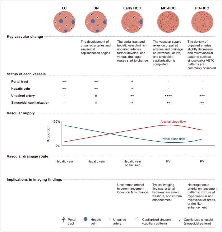
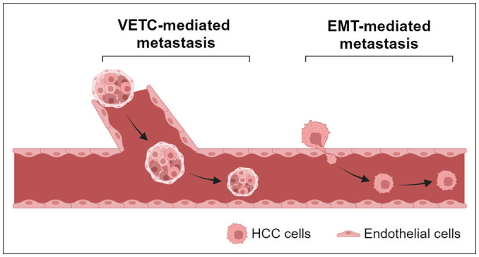
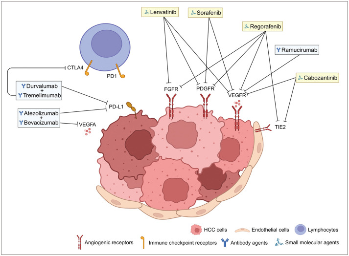
Hepatocellular carcinoma (HCC) progresses through multiple stages of hepatocarcinogenesis, with each stage characterized by specific changes in vascular supply, drainage, and microvascular structure. These vascular changes significantly influence the imaging findings of HCC, enabling non-invasive diagnosis. Vascular changes in HCC are closely related to aggressive histological characteristics and treatment responses. Venous drainage from the tumor toward the portal vein in the surrounding liver facilitates vascular invasion, and the unique microvascular pattern of vessels that encapsulate the tumor cluster (known as a VETC pattern) promotes vascular invasion and metastasis. Systemic treatments for HCC, which are increasingly being used, primarily target angiogenesis and immune checkpoint pathways, which are closely intertwined. By understanding the complex relationship between histopathological vascular changes in hepatocarcinogenesis and their implications for imaging findings, radiologists can enhance the accuracy of imaging diagnosis and improve the prediction of prognosis and treatment response. This, in turn, will ultimately lead to better patient care.

Keywords: Angiogenesis; Hepatocarcinogenesis; Hepatocellular carcinoma; Systemic therapy; Vascular invasion.


Conflict of interest statement

The authors have no potential conflicts of interest to disclose.

Figures

Fig. 1
Fig. 1. Summary of vascular changes and imaging implications in each stage of hepatocarcinogenesis. LC = liver cirrhosis, DN = dysplastic nodule, HCC = hepatocellular carcinoma, MD = moderately differentiated, PD = poorly differentiated, PV = portal vein, VETC = vessels that encapsulate tumor cluster
Fig. 2
Fig. 2. A 53-year-old male with alcoholic hepatitis and early HCC. A-F: Gadoxetic acid-enhanced MRI revealed an approximately 1.5-cm HCC in the right posterior liver (arrows). The lesion exhibits isointensity on the pre-contrast T1-weighted image (A), faint and partial hyperenhancement on the arterial phase (B), absence of washout on the portal phase (C), hypointensity on the hepatobiliary phase (D), mild hyperintensity on the diffusion-weighted image (b = 800) (E), and isointensity on the T2-weighted image (F). G: The lesion was identified as early HCC and displayed a vaguely nodular appearance in the gross specimen (arrows). H: A small number of unpaired arteries (arrowheads) were observed within the lesion (α-smooth muscle actin staining, ×100). I: Compared to the surrounding non-tumor liver (left side), partial sinusoidal capillarization occurred in the early HCC stage (right side) (CD34 staining, ×100). HCC = hepatocellular carcinoma
Fig. 3
Fig. 3. A 61-year-old male with C-viral hepatitis and moderately differentiated HCC. A-C: CT revealed an approximately 2-cm HCC in the dome of the right liver. The lesion demonstrates isoattenuation on the pre-contrast image (A), hyperenhancement in the arterial phase (B), and an enhancing capsule and equivocal washout in the portal phase (C). D: The lesion was identified as moderately differentiated-HCC and exhibited high density of unpaired arteries (arrowheads) within the lesion (α-smooth muscle actin staining, ×200). E: Compared to the surrounding non-tumor liver (left side), completely capillarized capillary pattern microvessels were observed in the HCC (right side) (CD34 staining, ×100). HCC = hepatocellular carcinoma
Fig. 4
Fig. 4. A 57-year-old male with B-viral hepatitis and poorly differentiated HCC. A-F: Gadoxetic acid–enhanced MRI revealed an approximately 4-cm HCC in liver segment 5. The lesion exhibits hypointensity in the pre-contrast T1-weighted image (A), rim enhancement in the arterial phase (B), hypointensity in the portal phase (C) and hepatobiliary phase (D), hyperintensity in the diffusion-weighted image (b = 800) (E), and hyperintensity on the T2-weighted image (F). G: The lesion was identified as poorly differentiated-HCC and displayed vessels that encapsulate the tumor cluster pattern and macrotrabecular patterns (CD34 staining, ±100). HCC = hepatocellular carcinoma
Fig. 5
Fig. 5. Schematic representation of two types of metastases of HCC. HCC = hepatocellular carcinoma, VETC = vessels that encapsulate tumor clusters, EMT = epithelial-mesenchymal transition
Fig. 6
Fig. 6. A 54-year-old man with moderately differentiated HCC with microvascular invasion and a satellite nodule. A-F: Gadoxetic acid-enhanced MRI revealed an approximately 3-cm HCC in liver segment 5/6. The lesion exhibits hypointensity on the pre-contrast T1-weighted image (A), hyperenhancement in the arterial phase (B), hypointensity in the portal phase (C) and hepatobiliary phase (D), hyperintensity in the diffusion-weighted image (b = 800) (E), and hyperintensity on the T2-weighted image (F). The lesion shows arterial phase peritumoral enhancement and peritumoral hypointensity in the hepatobiliary phase (arrows) and a non-smooth tumor margin in the portal and hepatobiliary phases, suggesting a high risk of microvascular invasion. A satellite nodule is observed in the area where the peritumoral change occurred (arrowheads). HCC = hepatocellular carcinoma
Fig. 7
Fig. 7. Inhibition of angiogenic and immune checkpoint pathways by systemic therapeutic agents for HCC. HCC = hepatocellular carcinoma, CTLA4 = cytotoxic T-lymphocyte associated protein 4, PD1 = programmed cell death protein 1, PD-L1 = programmed cell death-ligand 1, VEGFA = vascular endothelial growth factor A, FGFR = fibroblast growth factor receptor, PDGFR = platelet-derived growth factor receptor, VEGFR = vascular endothelial growth factor receptor

Similar articles

Cited by

References

    1. Rumgay H, Arnold M, Ferlay J, Lesi O, Cabasag CJ, Vignat J, et al. Global burden of primary liver cancer in 2020 and predictions to 2040. J Hepatol. 2022;77:1598–1606. - PMC - PubMed
    1. Kim DY. Changing etiology and epidemiology of hepatocellular carcinoma: Asia and worldwide. J Liver Cancer. 2024;24:62–70. - PMC - PubMed
    1. Rumgay H, Ferlay J, de Martel C, Georges D, Ibrahim AS, Zheng R, et al. Global, regional and national burden of primary liver cancer by subtype. Eur J Cancer. 2022;161:108–118. - PubMed
    1. Korean Liver Cancer Association (KLCA) and National Cancer Center (NCC) Korea. 2022 KLCA-NCC Korea practice guidelines for the management of hepatocellular carcinoma. Korean J Radiol. 2022;23:1126–1240. - PMC - PubMed
    1. Singal AG, Llovet JM, Yarchoan M, Mehta N, Heimbach JK, Dawson LA, et al. AASLD practice guidance on prevention, diagnosis, and treatment of hepatocellular carcinoma. Hepatology. 2023;78:1922–1965. - PMC - PubMed

Publication types

MeSH terms

Grants and funding