

Imaging characteristics of uterine smooth muscle tumors of uncertain malignant potential: a case report and literature review
- PMID: 39344823
- PMCID: PMC11459561
- DOI: 10.1177/03000605241279183
Imaging characteristics of uterine smooth muscle tumors of uncertain malignant potential: a case report and literature review
Abstract
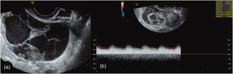
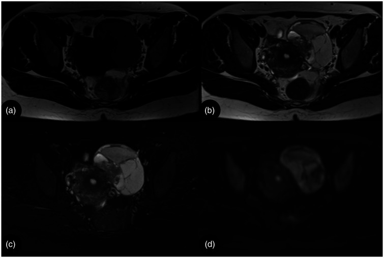
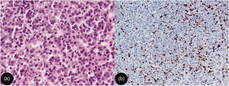
Uterine smooth muscle tumors of uncertain malignant potential (STUMPs) are rare tumors of the uterine myometrium that are often misdiagnosed, owing to limited knowledge of their characteristics on ultrasonography (US) and magnetic resonance imaging (MRI). We report a woman in her mid-30s who was hospitalized because of a pelvic tumor. A 6-cm mass was found in her lower left abdomen. US and MRI revealed a well-demarcated mass in the left adnexal area, with both cystic and solid elements, visible blood flow within the septa, a strong signal across >50% of the volume on T2-weighted imaging (T2WI), and a strong signal on diffusion-weighted imaging (DWI). After hysterectomy and bilateral salpingectomy, immunohistochemical examination confirmed STUMP. A review of the literature revealed characteristic imaging features of STUMP. Ultrasonography reveals STUMP as a solitary, well-circumscribed lesion with isoechoic or mixed echogenicity, the absence of posterior shadowing, and variations in blood flow. STUMP is characterized by strong signal intensity on T2WI, small areas of strong signal on T1WI, and non-enhancing cystic areas on contrast-enhanced MRI scans. Early diagnosis is crucial for the management and treatment of STUMP, and here we have summarized the imaging features of the lesion, thereby providing a valuable diagnostic reference.
Keywords: Smooth muscle tumor of uncertain malignant potential (STUMP); case report; imaging characteristic; magnetic resonance imaging; ultrasonography; uterus.
Conflict of interest statement
Declaration of conflicting interestsThe authors declare that there is no conflict of interest.
Figures
Similar articles
-
Value of ultrasonography and magnetic resonance imaging for the characterization of uterine mesenchymal tumors.Acta Obstet Gynecol Scand. 2014 Mar;93(3):261-8. doi: 10.1111/aogs.12325. Acta Obstet Gynecol Scand. 2014. PMID: 24372487
-
Ultrasound features of highly vascularized uterine myomas (uterine smooth muscle tumors) and correlation with histopathology.Ultrasound Obstet Gynecol. 2022 Aug;60(2):269-276. doi: 10.1002/uog.24855. Ultrasound Obstet Gynecol. 2022. PMID: 35018681
-
Uterine smooth muscle tumors of uncertain malignant potential: a case presentation.Int J Clin Oncol. 2011 Oct;16(5):592-5. doi: 10.1007/s10147-010-0172-4. Epub 2011 Jan 12. Int J Clin Oncol. 2011. PMID: 21225306
-
Smooth Muscle Tumor of Uncertain Malignant Potential (STUMP): A Systematic Review of the Literature in the Last 20 Years.Curr Oncol. 2024 Sep 5;31(9):5242-5254. doi: 10.3390/curroncol31090388. Curr Oncol. 2024. PMID: 39330016 Free PMC article.
-
Diversity of imaging features of ovarian sclerosing stromal tumors on MRI and PET-CT: a case report and literature review.J Ovarian Res. 2018 Dec 20;11(1):101. doi: 10.1186/s13048-018-0473-1. J Ovarian Res. 2018. PMID: 30572921 Free PMC article. Review.
References
-
- Picerno TM, Wasson MN, Gonzalez Rios AR, et al.. Morcellation and the incidence of occult uterine malignancy: a dual-institution review. Int J Gynecol Cancer 2016; 26: 149–155. - PubMed
-
- Amant F, Coosemans A, Debiec-Rychter M, et al.. Clinical management of uterine sarcomas. Lancet Oncol 2009; 10: 1188–1198. - PubMed
Publication types
MeSH terms
LinkOut - more resources
Full Text Sources
Medical
