

Autologous platelet concentrates in alveolar ridge preservation: A systematic review with meta-analyses
- PMID: 39345008
- PMCID: PMC11808431
- DOI: 10.1111/prd.12609
Autologous platelet concentrates in alveolar ridge preservation: A systematic review with meta-analyses
Abstract
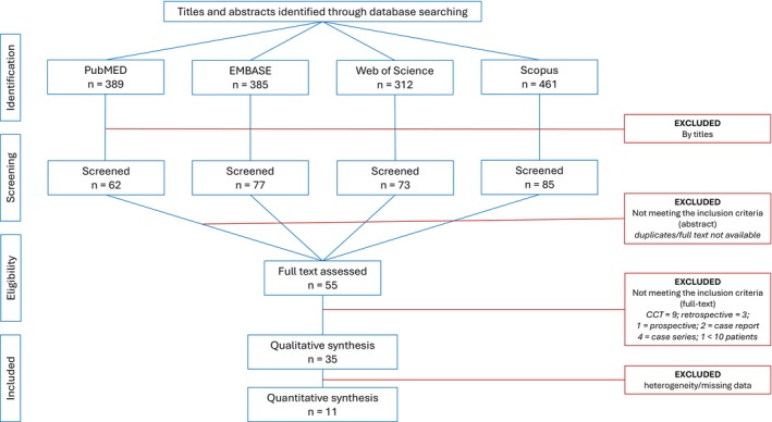
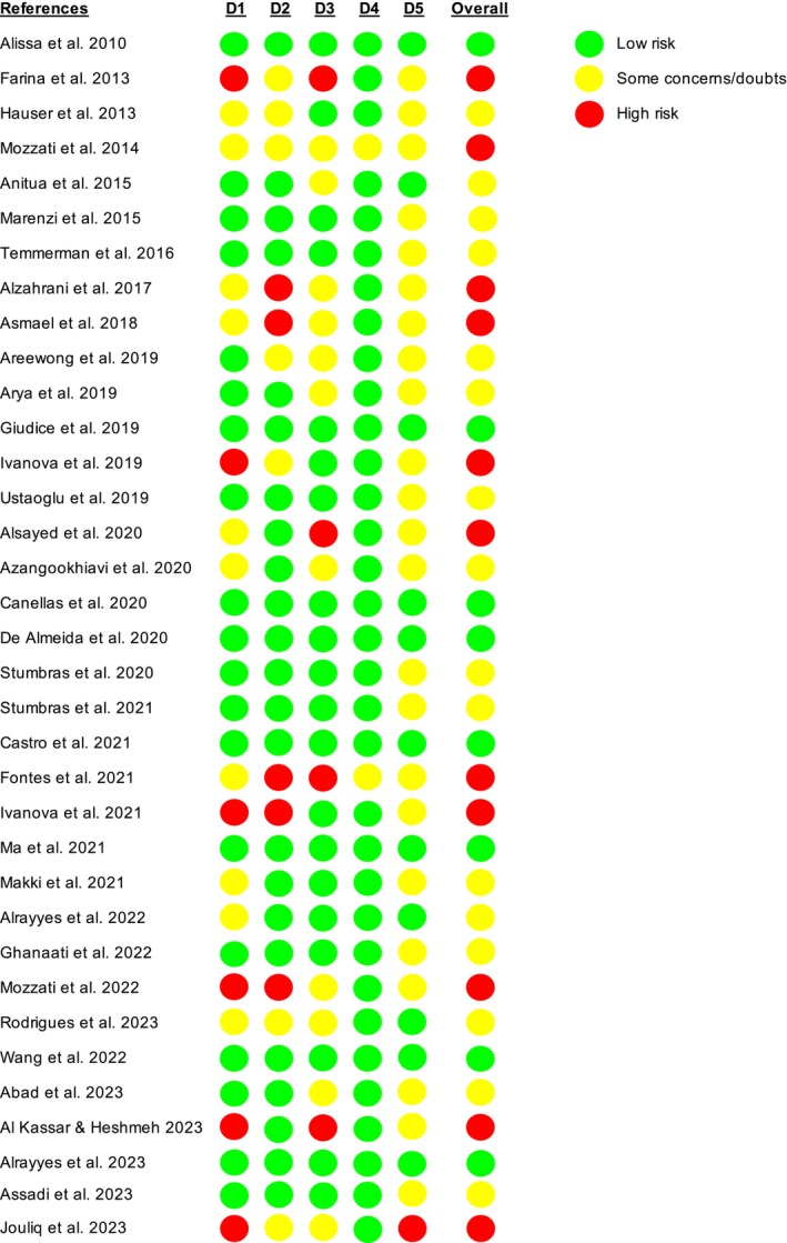
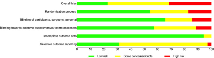
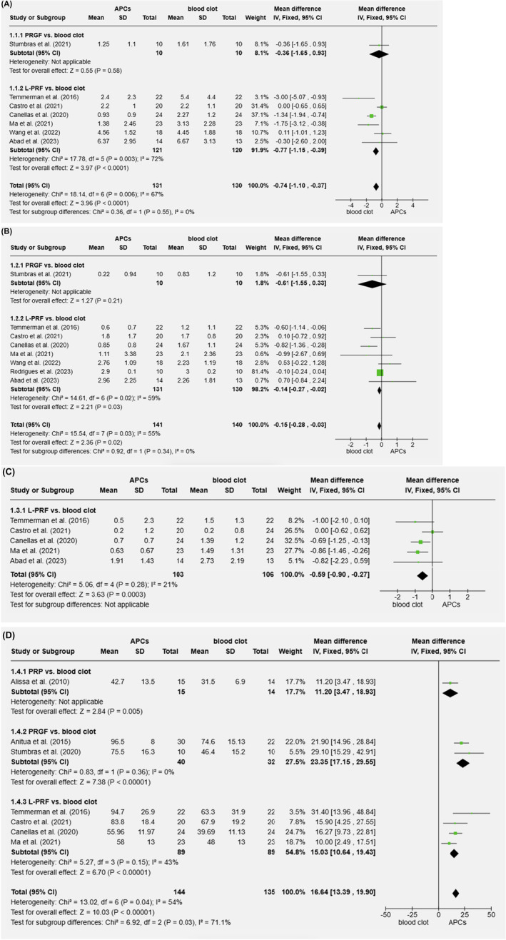
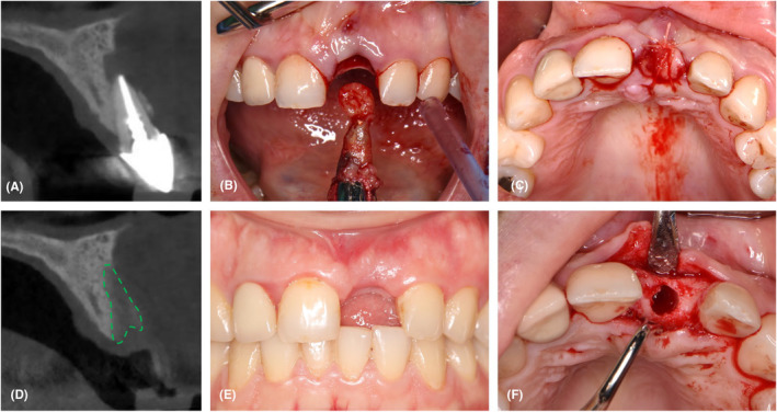
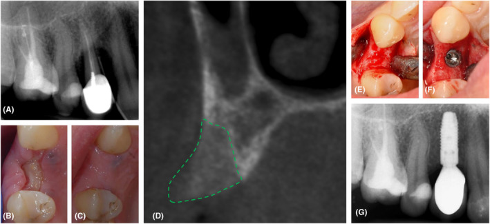
In order to evaluate the therapeutic advantages of various autologous platelet concentrates (APC) as a single biomaterial during alveolar ridge preservation (ARP), a systematic review with meta-analyses was conducted. PubMed, EMBASE, Web of Science, and Scopus were screened for randomized controlled trials (RCTs) that were released prior to 2024. The selected papers compared an APC with either unassisted healing (blood clot) or another biomaterial during ARP (third molars were not included). The outcome parameters included alveolar bone dimension alterations, soft tissue healing, and post-op pain intensity. The search yielded 35 papers (33 studies), one applying platelet-rich plasma (PRP), six using plasma rich in growth factors (PRGF), and 28 using leukocyte- and platelet-rich fibrin (L-PRF). These studies showed a large heterogeneity (e.g., outcome parameters, timing, surgical approach, and inclusion criteria), which hindered drawing strong conclusions. In most studies, however, ARP with PRP, PRGF, and L-PRF alone produced faster soft tissue healing, less post-extraction pain, less alveolar ridge resorption, more socket bone fill, and a higher bone density when compared to unassisted (spontaneous) healing. The ultimate benefit appears to be significantly influenced by the surgical approach. Limited literature exists comparing APC with other biomaterials for ARP, resulting in inconclusive data. APC application for ARP is a promising strategy to improve soft and hard tissue healing and reduce post-extraction pain.
Keywords: alveolar ridge preservation; autologous platelet concentrates; extraction socket; leukocyte‐ and platelet‐rich fibrin; plasma rich in growth factor; platelet rich fibrin; platelet rich plasma..
© 2024 The Author(s). Periodontology 2000 published by John Wiley & Sons Ltd.
Conflict of interest statement
All authors declare that they have no conflict of interest in relation to this chapter. The Department of Periodontology at the KU Leuven has received research support from different implant companies including Dentsply Sirona, Straumann and Henry Schein. Drs. Yu received support from the China Scholarship Council (File No. 202206170027).
Figures
Similar articles
-
Effect of the use of platelet concentrates on new bone formation in alveolar ridge preservation: a systematic review, meta-analysis, and trial sequential analysis.Clin Oral Investig. 2023 Aug;27(8):4131-4146. doi: 10.1007/s00784-023-05126-8. Epub 2023 Jul 13. Clin Oral Investig. 2023. PMID: 37439800 Free PMC article.
-
Hard and soft tissue changes following alveolar ridge preservation: a systematic review.Clin Oral Implants Res. 2017 Aug;28(8):982-1004. doi: 10.1111/clr.12911. Epub 2016 Jul 26. Clin Oral Implants Res. 2017. PMID: 27458031
-
Autologous platelet concentrates after third molar extraction: A systematic review.Periodontol 2000. 2025 Feb;97(1):131-152. doi: 10.1111/prd.12600. Epub 2024 Sep 24. Periodontol 2000. 2025. PMID: 39318055
-
Evaluation of Effectiveness of Nanocrystalline Hydroxyapatite and Demineralized Bone Matrix Combined with Titanium-platelet Rich Fibrin for Ridge Preservation: A Randomized Controlled Clinical Trial.J Contemp Dent Pract. 2024 Nov 1;25(11):1069-1076. doi: 10.5005/jp-journals-10024-3786. J Contemp Dent Pract. 2024. PMID: 39905614 Clinical Trial.
-
Clinical outcomes of using operating microscope for alveolar ridge preservation: A randomized controlled trial.J Periodontol. 2025 Mar;96(3):230-240. doi: 10.1002/JPER.24-0081. Epub 2024 Oct 15. J Periodontol. 2025. PMID: 39403776 Free PMC article. Clinical Trial.
Cited by
-
Applications of Platelet-Rich Fibrin (PRF) Membranes Alone or in Combination with Biomimetic Materials in Oral Regeneration: A Narrative Review.Biomimetics (Basel). 2025 Mar 11;10(3):172. doi: 10.3390/biomimetics10030172. Biomimetics (Basel). 2025. PMID: 40136826 Free PMC article. Review.
-
Instructions for the use of L-PRF in different clinical indications.Periodontol 2000. 2025 Feb;97(1):420-432. doi: 10.1111/prd.12564. Epub 2024 May 27. Periodontol 2000. 2025. PMID: 38803016 Free PMC article. Review.
-
Efficacy of collagenated bone substitutes for bone regeneration in two-wall-damaged extraction sockets without barrier membranes.Clin Oral Investig. 2025 Mar 22;29(4):201. doi: 10.1007/s00784-025-06281-w. Clin Oral Investig. 2025. PMID: 40119996
-
Titanium-prepared platelet-rich fibrin enhances alveolar ridge preservation: a randomized controlled clinical and radiographic study.Sci Rep. 2025 Jul 5;15(1):24065. doi: 10.1038/s41598-025-09528-4. Sci Rep. 2025. PMID: 40617944 Free PMC article. Clinical Trial.
-
Introduction and overview on Autogenous Platelet Concentrates.Periodontol 2000. 2025 Feb;97(1):7-15. doi: 10.1111/prd.12607. Epub 2024 Sep 11. Periodontol 2000. 2025. PMID: 39258791 Free PMC article.
References
-
- Tan WL, Wong TL, Wong MC, Lang NP. A systematic review of post‐extractional alveolar hard and soft tissue dimensional changes in humans. Clin Oral Implants Res. 2012;23(Suppl 5):1‐21. - PubMed
-
- Huynh‐Ba G, Pjetursson BE, Sanz M, et al. Analysis of the socket bone wall dimensions in the upper maxilla in relation to immediate implant placement. Clin Oral Implants Res. 2010;21(1):37‐42. - PubMed
-
- Couso‐Queiruga E, Stuhr S, Tattan M, Chambrone L, Avila‐Ortiz G. Post‐extraction dimensional changes: a systematic review and meta‐analysis. J Clin Periodontol. 2021;48(1):126‐144. - PubMed
-
- Schropp L, Wenzel A, Kostopoulos L, Karring T. Bone healing and soft tissue contour changes following single‐tooth extraction: a clinical and radiographic 12‐month prospective study. Int J Periodontics Restorative Dent. 2003;23(4):313‐323. - PubMed
-
- Van der Weijden F, Dell'Acqua F, Slot DE. Alveolar bone dimensional changes of post‐extraction sockets in humans: a systematic review. J Clin Periodontol. 2009;36(12):1048‐1058. - PubMed
Publication types
MeSH terms
LinkOut - more resources
Full Text Sources
Research Materials
Miscellaneous
