. 2024 Oct 29;9(10):e0065524.
doi: 10.1128/msphere.00655-24. Epub 2024 Sep 30.

Pneumolysin contributes to dysfunction of nasal epithelial barrier for promotion of pneumococcal dissemination into brain tissue

Affiliations

Pneumolysin contributes to dysfunction of nasal epithelial barrier for promotion of pneumococcal dissemination into brain tissue

Yuki Takahara et al. mSphere. .

Abstract

Streptococcus pneumoniae is one of the major pathogens responsible for bacterial meningitis and neurological sequelae. The present study was conducted to identify a non-hematogenous route used by S. pneumoniae to gain access to brain tissue without causing bacteremia or pneumonia, as well as bacterial and host factors involved in this process. To investigate the molecular mechanisms and dissemination pathways of pneumococcal infection in brain tissue, mice were intranasally inoculated with S. pneumoniae strain EF3030, a clinical isolate from a patient with otitis media. Pneumococci were isolated from the frontal olfactory bulb, caudal cerebrum, and cerebellum, with neither bacteremia nor pneumonia observed in the present model. Immunostaining imaging revealed the presence of S. pneumoniae organisms in olfactory nerve fibers. Knockout of the ply gene encoding pneumolysin (PLY) markedly compromised the ability of the bacterial organisms to disseminate into brain tissue, whereas the dissemination efficiency of the complemented strain was restored to nearly the same level as the wild type. Notably, distinct upregulation of Gli1 and Snail1, which are involved in the transcriptional repression of junctional proteins, along with downregulation of E-cadherin, was detected in nasal lavage samples from mice infected with the wild-type or complemented strain, but not in those from mice infected with the ply mutant. Taken together, the present findings indicate that PLY induces Gli1-Snail1-dependent dysfunction of the nasal epithelial barrier, thus allowing pneumococcal dissemination to brain tissue that occurs in a non-hematogenous manner.IMPORTANCEBacterial meningitis, considered to be caused by bacteremia, can lead to blood-brain barrier disruption and bacterial dissemination into the central nervous system. Despite the availability of intravenously administered antibiotics with cerebrospinal fluid transferability, bacterial meningitis remains associated with high rates of morbidity and mortality. Here, we utilized Streptococcus pneumoniae strain EF3030, clinically isolated from otitis media, for the construction of a murine infection model to investigate the molecular mechanisms by which nasally colonized pneumococci disseminate into brain tissue. The obtained findings indicate that pneumolysin (PLY) induces Gli1-Snail1-dependent dysfunction of the nasal epithelial barrier, which facilitates pneumococcal dissemination to brain tissue in a non-hematogenous manner. Our results support the existence of an alternative route by which S. pneumoniae can reach the central nervous system and indicate the need for the development of novel therapeutic strategies, which would be an important contribution to the clinical management of bacterial meningitis.

Keywords: Streptococcus pneumoniae; bacterial meningitis; non-hematogenous route; pneumolysin.


Conflict of interest statement

The authors declare no conflict of interest.

Figures
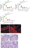
Fig 1
Fig 1
S. pneumoniae colonized in nasopharynx disseminates into brain tissue through a non-hematogenous route. Mice were intranasally infected with S. pneumoniae EF3030 (WT, 1 × 10⁷ CFU in 10 μL of phosphate-buffered saline [PBS]). (A) Bacterial burden in the nasopharynx (nose), olfactory bulb (OB), cerebrum (CB), cerebellum (CE), lung tissues, and blood was assessed at days 1, 3, and 7 following infection. Dots indicate individual mice. Horizontal lines show median values and interquartile range. Dotted line shows the limit of detection (LOD). Data shown are representative of at least three separate experiments, with 10 mice used per group. Statistically significant differences were evaluated using one-way analysis of variance (ANOVA), followed by Tukey’s multiple-comparison test. *P < 0.0001, **P < 0.001. (B) Brain section obtained from mice nasally infected with EF3030 were subjected to immunofluorescence staining at day 3 following infection. Pneumococci were labeled with an anti-serotype 19 capsule antibody, followed by incubation with an Alexa Fluor 488-conjugated antibody. The olfactory nerve was labeled with an anti-olfactory marker protein and Alexa Fluor 594-conjugated antibodies, and cell nuclei were stained with 4′,6-diamidino-2-phenylindole (DAPI). Obtained tissue sections were analyzed using confocal laser microscopy. Arrowheads indicate pneumococcal association with olfactory neurons. Nasal cavity (NC) and olfactory epithelium (OE). Data shown are representative of at least three separate experiments. (C) Lung tissues obtained from non-infected and infected mice at day 3 following infection were subjected to hematoxylin and eosin (HE) staining. Magnified images of boxed areas in upper panels are shown in lower panels. Data shown are representative of at least three separate experiments, with three mice used per group.
Fig 2
Fig 2
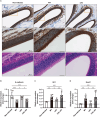
PLY induces Gli1- and Snail1-dependent dysfunction of the nasal epithelial barrier. (A) Brain tissues were obtained from mice infected with S. pneumoniae EF3030 (WT, 1 × 10⁷ CFU in 10 μL of PBS), or an isogenic ply knockout strain (Δply), then subjected to immunohistochemistry using an anti-E-cadherin antibody and HE staining performed on day 3 following infection. Magnified images of boxed areas in the upper panels are presented in the middle panels. Data shown are representative of at least three separate experiments, with five mice per group. (B–D) Transcriptional levels of genes encoding (B) E-cadherin, (C) Gli1, and (D) Snail1 in nasal lavage fluid were determined at 3 days following infection using real-time RT-PCR. A gapdh transcript served as the internal control. Dots indicate individual mice. Values for expression ratios pooled from three independent examinations are presented as the mean ± SD. Transcriptional levels are presented as relative expression normalized to that seen in non-infected tissues. *P < 0.05, **P < 0.01, ***P < 0.001.
Fig 3
Fig 3
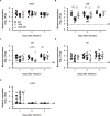
PLY contributes to pneumococcal dissemination into the brain tissue. Mice were intranasally infected with S. pneumoniae strains (1 × 10⁷ CFU in 10 μL of PBS), then bacterial burden in the (A) nasopharynx (nose), (B) OB, (C) CB, (D) CE, and (E) lung tissues was assessed at 3 days following infection. Dots indicate individual mice. Horizontal lines show median values and interquartile range. Dotted line shows the limit of detection (LOD). Data shown are representative of at least three separate experiments, with 10 mice used per group. Statistically significant differences were evaluated using one-way ANOVA, followed by Tukey’s multiple-comparison test. *P < 0.05, **P < 0.01, ***P < 0.001.
Fig 4
Fig 4
Pore-forming activity of PLY critical for pneumococcal dissemination into the brain tissue. Mice were intranasally administered with the vehicle, recombinant PLY (rPLY-WT), or a non-pore-forming toxoid with the W433F point mutation (rPLY-W433F) 24 h before infection with the ply mutant. On day 3 following infection, bacterial colonization in the (A) nasopharynx (nose), (B) OB, (C) CB, and (D) CE tissues was assessed. Dots indicate individual mice. Horizontal lines show median values and interquartile range. Dotted line shows the limit of detection (LOD). Data shown are representative of at least three separate experiments, with 10 mice used per group. Statistically significant differences were evaluated using one-way ANOVA, followed by Tukey’s multiple-comparison test. *P < 0.05, **P < 0.001.
Fig 5
Fig 5
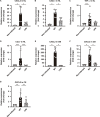
PLY-dependent inflammatory response in brain tissues. (A–D) Real-time RT-PCR assays were performed on day 3 following infection to analyze transcriptional levels of genes encoding (A) CXCL2, (B) CXCL1, (C) TNF-α, and (D) CCL7 in the nasal lavage fluid (NL) of mice infected with S. pneumoniae strains. (E–G), Transcriptional levels of the CXCL2 gene in (E) OB, (F) CB, and (G) CE were assessed at 3 days following infection. A gapdh transcript served as the internal control. Dots indicate individual mice. Values for expression ratios pooled from three independent examinations are presented as the mean ± SD. Transcriptional levels are presented as relative expression normalized to that of non-infected tissues. *P < 0.0001, **P < 0.001, ***P < 0.05.

Similar articles

References

    1. Brouwer MC, Tunkel AR, van de Beek D. 2010. Epidemiology, diagnosis, and antimicrobial treatment of acute bacterial meningitis. Clin Microbiol Rev 23:467–492. doi:10.1128/CMR.00070-09 - DOI - PMC - PubMed
    1. Oordt-Speets AM, Bolijn R, van Hoorn RC, Bhavsar A, Kyaw MH. 2018. Global etiology of bacterial meningitis: a systematic review and meta-analysis. PLoS One 13:e0198772. doi:10.1371/journal.pone.0198772 - DOI - PMC - PubMed
    1. Butler JC, Schuchat A. 1999. Epidemiology of pneumococcal infections in the elderly. Drugs Aging 15 Suppl 1:11–19. doi:10.2165/00002512-199915001-00002 - DOI - PubMed
    1. Fedson DS, Anthony J, Scott G. 1999. The burden of pneumococcal disease among adults in developed and developing countries: what is and is not known. Vaccine (Auckl) 17:S11–S18. doi:10.1016/S0264-410X(99)00122-X - DOI - PubMed
    1. WHO . 2014. The world health report 2014. Geneva

Substances

LinkOut - more resources