. 2024 Oct 22;98(10):e0049724.
doi: 10.1128/jvi.00497-24. Epub 2024 Sep 30.

Duck circovirus regulates the expression of duck CLDN2 protein by activating the MAPK-ERK pathway to affect its adhesion and infection

Affiliations

Duck circovirus regulates the expression of duck CLDN2 protein by activating the MAPK-ERK pathway to affect its adhesion and infection

Mingyue Shen et al. J Virol. .

Abstract

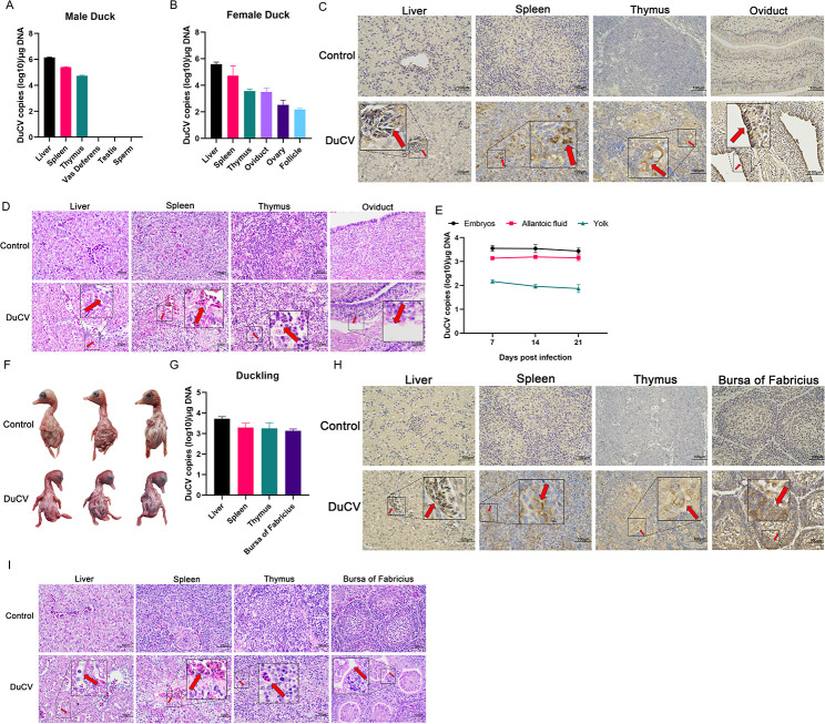
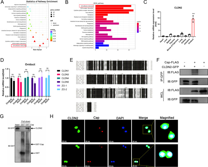
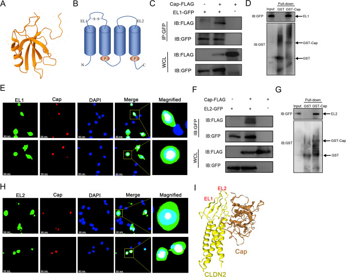
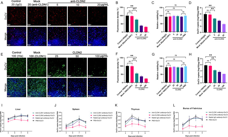
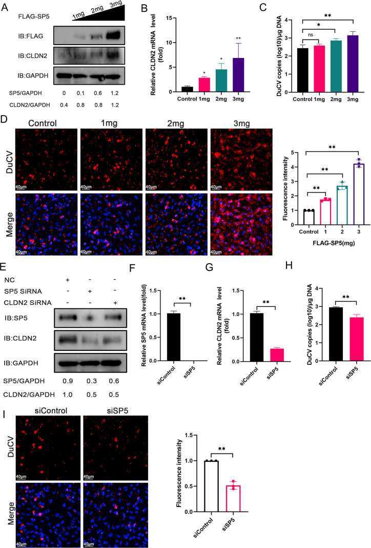
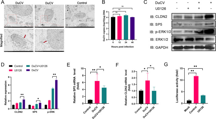
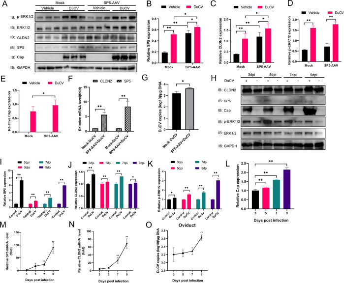
Duck circovirus (DuCV) is widely recognized as a prominent virus in China's duck farming industry, known for its ability to cause persistent infections and significant immunosuppression, which can lead to an increased susceptibility to secondary infections, posing a significant threat to the duck industry. Moreover, clinical evidence also indicates the potential vertical transmission of the virus through duck embryos to subsequent generations of ducklings. However, the limited availability of suitable cell lines for in vitro cultivation of DuCV has hindered further investigation into the molecular mechanisms underlying its infection and pathogenicity. In this study, we observed that oral DuCV infection in female breeding ducks can lead to oviduct, ovarian, and follicular infections. Subsequently, the infection can be transmitted to the fertilized eggs, resulting in the emergence of virus-carrying ducklings upon hatching. In contrast, the reproductive organs of male breeding ducks were unaffected by the virus, thus confirming that vertical transmission of DuCV primarily occurs through infection in female breeding ducks. By analyzing transcriptome sequencing data from the oviduct, we focused on claudin-2, a gene encoding the tight junction protein CLDN2 located on the cell membrane, which showed significantly increased expression in DuCV-infected oviducts of female breeding ducks. Notably, CLDN2 was confirmed to interact with the unique structural protein of DuCV, namely capsid protein (Cap), through a series of experimental approaches including co-immunoprecipitation (co-IP), GST pull-down, immunofluorescence, and adhesion-blocking assays. Furthermore, we demonstrated that the Cap protein binds to the extracellular loop structural domains EL1 and EL2 of CLDN2. Subsequently, by constructing a series of truncated bodies of the CLDN2 promoter region, we identified the transcription factor SP5 for CLDN2. Moreover, we found that DuCV infection triggers the activation of the MAPK-ERK signaling pathway in DEF cells and ducks, leading to an upregulation of SP5 and CLDN2 expression. This process ultimately leads to the transportation of mature CLDN2 to the cell surface, thereby facilitating increased virus adherence to the target organs. In conclusion, we discovered that DuCV utilizes host CLDN2 proteins to enhance adhesion and infection in oviducts and other target organs. Furthermore, we elucidated the signaling pathways involved in the interaction between DuCV Cap proteins and CLDN2, which provides valuable insights into the molecular mechanism underlying DuCV's infection and vertical transmission.

Importance: Although duck circovirus (DuCV) poses a widespread infection and a serious hazard to the duck industry, the molecular mechanisms underlying DuCV infection and transmission remain elusive. We initially demonstrated vertical transmission of DuCV through female breeding ducks by simulating natural infection. Furthermore, a differentially expressed membrane protein CLDN2 was identified on the DuCV-infected oviduct of female ducks, and its extracellular loop structural domains EL1 and EL2 were identified as the interaction sites of DuCV Cap proteins. Moreover, the binding of DuCV Cap to CLDN2 triggered the intracellular MAPK-ERK pathway and activated the downstream transcription factor SP5. Importantly, we demonstrated that intracellular Cap also interacts with SP5, leading to upregulation of CLDN2 transcription and facilitating enhanced adherence of DuCV to target tissue, thereby promoting viral infection and transmission. Our study sheds light on the molecular mechanisms underlying vertical transmission of DuCV, highlighting CLDN2 as a promising target for drug development against DuCV infection.

Keywords: MAPK-ERK pathway; adhesion; claudin-2; duck circovirus; vertical transmission.


Conflict of interest statement

The authors declare no conflict of interest.

Figures

Fig 1
Fig 1
Identification of vertical transmission of DuCV to ducklings through female breeding ducks. The infection process in breeding ducks is described in the Materials and Methods section. (A) Viral copies in the liver, spleen, thymus, vas deferens, testes, and sperm of male breeding ducks were determined using Q-PCR. (B) Viral copies in the liver, spleen, thymus, ovary, oviduct, and follicle of female breeding ducks were determined using Q-PCR. (C) IHC of pathological tissue sections of liver, spleen, thymus, and oviduct in female breeding ducks. The DuCV and Control groups used rabbit anti-Cap protein polyclonal antibody as the primary antibody. A mock group using negative rabbit serum as the primary antibody was placed in the supplemental material. (D) Hematoxylin and eosin (H&E) staining of pathological tissue sections of liver, spleen, thymus, and oviduct of female breeding ducks. (E) Dynamics of viral copy numbers in allantoic fluid, yolk, and embryo body of 21 virus-positive duck embryos determined by Q-PCR. (F) Pathologic changes in duck embryos infected with DuCV. (G) Viral copies in the liver, spleen, thymus, and bursa of 1-day-old ducklings hatched from duck embryos determined by Q-PCR. (H) IHC of pathological tissue sections of liver, spleen, thymus, and bursa in 1-day-old ducklings hatched from duck embryos. The DuCV and Control groups used rabbit anti-Cap protein polyclonal antibody as the primary antibody. A mock group using negative rabbit serum as the primary antibody was placed in the supplemental material. (I) Hematoxylin and eosin (H&E) staining of pathological tissue sections of liver, spleen, thymus, and bursa of 1-day-old ducklings hatched from duck embryos.
Fig 2
Fig 2
Identification of the interaction between DuCV Cap protein and transmembrane protein CLDN2. (A and B) Transcriptome sequencing data from the oviducts of female breeding ducks were analyzed to identify KEGG-enriched pathways associated with cell adhesion molecules. (C) mRNA levels of CLDN2 in different tissues of female breeding ducks quantified by Q-PCR. (D) mRNA levels of CLDN1, CLDN3, CLDN4, CLDN5, ZO-1, and ZO-2 in the oviduct of female breeding ducks quantified by Q-PCR. (E) Comparison of duck CLDN2 amino acid sequences with those of other species (pig, mouse, human, chicken, bird, and rat). (F) Cloning of Cap sequence into pCAGGS vector and CLDN2 sequence into pEGFP-C1 vector. Cap-FLAG and CLDN2-GFP recombinant plasmids were transfected in HEK 293T cells, whole cell lysates (WCL) were precipitated with anti-GFP monoclonal antibody and separated using magnetic beads, and Western blotting was performed using anti-FLAG and anti-GFP antibodies. (G) CLDN-GFP recombinant plasmid was transfected into HEK 293T cells, and the Cap sequence was cloned into the GST6P-1 vector. The Cap-GST protein was expressed in BL21 (DE3) cells, followed by cell lysate separation using GST affinity. Eluted proteins were subsequently analyzed by Western blotting using anti-GST and anti-GFP antibodies. (H) Co-localization of Cap and CLDN2 in HEK 293T cells detected by confocal laser microscopy.
Fig 3
Fig 3
Identification of interaction between the DuCV cap protein and two extracellular loop structures derived from the CLDN2 protein. (A and B) Prediction of the spatial architecture of the CLDN2 protein utilizing the InterPro network. (C) Cap-FLAG and EL1-GFP recombinant plasmids were transfected in HEK 293T cells. Co-IP assay was performed after precipitation of whole cell lysates (WCL) with the anti-GFP monoclonal antibody, and Western blotting was performed using anti-FLAG and anti-GFP antibodies. (D) EL1-GFP recombinant plasmid was transfected into HEK 293T cells to express Cap-GST protein in BL21 (DE3). Cell lysates were separated by GST affinity, and eluted proteins were subsequently analyzed by Western blotting using anti-GST and anti-GFP antibodies. (E) Co-localization of Cap and EL1 in HEK 293T cells, revealed through confocal laser microscopy. (F) Cap-FLAG and EL2-GFP recombinant plasmids were transfected in HEK 293T cells, whole cell lysates (WCL) were precipitated with anti-GFP monoclonal antibody for co-IP assay, and Western blotting was performed using anti-FLAG and anti-GFP antibodies. (G) EL2-GFP recombinant plasmid was transfected into HEK 293T cells. Cap-GST protein was expressed in BL21 (DE3), followed by cell lysate separation using GST affinity. Eluted proteins were subsequently analyzed by Western blotting using anti-GST and anti-GFP antibodies. (H) Co-localization of Cap and EL2 in HEK 293T cells, revealed through confocal laser microscopy. (I) Prediction of the interaction between Cap and two structural domains, EL1 and EL2, of CLDN2 using the PPI network (http://hdock.phys.hust.edu.cn/). The error line represents the SD of the three test replicates. Asterisks denote statistically significant differences compared to the control group: *, P < 0.05; **, P < 0.01; ns, no significant difference.
Fig 4
Fig 4
Identification that blocking DuCV adhesion to CLDN2 reduces viral infection. (A) Viral fluorescence densities on DEF cells incubated with different concentrations of rabbit anti-CLDN2 polyclonal antibodies (5, 10, and 20 µg/mL) were determined by fluorescence microscopy. The rabbit IgG monoclonal antibody (20 µg/mL) was used as the Control group, and the anti-CLDN1 monoclonal antibody (20 µg/mL) was used as the Mock group. (B) Viral fluorescence density against DEF cells incubated with different concentrations of antibodies quantified by ImageJ software. (C) Effect of different concentrations of antibodies on DEF cell viability analyzed by the CCK-8 assay. (D) Changes in viral copy number in adherent DEF cells determined by Q-PCR. (E) Viral fluorescence density on DEF cells incubated with various concentrations of recombinant CLDN2 soluble proteins (25, 50, and 100 µg/mL) was determined by fluorescence microscopy. Recombinant His (100 µg/mL) was used as the Control group, and the recombinant CLDN1-His (100 µg/mL) was used as the Mock group. (F) Viral fluorescence density in DEF cells incubated with different concentrations of soluble proteins quantified by ImageJ for analysis. (G) The impact of varying concentrations of soluble proteins on DEF cell viability was assessed by the CCK-8 assay. (H) Changes in viral copy number in adherent DEF cells were detected using Q-PCR. (I to L) Changes in virus copy number in the liver, spleen, thymus, and bursa of DuCV-infected ducklings from the different antibody treatment groups determined by Q-PCR. PBS-treated DuCV-infected ducklings served as the Mock group. The error line represents the SD of the three test replicates. Asterisks denote statistically significant differences compared to the control group: *, P < 0.05; **, P < 0.01; ns, no significant difference.
Fig 5
Fig 5
Identification of transcription factor SP5 interacting with cap proteins to upregulate CLDN2 expression. (A) DEF cells were transfected with a series of truncated CLDN2 promoter sequence constructs, along with the sea kidney luciferase reporter vector pRL-TK, to quantitate dual luciferase activity. (B) Prediction of the transcription factors regulating CLDN2 using JASPAR database. (C) mRNA levels of ATM, SP3, SP4, and SP5 determined by Q-PCR in both DuCV-infected and Mock groups. (D) DEF cells were transfected with FLAG-SP5, FLAG-ATM, and CLDN2 promoter vectors, along with the sea kidney luciferase reporter vector pRL-TK, to quantitate dual luciferase activity. (E) FLAG-ATM, FLAG-SP3, FLAG-SP4, and FLAG-SP5 recombinant plasmids were individually transfected into DEF cells, followed by the detection of CLDN2 mRNA levels using Q-PCR. (F) DEF cells were transfected with recombinant plasmids encoding FLAG-Cap and FLAG-Rep, along with the CLDN2 promoter plasmid, respectively, and the dual luciferase activity of the cells was determined. (G) Cap-GFP and SP5-FLAG recombinant plasmids were transfected into HEK 293T cells, followed by precipitation of whole cell lysates (WCL) using an anti-GFP monoclonal antibody for co-IP analysis. Western blotting was performed using both anti-FLAG and anti-GFP antibodies. (H) SP5-FLAG recombinant plasmid was transfected into HEK 293T cells. Cap-GST protein was expressed in BL21 (DE3) cells, followed by the separation of cell lysates using GST affinity. Eluted proteins were subsequently analyzed by Western blotting using anti-FLAG and anti-GST antibodies. (I) Co-localization of Cap and SP5 in HEK 293T cells determined by confocal laser microscopy. (J) Prediction of the interaction between Cap and SP5 using the PPI network (http://hdock.phys.hust.edu.cn/). The error line represents the SD of the three test replicates. Asterisks denote statistically significant differences compared to the control group: *, P < 0.05; **, P < 0.01; ns, no significant difference.
Fig 6
Fig 6
Identification of SP5 affecting DuCV adsorption by regulating CLDN2 expression. (A) Elevated concentrations of FLAG-SP5 recombinant plasmid were transfected into DEF cells, followed by Western blotting using rabbit anti-CLDN2 polyclonal antibody and anti-FLAG antibody. (B) mRNA levels of CLDN2 determined by Q-PCR. (C) DuCV copy number determined by Q-PCR. (D) The cell nucleus was stained using DAPI, and a homemade rabbit anti-Cap protein polyclonal antibody was utilized as the primary antibody. Immunofluorescence microscopy was used to detect viral fluorescence density. (E) Protein levels for SP5 and CLDN2 in DEF cells transfected with siRNA targeting SP5 and CLDN2 detected by Western blotting. The mRNA levels of SP5 (F), CLDN2 (G), and DuCV copy number (H) in DEF cells transfected with siRNA against SP5 were quantified using Q-PCR. (I) Viral fluorescence density in DEF cells transfected with siRNA against SP5 detected by immunofluorescence microscopy. The error line represents the SD of the three test replicates. Asterisks denote statistically significant differences compared to the control group: *, P < 0.05; **, P < 0.01; ns, no significant difference.
Fig 7
Fig 7
DuCV regulates the expression of SP5 and CLDN2 by activating the MAPK-ERK signaling pathway in DEF cells. (A) Observation of DuCV adhesion to the surface of DEF cells by using transmission electron microscopy. (B) Viral copy number at different time points of DuCV-infected DEF cells determined by Q-PCR. (C) Protein levels of p-ERK1/2, ERK1/2, CLDN2, and SP5 in whole cell lysates from DuCV-infected cells detected by Western blotting. (D) Immunoblots were analyzed by utilizing ImageJ software. (E) mRNA levels of SP5 determined by Q-PCR. (F) mRNA levels of CLDN2 determined by Q-PCR. (G) The DEF cells were transfected with the CLDN2 promoter luciferase plasmid and the sea kidney luciferase plasmid to assess dual luciferase activity, while concurrently treating the DEF cells with the MEK inhibitor U0126. The error line represents the SD of the three test replicates. Asterisks denote statistically significant differences compared to the control group: *, P < 0.05; **, P < 0.01; ns, no significant difference.
Fig 8
Fig 8
SP5 regulates the CLDN2 expression and DuCV replication by activating the MAPK-ERK signaling pathway in ducks. Purified and concentrated adeno-associated virus liquid was injected intramuscularly into ducklings. The process of animal infection was described in the Materials and Methods section. (A) Expression levels of p-ERK1/2, ERK1/2, CLDN2, SP5, and Cap proteins in liver lysates were assessed by Western blotting. (B–E) Gray values of the immunoblots were quantified by using ImageJ software. (F) mRNA levels of CLDN2 and SP5 determined by Q-PCR. (G) DuCV copy numbers in the Mock-DuCV group and SP5-AAV + DuCV group determined by Q-PCR. (H) Protein levels of p-ERK1/2, ERK1/2, CLDN2, and SP5 in oviduct tissue lysates of DuCV-infected female breeding ducks at four time points determined by Western blotting. Immunoblotting analysis of SP5 protein (I), CLDN2 protein (J), p-ERK1/2 protein (K), and Cap protein (L) in oviduct tissue lysates from DuCV-infected female breeding ducks at four time points determined by ImageJ software. The mRNA levels of SP5 (M), CLDN2 (N), and DuCV copies (O) in the oviduct of DuCV-infected female breeding ducks were quantified using Q-PCR at four time points. The error line represents the SD of the three test replicates. Asterisks denote statistically significant differences compared to the control group: *, P < 0.05; **, P < 0.01; ns, no significant difference.
Fig 9
Fig 9
Complete schematic of the hypothesized DuCV and CLDN2 interaction. After infecting DEF cells with DuCV, icosahedral structured viral particles activate the intracellular MAPK-ERK pathway and its downstream transcription factor SP5 in the nucleus through the interaction of its capsid protein Cap with the EL1 and EL2 structural domains of CLDN2. Simultaneous interaction between intracellular Cap proteins and the transcription factor SP5 leads to an upregulation of CLDN2 transcription. Consequently, mature CLDN2 is targeted to the cell surface, thereby enhancing viral adhesion for efficient vertical transmission of DuCV.

Similar articles

References

    1. Zhu F, Yin Z-T, Wang Z, Smith J, Zhang F, Martin F, Ogeh D, Hincke M, Lin F-B, Burt DW, Zhou Z-K, Hou S-S, Zhao Q-S, Li X-Q, Ding S-R, Li G-S, Yang F-X, Hao J-P, Zhang Z, Lu L-Z, Yang N, Hou Z-C. 2021. Three chromosome-level duck genome assemblies provide insights into genomic variation during domestication. Nat Commun 12:1–11. doi:10.1038/s41467-021-26272-1 - DOI - PMC - PubMed
    1. El Sabry MI, Almasri O. 2023. Global waterfowl production: stocking rate is a key factor for improving productivity and well-being—a review. Trop Anim Health Prod 55:1–10. doi:10.1007/s11250-023-03835-6 - DOI - PMC - PubMed
    1. FAO . 2021. Crops and livestock products. Available from: https://www.fao.org/faostat/en/#data.last access. Retrieved Nov 2021.
    1. Li X, Yang B, Dong Z, Geng D, Wang C, Guo Q, Jiang Y, Chen G, Chang G, Bai H. 2023. Growth performance, carcass traits, meat quality, and blood variables of small-sized meat ducks with different feed efficiency phenotypes. Poult Sci 102:102818. doi:10.1016/j.psj.2023.102818 - DOI - PMC - PubMed
    1. Lei X, Wang A, Zhu S, Wu S. 2024. From obscurity to urgency: a comprehensive analysis of the rising threat of duck circovirus. Vet Res 55:12. doi:10.1186/s13567-024-01265-2 - DOI - PMC - PubMed

Supplementary concepts

LinkOut - more resources