. 2024 Oct 22;98(10):e0080624.
doi: 10.1128/jvi.00806-24. Epub 2024 Sep 30.

Tetracistronic minigenomes elucidate a functional promoter for Ghana virus and unveils Cedar virus replicase promiscuity for all henipaviruses

Affiliations

Tetracistronic minigenomes elucidate a functional promoter for Ghana virus and unveils Cedar virus replicase promiscuity for all henipaviruses

Griffin D Haas et al. J Virol. .

Abstract

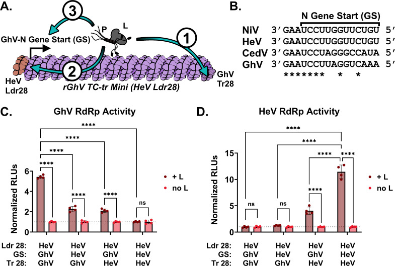
Batborne henipaviruses, such as Nipah and Hendra viruses, represent a major threat to global health due to their propensity for spillover, severe pathogenicity, and high mortality rate in human hosts. Coupled with the absence of approved vaccines or therapeutics, work with the prototypical species and uncharacterized, emergent species is restricted to high biocontainment facilities. There is a scarcity of such specialized spaces for research, and often, the scope and capacity of research, which can be conducted at BSL-4, is limited. Therefore, there is a pressing need for innovative life-cycle modeling systems to enable comprehensive research within lower biocontainment settings. This work showcases tetracistronic, transcription, and replication-competent minigenomes for the Nipah, Hendra, and Cedar viruses, which encode viral proteins facilitating budding, fusion, and receptor binding. We validate the functionality of all encoded viral proteins and demonstrate a variety of applications to interrogate the viral life cycle. Notably, we found that the Cedar virus replicase exhibits remarkable promiscuity, efficiently driving replication and transcription of minigenomes from all tested henipaviruses. We also apply this technology to Ghana virus (GhV), an emergent species that has so far not been isolated in culture. We demonstrate that the reported sequence of GhV is incomplete, but that this missing sequence can be substituted with analogous sequences from other henipaviruses. The use of our GhV system establishes the functionality of the GhV replicase and identifies two antivirals that are highly efficacious against the GhV polymerase.

Importance: Henipaviruses are recognized as significant global health threats due to their high mortality rates and lack of effective vaccines or therapeutics. Due to the requirement for high biocontainment facilities, the scope of research which may be conducted on henipaviruses is limited. To address this challenge, we developed innovative tetracistronic, transcription, and replication-competent minigenomes. We demonstrate that these systems replicate key aspects of the viral life cycle, such as budding, fusion, and receptor binding, and are safe for use in lower biocontainment settings. Importantly, the application of this system to the Ghana virus revealed that its known sequence is incomplete; however, substituting the missing sequences with those from other henipaviruses allowed us to overcome this challenge. We demonstrate that the Ghana virus replicative machinery is functional and can identify two orally efficacious antivirals effective against it. Our research offers a versatile system for life-cycle modeling of highly pathogenic henipaviruses at low biocontainment.

Keywords: Ghana virus; Nipah virus; antivirals; emerging pathogens; henipavirus; high biocontainment; minigenome; paramyxovirus; reverse genetics; viral RNA dependent RNA polymerase.


Conflict of interest statement

The authors declare no conflict of interest.

Figures

Fig 1
Fig 1
Establishment and implementation of tetracistronic, transcription, and replication-competent (TC-tr) minigenomes. (A) Design of rHNV TC-tr minigenomes that encode a HiBiT-mCherry reporter gene in addition to HNV-M, -F, and -RBP. Full-length virus genome structure is shown above for reference. Detailed descriptions of each minigenome design are available in Fig. S1. (B) Schematic describing the transfection approach for TC-tr minigenomes in which BSRT7 cells are co-transfected with plasmid encoding the rHNV minigenome, T7-driven HNV-N, -P, -L, and codon-optimized T7 polymerase. A more detailed depiction of the process is available in Fig. S2. (C) Microscopy demonstrating replication and transcription of rNiV, rHeV, and rCedV TC-tr minigenomes in the presence (+) or absence (-) of their respective HNV-L proteins. For all images, Hoechst is shown in blue, whereas mCherry is shown in red. (D) Quantification of mCherry-positive events produced by rHNV TC-tr minigenome replication and transcription and (E) matched quantification of normalized nanoluciferase signal. To measure vRdRp activity, RLUs from cells in the presence of HNV-L were normalized to RLUs from cells in the absence of HNV-L. Statistical significance was assessed by multiple unpaired t tests in GraphPad Prism to compare counts or normalized RLUs in the presence of HNV-L with counts or normalized RLUs in the absence of HNV-L for each respective minigenome. Transfections were conducted in biological quadruplicate. Error bars depict standard deviation. For all graphs: ns, P > 0.05; *, P ≤ 0.05; **, P ≤ 0.01; ***, P ≤ 0.001; ****, P ≤ 0.0001.
Fig 2
Fig 2
Matrix protein encoded by rHNV TC-tr VLPs is functionally competent. (A) Diagram depicting the experimental approach used for the BiFC assay. HeLa cells transfected with VN173-fused NHP2L1 or VN173-fused NOP56 were co-cultured with BSRT7 cells infected with rNiV TC-tr minigenomes encoding WT NiV-M or NiV-M fused with VC155. Fusion mediated by NiV-F and NiV-RBP facilitates cytoplasmic mixing and results in BiFC within mCherry-positive syncytia. Inset cartoon demonstrates reconstitution of Venus fluorescent protein if both VC155-fused NiV-M and VN173-fused host box C/D snoRNP proteins interact. (B) Microscopic images capturing instances of BiFC within mCherry-positive syncytia. For all images, mCherry is colored in red, BiFC (venus reconstitution) is colored in yellow, and Hoechst stain is colored in blue. Scale bars represent 100 μm. (C) Detection of TC-tr minigenome vRNA copies in supernatant from rescue cells by RT-qPCR. To ensure accurate quantification of vRNA, genome copies per mL were calculated by subtracting the signal obtained in the no-RT control from the RT condition. (D) Iterative passaging of rNiV and rHeV TC-tr VLPs captured by quantification of mCherry-positive events at each passage (P0 = rescue; P1 = passage 1; P2 = passage 2). Passaging experiments were conducted in biological triplicate. Error bars depict standard deviation.
Fig 3
Fig 3
Constrained sequence alignment of the GhV genome reveals that GhV is missing 28 nucleotides from its genomic promoter. (A) Sequence alignment of the reported genomic vRNA sequences of NiV, HeV, CedV, and GhV with constraint to proper phasing of the genomic promoter element II (PrE-II) sequence. (B) Sequence alignment of the reported antigenomic vRNA sequences of NiV, HeV, CedV, and GhV with constraint to proper phasing of the antigenomic PrE-II sequence. All alignments are shown in a 3′ to 5′ orientation, reflective of the biologically relevant sequences. Bases that are outwards facing towards the solvent are shown in full case, whereas bases buried within the nucleocapsid protein are shown in superscript text. Dashes (-) are used to denote unmapped nucleotides missing in the GhV sequence. The consensus sequence is shown below each hexamer, with absolutely conserved residues colored in red. All nucleotides follow the International Union of Pure and Applied Chemistry nomenclature. (C) Cartoon depicting a vRNP with properly phased bipartite promoters on each end. The inset panel shows the relative localization of PrE-I and PrE-II within the genomic promoter, with numbers detailing each hexamer position relative to the 3′ end of the vRNA. This model reflects phasing observed in the cryoEM structure of the NiV helical nucleocapsid assembly (PDB 7NT5). (D) Cartoon depicting hexamers and their relative positions in the genomic promoter of the GhV vRNP, which correspond to incompletely mapped GhV sequence. Missing hexamers/sequence are shown as a transparent outline. For all figures, monomers of nucleocapsid containing a hexamer of vRNA are denoted in brown, except for hexamers 1–3 and 14–16; hexamers encoding elements of the bipartite promoter elements are colored in teal. The antigenomic bipartite promoter elements are depicted with additional shaded pattern.
Fig 4
Fig 4
Restoration of a functional PrE-I facilitates replication and transcription of a rGhV TC-tr minigenome. (A) Cartoon depicting the design of chimeric rGhV TC-tr minigenomes in which the unmapped terminal 28 nucleotides of the GhV 3′ Ldr sequence are replaced with equivalent sequences derived from NiV (NiV Ldr28), HeV (HeV Ldr28), CedV (CedV Ldr28), or the GhV antigenomic promoter (GhV Tr28). The minigenome design of rGhV is further detailed in Fig. S1D. (B) Quantification of mCherry-positive events and (C) matched vRdRp activity resulting from the transcription and replication of each rGhV TC-tr minigenome in the presence (+) or absence (-) of GhV-L. (D) Microscopy depicting rGhV (HeV Ldr28) TC-tr minigenome events in the presence or absence of GhV-L. Red depicts mCherry signal, whereas blue depicts Hoechst stain. (E) Comparison of GhV vRdRp activity resulting from the transcription and replication of rGhV TC-tr minigenomes encoding either the HeV Ldr28 sequence or the GhV Tr28 sequence in the presence or absence of GhV-L. (F) Quantification of HeV vRdRp activity resulting from the transcription and replication of rHeV TC-tr minigenomes encoding either the WT (HeV Ldr28) sequence or the HeV Tr28 sequence in the presence or absence of HeV-L. To calculate vRdRp activity, RLUs from cells in the presence of HNV-L were normalized to respective RLUs generated by each minigenome in the absence of HNV-L. Statistical significance was assessed using a two-way ANOVA analysis in GraphPad Prism to compare counts or normalized RLUs in the presence of HNV-L with respective counts or normalized RLUs in the absence of HNV-L. For (E) and (F), additional comparisons were conducted to determine the significance between respective minigenome mutants in the presence of HNV-L with Šídák test for multiple comparisons. All experiments were conducted in at least biological triplicate. Error bars depict standard deviation. For all graphs: ns, P > 0.05; *, P ≤ 0.05; **, P ≤ 0.01; ***, P ≤ 0.001; ****, P ≤ 0.0001.
Fig 5
Fig 5
Heterotypic combinations of replicase and minigenomes from diverse henipavirus species uncovers a remarkable plasticity in template recognition by the CedV vRdRp. vRdRp activity above the background from the co-transfection of (A) rNiV, (B) rHeV, (C) rGhV, or (D) rCedV TC-tr minigenomes by NiV-N/-P/-L, HeV-N/-P/-L, CedV-N/-P/-L, or GhV-N/-P/-L, respectively. RLUs from each condition were normalized to RLUs from respective minigenome transfections conducted in parallel but in the absence of HNV-L. (E) Schematic summarizing the capability of each HNV replicase to support replication and transcription of various rHNV TC-tr minigenomes. Statistical significance was determined by ordinary, one-way ANOVA analysis in GraphPad Prism comparing the normalized RLUs yielded by each HNV replicase species to normalized RLUs from the no-L control. All experiments were conducted in biological triplicate. Error bars demonstrate standard deviation. For all graphs: ns, P > 0.05; *, P ≤ 0.05; **, P ≤ 0.01; ***, P ≤ 0.001; ****, P ≤ 0.0001.
Fig 6
Fig 6
Incompatibilities exist between the respective replicase, gene starts, and 5′ Tr sequences of GhV and HeV. (A) Cartoon depicting the rGhV (HeV Ldr28) TC-tr minigenome and the order in which elements required for both replication and transcription must be recognized by the vRdRp (1). The antigenomic promoter in the 5′ Tr must be recognized by the vRdRp to drive synthesis of genomic vRNA (2); the genomic promoter in the 3′ Ldr of genomic vRNA must be recognized and must prompt the vRdRp to enter scanning mode; and (3) the N gene start must be recognized by the vRdRp to initiate transcription of viral mRNAs. (B) Alignment of the N gene start (GS) sequences from NiV, HeV, CedV, and GhV. Alignments are shown in the genomic vRNA sense, 3′ to 5′. Asterisks depict complete conservation. (C) GhV vRdRp activity above background resulting from the co-transfection of the GhV replicase with chimeric rGhV TC-tr minigenomes with systematic replacement of the GhV-N GS or Tr28 sequences with analogous HeV sequences. (D) HeV vRdRp activity above background resulting from the co-transfection of HeV replicase with each chimeric rGhV TC-tr minigenome. RLUs from each condition were normalized to RLUs derived from the transfection of respective minigenomes in the absence of HNV-L. Statistical significance was determined by a two-way ANOVA analysis with Šídák test for multiple comparisons in GraphPad Prism comparing normalized RLUs from transfected cells in the presence of HNV-L with normalized RLUs from cells in the absence of HNV-L. Additional comparisons were conducted to determine significance between the parental rGhV (HeV Ldr28) TC-tr minigenome with the other minigenome mutants in the presence of HNV-L. All experiments were conducted in biological quadruplicate. Error bars depict standard deviation. For all graphs: ns, P > 0.05; *, P ≤ 0.05; **, P ≤ 0.01; ***, P ≤ 0.001; ****, P ≤ 0.0001.
Fig 7
Fig 7
The GhV-L protein is susceptible to two vRdRp inhibitors. Inhibition curves conducted on a panel of authentic viruses or TC-tr minigenome systems treated with (A) EIDD-2749 or (B) GHP-88309. IC50 values were determined in GraphPad Prism by nonlinear regression of [inhibitor] vs. normalized response, and are listed to the right of each system implemented. The 95% CI estimate for each IC50 is shown in parentheses. Error bars depict standard error of the mean. All inhibition experiments were conducted in at least biological triplicate. (C) Overview of a GhV-L homology model generated using PIV5-L as template (pdb: 6v85).The vRdRP, capping, connector, MTase, and CTD domains are colored blue, green, yellow, orange, and red, respectively. GHP-88309 is shown as orange spheres. The magnified inset depicts the locations where analogous residue mutations are known to induce resistance to GHP-88309. The third inset depicts alignment of a GHP-88309-MeV L complex with the GhV-L model (rms = 0.373). Homologous residues to resistance sites in HPIV3 L are shown in red and labeled. GHP-88309 is shown as orange sticks. (D) Sequence alignment of HPIV3, MeV, and members of the HNV genus. All known residues shown to induce resistance (boxed) to GHP-88309 are conserved across genera except for NiV-L and HeV-L at position H1165 (red; H1165).

Update of

References

    1. Drexler JF, Corman VM, Müller MA, Maganga GD, Vallo P, Binger T, Gloza-Rausch F, Cottontail VM, Rasche A, Yordanov S, et al. . 2012. Bats host major mammalian paramyxoviruses. Nat Commun 3:796. doi:10.1038/ncomms1796 - DOI - PMC - PubMed
    1. Barr J, Smith C, Smith I, de Jong C, Todd S, Melville D, Broos A, Crameri S, Haining J, Marsh G, Crameri G, Field H, Wang L-F. 2015. Isolation of multiple novel paramyxoviruses from pteropid bat urine. J Gen Virol 96:24–29. doi:10.1099/vir.0.068106-0 - DOI - PubMed
    1. Larsen BB, Gryseels S, Otto HW, Worobey M. 2022. Evolution and diversity of bat and rodent paramyxoviruses from North America. J Virol 96:e0109821. doi:10.1128/JVI.01098-21 - DOI - PMC - PubMed
    1. Wells HL, Loh E, Nava A, Solorio MR, Lee MH, Lee J, Sukor JRA, Navarrete-Macias I, Liang E, Firth C, Epstein JH, Rostal MK, Zambrana-Torrelio C, Murray K, Daszak P, Goldstein T, Mazet JAK, Lee B, Hughes T, Durigon E, Anthony SJ. 2022. Classification of new morbillivirus and jeilongvirus sequences from bats sampled in Brazil and Malaysia. Arch Virol 167:1977–1987. doi:10.1007/s00705-022-05500-z - DOI - PMC - PubMed
    1. Eaton BT, Broder CC, Middleton D, Wang L-F. 2006. Hendra and Nipah viruses: different and dangerous. Nat Rev Microbiol 4:23–35. doi:10.1038/nrmicro1323 - DOI - PMC - PubMed

Substances

Supplementary concepts

LinkOut - more resources