. 2024 Oct 22;9(10):e0096424.
doi: 10.1128/msystems.00964-24. Epub 2024 Sep 30.

PhoPQ-mediated lipopolysaccharide modification governs intrinsic resistance to tetracycline and glycylcycline antibiotics in Escherichia coli

Affiliations

PhoPQ-mediated lipopolysaccharide modification governs intrinsic resistance to tetracycline and glycylcycline antibiotics in Escherichia coli

Byoung Jun Choi et al. mSystems. .

Abstract

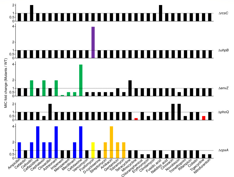
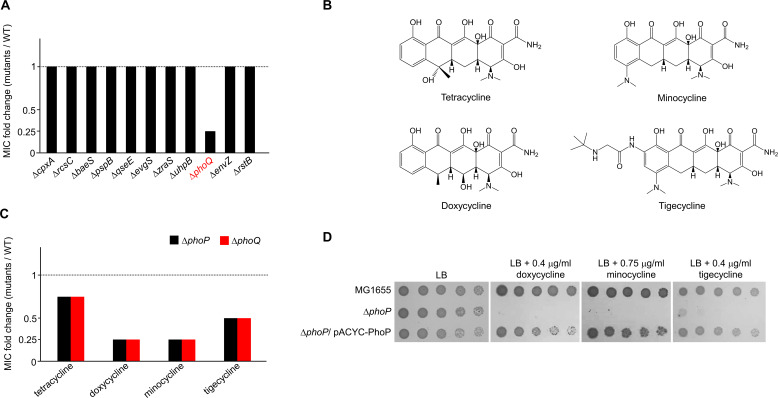
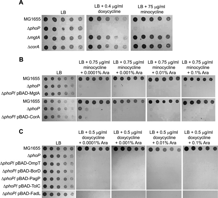
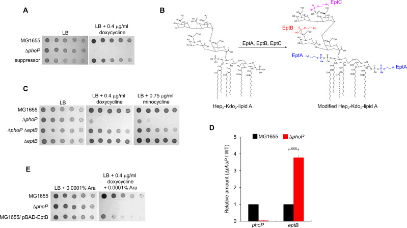
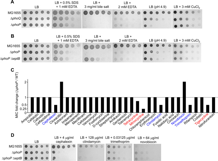
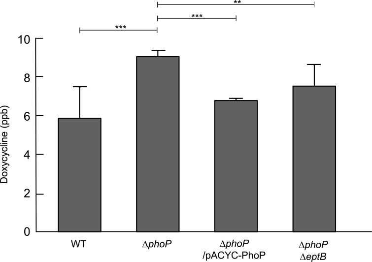
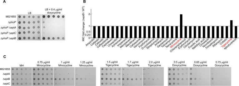
Tetracyclines and glycylcycline are among the important antibiotics used to combat infections caused by multidrug-resistant Gram-negative pathogens. Despite the clinical importance of these antibiotics, their mechanisms of resistance remain unclear. In this study, we elucidated a novel mechanism of resistance to tetracycline and glycylcycline antibiotics via lipopolysaccharide (LPS) modification. Disruption of the Escherichia coli PhoPQ two-component system, which regulates the transcription of various genes involved in magnesium transport and LPS modification, leads to increased susceptibility to tetracycline, minocycline, doxycycline, and tigecycline. These phenotypes are caused by enhanced expression of phosphoethanolamine transferase EptB, which catalyzes the modification of the inner core sugar of LPS. PhoPQ-mediated regulation of EptB expression appears to affect the intracellular transportation of doxycycline. Disruption of EptB increases resistance to tetracycline and glycylcycline antibiotics, whereas the other two phosphoethanolamine transferases, EptA and EptC, that participate in the modification of other LPS residues, are not associated with resistance to tetracyclines and glycylcycline. Overall, our results demonstrated that PhoPQ-mediated modification of a specific residue of LPS by phosphoethanolamine transferase EptB governs intrinsic resistance to tetracycline and glycylcycline antibiotics.

Importance: Elucidating the resistance mechanisms of clinically important antibiotics helps in maintaining the clinical efficacy of antibiotics and in the prescription of adequate antibiotic therapy. Although tetracycline and glycylcycline antibiotics are clinically important in combating multidrug-resistant Gram-negative bacterial infections, their mechanisms of resistance are not fully understood. Our research demonstrates that the E. coli PhoPQ two-component system affects resistance to tetracycline and glycylcycline antibiotics by controlling the expression of phosphoethanolamine transferase EptB, which catalyzes the modification of the inner core residue of lipopolysaccharide (LPS). Therefore, our findings highlight a novel resistance mechanism to tetracycline and glycylcycline antibiotics and the physiological significance of LPS core modification in E. coli.

Keywords: PhoPQ; antibiotic resistance; doxycycline; glycylcycline; lipopolysaccharide modification; minocycline; tetracycline; tigecycline.


Conflict of interest statement

The authors declare no conflict of interest.

Figures

FIG 1
FIG 1
The inactivation of two-component system sensor kinases affects intrinsic antibiotic resistance. The MICs of various antibiotics were measured against the wild-type and indicated mutant strains in MH medium. The relative MIC values for the indicated mutant cells compared to those for the wild-type cells are presented. Colored bars indicate the MIC values of important antibiotics, which increase or decrease in the indicated mutant cells.
FIG 2
FIG 2
The loss of the PhoPQ two-component system confers increased susceptibility to tetracycline and glycylcycline antibiotics. (A) Increased susceptibility of the ΔphoQ mutant to minocycline. The MICs of minocycline were measured against the wild-type and indicated mutant strains in MH medium. The relative MIC values for the indicated mutant cells compared to those for the wild-type cells are presented. (B) Structures of tetracycline and glycylcycline antibiotics. (C) Increased susceptibility of the ΔphoP or ΔphoQ mutant to tetracycline and glycylcycline antibiotics. The MICs of indicated antibiotics were measured against the wild-type and ΔphoP or ΔphoQ mutant strains in MH medium. The relative MIC values for the ΔphoP (black bars) or ΔphoQ (red bars) mutant cells compared to those for the wild-type cells are presented. (D) Complementation of antibiotic sensitivities of the ΔphoP mutant. The cells of the indicated strains were serially diluted from 108 to 104 cells/mL in 10-fold steps and spotted onto LB plates with or without the indicated concentration of each antibiotic. The experiments were performed in triplicate, and a representative image is presented.
FIG 3
FIG 3
The effect of several known PhoPQ regulon genes on the resistance to tetracycline antibiotics. (A) The effect of depletion of magnesium transporter genes on doxycycline and minocycline resistance. The cells of the indicated strains were serially diluted from 108 to 104 cells/mL in 10-fold steps and spotted onto LB plates with or without the indicated concentration of each antibiotic. (B) The effect of expression of magnesium transporter genes on minocycline resistance in the ΔphoP mutant strain. The cells of the indicated strains were serially diluted from 108 to 104 cells/mL in 10-fold steps and spotted onto LB plates with or without the indicated concentrations of minocycline and arabinose (Ara). (C) The effect of expression of several known PhoPQ regulon genes on doxycycline resistance in the ΔphoP mutant strain. The cells of the indicated strains were serially diluted from 108 to 104 cells/mL in 10-fold steps and spotted onto LB plates with or without the indicated concentrations of doxycycline and arabinose (Ara). (A–C) The experiments were performed in triplicate, and a representative image is presented.
FIG 4
FIG 4
The effect of the phosphoethanolamine transferase EptB on the resistance to tetracycline antibiotics. (A) Isolation of the suppressor mutant of the ΔphoP mutant. The cells of the indicated strains were serially diluted from 108 to 104 cells/mL in 10-fold steps and spotted onto LB plates with or without the indicated concentration of doxycycline. (B) Schematic representation depicting the roles of phosphoethanolamine transferases in LPS modification. EptA, EptB, and EptC catalyze the addition of phosphoethanolamine to the phosphate group of the glucosamine disaccharide of lipid A, the KdoII sugar in the inner core, and the phosphate group of the heptose I residue in the inner core, respectively. (C) The depletion of EptB suppresses the sensitivity of the ΔphoP mutant to tetracycline antibiotics. The cells of the indicated strains were serially diluted from 108 to 104 cells/mL in 10-fold steps and spotted onto LB plates with or without the indicated concentration of doxycycline or minocycline. (D) Relative mRNA levels of the phoP and eptB genes in the wild-type and ΔphoP mutant strains. Total mRNA was extracted from the wild-type (black bars) and ΔphoP mutant (red bars) cells cultured up to the early exponential phase [the optical density at 600 nm (OD600) = 0.4]. mRNA levels of the phoP and eptB genes were normalized to the levels of 16S rRNA. Data were produced from three independent experiments. Statistical significance was determined using the student’s t-test. ***P < 0.001. (E) The effect of overexpression of EptB on doxycycline resistance. The cells of the indicated strains were serially diluted from 108 to 104 cells/mL in 10-fold steps and spotted onto LB plates with or without the indicated concentrations of doxycycline and arabinose (Ara). (A, C, and E) The experiments were performed in triplicate, and a representative image is presented.
FIG 5
FIG 5
The effect of EptB on various phenotypes of the ΔphoP mutant. (A) Growth defect of the ΔphoP and ΔphoQ mutant strains under various envelope stress conditions. The cells of the indicated strains were serially diluted from 108 to 104 cells/mL in 10-fold steps and spotted onto LB plates with or without the indicated chemicals, or an acidic LB plate. (B) The effect of EptB inactivation on the sensitivity of the ΔphoP mutant to envelope stress. The cells of the indicated strains were serially diluted from 108 to 104 cells/mL in 10-fold steps and spotted onto LB plates with or without the indicated chemicals, or an acidic LB plate. (C) The effect of PhoP on the MICs of antibiotics. The MICs of various antibiotics were measured against the wild-type and ΔphoP mutant strains in MH medium. The relative MIC values for the ΔphoP mutant cells compared to those for the wild-type cells are presented. (D) The effect of EptB inactivation on the altered susceptibility of the ΔphoP mutant against antibiotics. The cells of the indicated strains were serially diluted from 108 to 104 cells/mL in 10-fold steps and spotted onto LB plates with or without the indicated concentrations of antibiotics. (A, B, and D) The experiments were performed in triplicate, and a representative image is presented.
FIG 6
FIG 6
The intracellular accumulated levels of doxycycline. At the early exponential phase, 0.5 mg/mL of doxycycline was added to LB medium and the cells were harvested after additional incubation for 20 min at 37°C. After washing, the harvested cells were disrupted and cell debris was removed by centrifugation. After removing soluble proteins using acetonitrile, the doxycycline level in the supernatant was determined using a Doxycycline ELISA Kit. The doxycycline level was estimated by measuring the absorbance at 450 nm. The exact concentration of doxycycline was estimated based on the standard curve made using the standard concentrations of doxycycline. Data were produced from three independent experiments. Statistical significance was determined using the student’s t-test. **P < 0.01; ***P < 0.001.
FIG 7
FIG 7
The depletion of EptB induces tetracycline and glycylcycline resistance. (A) The effect of inactivation of the phosphoethanolamine transferases on the sensitivity of the ΔphoP mutant to doxycycline. The cells of the indicated strains were serially diluted from 108 to 104 cells/mL in 10-fold steps and spotted onto LB plates with or without doxycycline. (B) The effect of EptB depletion on the MICs of antibiotics. The MICs of various antibiotics were measured against the wild-type and ΔeptB mutant strains in MH medium. The relative MIC values for the ΔeptB mutant cells compared to those for the wild-type cells are presented. (C) The resistance of the ΔeptB mutant to tetracycline and glycylcycline antibiotics. The cells of the indicated strains were serially diluted from 108 to 104 cells/mL in 10-fold steps and spotted onto LB plates with or without the indicated concentration of each antibiotic. The experiments were performed in triplicate, and a representative image is presented.

Similar articles

Cited by

References

    1. Lee JW. 2023. Bacterial regulatory mechanisms for the control of cellular processes: simple organisms' complex regulation. J Microbiol 61:273–276. doi:10.1007/s12275-023-00036-6 - DOI - PubMed
    1. Zschiedrich CP, Keidel V, Szurmant H. 2016. Molecular mechanisms of two-component signal transduction. J Mol Biol 428:3752–3775. doi:10.1016/j.jmb.2016.08.003 - DOI - PMC - PubMed
    1. García Véscovi E, Soncini FC, Groisman EA. 1996. Mg2+ as an extracellular signal: environmental regulation of Salmonella virulence. Cell 84:165–174. doi:10.1016/s0092-8674(00)81003-x - DOI - PubMed
    1. Prost LR, Daley ME, Le Sage V, Bader MW, Le Moual H, Klevit RE, Miller SI. 2007. Activation of the bacterial sensor kinase PhoQ by acidic pH. Mol Cell 26:165–174. doi:10.1016/j.molcel.2007.03.008 - DOI - PubMed
    1. Bader MW, Sanowar S, Daley ME, Schneider AR, Cho U, Xu W, Klevit RE, Le Moual H, Miller SI. 2005. Recognition of antimicrobial peptides by a bacterial sensor kinase. Cell 122:461–472. doi:10.1016/j.cell.2005.05.030 - DOI - PubMed

MeSH terms

LinkOut - more resources