. 2024 Nov 13;15(11):e0240124.
doi: 10.1128/mbio.02401-24. Epub 2024 Sep 30.

Chlamydia-driven ISG15 expression dampens the immune response of epithelial cells independently of ISGylation

Affiliations

Chlamydia-driven ISG15 expression dampens the immune response of epithelial cells independently of ISGylation

Yongzheng Wu et al. mBio. .

Abstract

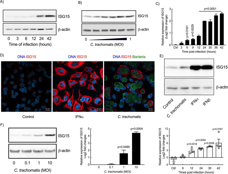
Excessive inflammation upon Chlamydia trachomatis infection can cause severe damages in the female genital tract. This obligate intracellular bacterium develops mainly in epithelial cells, whose innate response contributes to the overall inflammatory response to infection. The ubiquitin-like protein interferon-stimulated gene 15 (ISG15) stimulates interferon γ (IFNγ) production and is required for bacterial clearance in several infectious contexts. Here, we describe and investigate the consequences of the increase in ISG15 expression by epithelial cells infected with C. trachomatis. Infection of HeLa cells and primary ecto-cervical epithelial cells resulted in a transcriptional upregulation of ISG15 expression. This did not involve the canonical type I interferon (IFN-I) signaling pathway and depended instead on the activation of the STING/TBK1/IRF3 pathway. The absence or reduction of ISG15 synthesis led to increased production of several cytokines and chemokines, including interleukin (IL) 6 and IL8. This implicates that ISG15 normally dampens the immune response induced by C. trachomatis infection in epithelial cells. ISG15 exerted its control from an intracellular location, but without involving ISGylation. Finally, higher levels of inflammation and delayed bacterial clearance were observed in the genital tracts of ISG15-KO mice infected by C. trachomatis compared with wild-type animals; however, IFNγ production was unchanged. Altogether, our data show that ISG15 expression acts as a brake on the immune response to C. trachomatis infection in epithelial cells and limits bacterial burden and inflammation in mice.IMPORTANCEInfection of epithelial cells by Chlamydia trachomatis elicits an innate immune response by these cells. The signaling pathways involved, and their outcomes, are still very poorly understood. In this paper, we described how Chlamydia infection triggered the expression of ISG15, a small molecule normally associated to type I interferon (IFN-I) signaling and control of INF-γ production. ISG15 synthesis by epithelial cells attenuated their immune response to Chlamydia infection. In mice, we observed that ISG15 displayed a marginal role in modulating the production of IFN-γ, a key component of the host immune response to infection, but facilitated bacterial clearance. Overall, our study strengthens the importance of ISG15 not only in the resolution of viral but also of bacterial infection and document its role of "immune brake" in the context of Chlamydia infection.

Keywords: C. trachomatis; ISGylation; epithelial cells; inflammation; innate defense; interferon stimulated gene 15 (ISG15).


Conflict of interest statement

The authors declare no conflict of interest.

Figures

Fig 1
Fig 1
C. trachomatis infection induces ISG15 synthesis in epithelial cells. (A and C) Time course of infection of HeLa cells with C. trachomatis (A and C, MOI = 1) or with increasing MOI (B, 42 hpi). After infection, ISG15 transcript (C) and protein (A-B) levels were examined by RT-qPCR and Western blot, respectively. (D) HeLa cells were incubated with IFNα (10 ng/mL) or C. trachomatis (MOI = 1) for 30 h followed by immunostaining. (E) HeLa cells were incubated with C. trachomatis (MOI = 1) for 42 h or IFN-I (IFNα/β, 10 ng/mL for each) for 24 h before determining the expression of ISG15. (F) Primary epithelial cells from patients were incubated with C. trachomatis at the indicated MOI. ISG15 expression was examined 42 h post-infection (left and middle panels) or at the indicated times post-infection (right panel). The Western blots and immunofluorescence data are representative of at least two independent experiments. All other data represent three independent experiments. One-way ANOVA test (time course) and unpaired t-test (dose course in primary cells) were performed, and the p-value of the comparison with uninfected control is shown.
Fig 2
Fig 2
ISG15 exerts a negative control on IL6 and IL8 expression. (A) Cells were transfected with siRNA targeting human ISG15 or non-relevant oligonucleotides for 48 h before infection with C. trachomatis (MOI = 1). Cells were collected at 42 hpi. ISG15 levels were determined by Western blot (left panel). HSP60, a bacterial protein, control for bacterial load, and actin serves as a loading control. The IL6 and IL8 transcript (top histograms) and protein (bottom histograms) levels were determined by RT-qPCR and ELISA, respectively. (B) MEFs lacking ISG15 or not were infected with C. trachomatis (MOI = 1), and the amount of IL6 and mouse IL8 (KC) transcripts were determined at 42 hpi by RT-qPCR. (C) ISG15-deficient clones (C2 and C4) were complemented with ISG15 (left panel) and infected with C. trachomatis (MOI = 1). Transcript levels for IL6 and IL8 were measured at 42 hpi (middle and right panels). (D) Quantification of infectious bacteria collected in the indicated cellular backgrounds at 48 hpi. Each dot in panel D represents an individual experiment. The rest of the data are representative of at least three independent experiments. Unpaired t test was conducted in (A) and (B), and one-way ANOVA was performed in (C), and paired t-test was used in (D).
Fig 3
Fig 3
Autocrine IFN signaling is not implicated in Chlamydia-induced ISG15 expression by epithelial cells. (A) Two clones of ISG15-KO HeLa cells and wild-type cells were infected with C. trachomatis (MOI = 1) for 42 h or incubated with IFNα (10 ng/mL) for 24 h. After treatment, the transcript levels of the ISGs including IFIT3, RIG1, ISG56, and MXA normalized to actin were determined and expressed relative to non-infected WT cells. (B) HeLa cells were infected with C. trachomatis (MOI = 1) for 42 h. The expression of ISGs were normalized to actin and are expressed relative to non-infected cells. (C) U5A epithelial cells with mutated IFNa/b receptor and the parental 2fTGH cells were infected by C. trachomatis at the indicated MOI, and the ISG15 transcript level normalized to actin was determined at 42 hpi. The data represent three independent experiments. Unpaired t test in A and one-way ANOVA test in B were performed. The p-values of the comparison with WT cells (A), Chlamydia-induced ISG15 (B), or as indicated are shown.
Fig 4
Fig 4
The TBK1/Sting/IRF3 pathway lies upstream of ISG15 synthesis by epithelial cells in response to Chlamydia infection. (A-B) The indicated genes were silenced by siRNA (30 nM) for 48 h before Chlamydia infection. At 42 h post-infection, ISG15 expression was examined either by RT-qPCR (A) or by immunoblot (B). (C) HeLa cells were incubated with c-di-AMP at the indicated concentration for 24 h before quantification of the ISG15 levels in whole cell lysates by Western blot. (D) c-di-AMP (10 µM) was introduced into HeLa cells using transfection reagent at 37°C for the indicated times. The cells were then washed and were incubated for an additional 24 h before examining ISG15 level in whole cell lysates by Western blot. Incubation of HeLa cells with IFNα (10 ng/mL) for 24 h was used as positive control (C and D). (E-F) siRNA against cGAS or irrelevant oligonucleotides were transfected into HeLa cells for 48 h before Chlamydia infection. At 42 h post-infection, the expression of cGAS (E, left panel), ISG15 (E, middle and right panels), IL6 and IL8 (F), and actin were measured. All data represent three independent experiments. The p-values of unpaired t test with the condition “control siRNA” (A) or between the indicated conditions (E, F) are shown.
Fig 5
Fig 5
ISG15 regulates inflammation from an intracellular location and independently of ISGylation. (A) Culture medium was collected at different times post-infection for ISG15 detection by dot blot. First row: 0, 3, 6, and 9 hpi; second row: 12, 18, 24, and 30 hpi; third row: 36, 42, and 48 hpi; fourth row: 25, 50, 100, and 200 ng/mL of rhISG15. (B) ISG15-KO HeLa cells were pre-treated with the indicated concentration of rhISG15 for 30 min before Chlamydia infection (MOI = 1). The cells were further incubated for 42 h in the presence of rhISG15 before RNA extraction and quantification of IL6 and IL8 expression by RT-qPCR. (C) HeLa cells were incubated with C. trachomatis (MOI = 1) for 42 h or with IFN-α/β (10 ng/mL) for 24 h, lysed, and the amount of ISG15 (free form & conjugates) was analyzed by immunoblot on whole cell lysates. (D) Primary cervical epithelial cells were treated with IFNα (10 ng/mL) for 24 h or with C. trachomatis (MOI = 10) for 42 h followed by ISG15 detection. Top panels of C and D show overexposed images of the upper part of the membranes to visualize ISGylated proteins. (E) ISG15-KO cells (C2 and C4) or complemented cells (with ISG15 WT or ΔGG) were infected or not with C. trachomatis (MOI = 1) for 42 h before measuring the transcription of IL6 and IL8. The panel on the left shows ISG15 expression in the complemented cells. The images displayed are representative of two independent experiments in A and of three independent experiments in D using primary cells from three individuals. All other data correspond to three independent experiments. The p-values of unpaired t test between the indicated groups are shown.
Fig 6
Fig 6
ISG15 limits the inflammatory response in the upper FGT of mice infected with C. trachomatis. ISG15-KO and ISG15-WT mice were infected by introducing C. trachomatis into the uterine horn trans-cervically. The upper FGTs were collected at the indicated times post-infection. The upper FGTs from non-infected mice were collected as negative control. Bacterial loads (A) and inflammatory cytokines (B) in FGT homogenates were determined using quantitative PCR and multi-plex ELISA assays, respectively. Each dot represents one animal. (C) Representative FGTs (left) and hydrosalpinx index for six infected animals (right). The smallest interval of the scale in this image represents 0.05 cm. The p-value of unpaired t test between indicated groups are shown. (D) Graphical summary. Chlamydia infection of epithelial cells is sensed by cGas, which activates the STING/TBK1/IRF3 signaling cascade. Direct release of c-di-AMP by the bacteria may also be involved. IRF3 activates transcription of ISG15. Increase in intracellular free ISG15 restricts bacterial proliferation and the transcription of the host inflammatory cytokines IL6 and IL8, independently of ISGylation.

Similar articles

Cited by

References

    1. Knight E, Fahey D, Cordova B, Hillman M, Kutny R, Reich N, Blomstrom D. 1988. A 15-kDa interferon-induced protein is derived by COOH-terminal processing of A 17-kDa precursor. J Biol Chem 263:4520–4522. - PubMed
    1. Bogunovic D, Boisson-Dupuis S, Casanova JL. 2013. ISG15: leading a double life as a secreted molecule. Exp Mol Med 45:e18. doi:10.1038/emm.2013.36 - DOI - PMC - PubMed
    1. Swaim CD, Scott AF, Canadeo LA, Huibregtse JM. 2017. Extracellular ISG15 signals cytokine secretion through the LFA-1 integrin receptor. Mol Cell 68:581–590. doi:10.1016/j.molcel.2017.10.003 - DOI - PMC - PubMed
    1. Zhang X, Bogunovic D, Payelle-Brogard B, Francois-Newton V, Speer SD, Yuan C, Volpi S, Li Z, Sanal O, Mansouri D, et al. . 2015. Human intracellular ISG15 prevents interferon-α/β over-amplification and auto-inflammation. Nature New Biol 517:89–93. doi:10.1038/nature13801 - DOI - PMC - PubMed
    1. Perng YC, Lenschow DJ. 2018. ISG15 in antiviral immunity and beyond. Nat Rev Microbiol 16:423–439. doi:10.1038/s41579-018-0020-5 - DOI - PMC - PubMed

MeSH terms