[Preprint]. 2024 Sep 19:2024.09.17.611698.
doi: 10.1101/2024.09.17.611698.

The Gq/11 family of Gα subunits is necessary and sufficient for lower jaw development

Affiliations

The Gq/11 family of Gα subunits is necessary and sufficient for lower jaw development

Stanley M Kanai et al. bioRxiv. .

Update in

Abstract

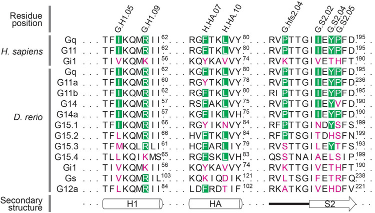
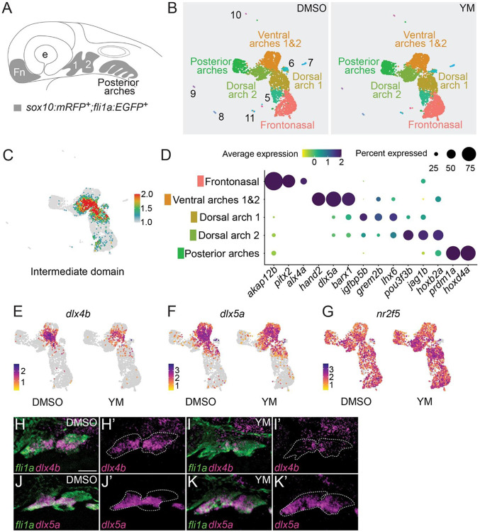
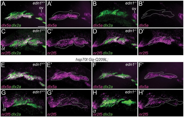
Vertebrate jaw development is coordinated by highly conserved ligand-receptor systems such as the peptide ligand Endothelin 1 (Edn1) and Endothelin receptor type A (Ednra), which are required for patterning of lower jaw structures. The Edn1/Ednra signaling pathway establishes the identity of lower jaw progenitor cells by regulating expression of numerous patterning genes, but the intracellular signaling mechanisms linking receptor activation to gene regulation remain poorly understood. As a first step towards elucidating this mechanism, we examined the function of the Gq/11 family of Gα subunits in zebrafish using pharmacological inhibition and genetic ablation of Gq/11 activity and transgenic induction of a constitutively active Gq protein in edn1 -/- embryos. Genetic loss of Gq/11 activity fully recapitulated the edn1 -/- phenotype, with genes encoding G11 being most essential. Furthermore, inducing Gq activity in edn1 -/- embryos not only restored Edn1/Ednra-dependent jaw structures and gene expression signatures but also caused homeosis of the upper jaw structure into a lower jaw-like structure. These results indicate that Gq/11 is necessary and sufficient to mediate the lower jaw patterning mechanism for Ednra in zebrafish.

Keywords: CRISPR/Cas9; craniofacial development; heterotrimeric G protein; single cell RNA-sequencing; small molecule inhibitor; zebrafish.


Figures

Figure 1.
Figure 1.. Sensitivity to YM is determined by eight conserved residues in a subset of Gq/11 family members.
In zebrafish, Gq/11 family members are encoded by gnaq (Gq), gna11a (G11a), gna11b (G11b), gna14 (G14), gna14a (G14a) and gna15.1-15.4 (G15.1-15.4). Residue positions in Gq/11 family members that interact with YM are annotated with the Common Gα Numbering (CGN) system (Flock et al. 2015). YM-binding residues that are conserved in a subset of Gq/11 family members are shown in green boxes, while diverged residues are labeled in magenta. Superscript numbers indicate the amino acid position for the respective Gα protein. H1 and HA are α-helices of the Ras-like domain and the α helical domain, respectively, and S2 is a β-sheet of the Ras-like domain. These structures constitute the hydrophobic binding pocket for YM in a subset of Gq/11 family members.
Figure 2.
Figure 2.. YM causes defects to a subset of Edn1/Ednra-dependent lower jaw skeletal elements.
(A-D) Lateral views of 6 dpf embryos treated with DMSO (A) or YM (B), shown in gross (A,B) or whole-mount skeletal preparations of larvae (C,D). The asterisk in (B) indicates the absence of inflated swim bladder. (E-J) Ventral (E-G) or lateral (H-J) views of flat-mounts for the viscerocranium from 6 dpf wild-type larvae treated with DMSO (E,H) or YM (F,I), or an untreated edn1−/− larvae (G,J). In E,F,H,I, white outlines highlight the symplectic cartilage (sy). In F,I, black arrowheads indicate fusion of the jaw joint (jj), white arrowhead indicates fusion of the hyomandibular joint (hj) and the arrow indicates fusion of the opercle (op) and branchiostegal ray (bsr). In G,J, the white asterisk indicates absence of the ceratohyal (ch). (K) Frequency of defects in six Edn1/Ednra-dependent skeletal elements in larvae treated with YM from 16-36 hpf. Absence of structure, hypoplasia, or fusion were scored as defects (accounting for sidedness). A total of 28 larvae were examined across two independent experiments. The effect of YM was determined to be statistically significant with a chi-square test (p=0.0001), comparing the number of defects in DMSO-treated larvae (0) to YM-treated larvae. (L) A schematic illustrating the four overlapping time intervals of YM application (grey bars). Numbers within bars represent the percentages of larvae exhibiting at least one defect to a Edn1/Ednra-dependent lower jaw structure. The total number of larvae examined is given in parentheses. Scale bars are 500 μm (A,C) and 100 μm (E, H). hm; hyomandibular, Mc; Meckel’s cartilage, pq; palatoquadrate ptp; pterygoid process of the palatoquadrate.
Figure 3.
Figure 3.. YM reduces expression of intermediate patterning genes and increases expression of dorsal patterning genes.
(A) Schematic of a zebrafish embryo at 36 hpf. Cells double-labeled with sox10:mRFP and fli1a:EGFP transgenic reporters (highlighted in grey) represent cranial neural crest populations from the frontonasal region (Fn), anterior pharyngeal arches 1 (1) and 2 (2) and posterior pharyngeal arches. (B) UMAP plots for DMSO or YM-treated samples. Clusters analyzed in this study are labeled with the NCC populations they represent. Clusters 5-11 are described further in Supplementary Information (Fig. S3, Table S1). Equivalent clusters between DMSO and YM-treated samples are labeled with the same colors. (C) Feature map highlighting approximate cell populations in the intermediate domains of pharyngeal arches 1 and 2, shown overlayed on combined UMAP plots of control and YM-treated samples. The feature map represents the composite average expression level for 14 experimentally verified intermediate domain patterning genes, ccn2b, dlx3b, dlx4a, dlx4b, emx2, fgfbp2a, foxc1b, foxd1, fsta, grem2b, igfbp5b, msx1a, nkx3.2 and shox (Fig. S4). The scale is average expression. (D) Dot plot of selected marker genes and their respective cluster identity (Fig. S3). (E-G) Feature maps highlighting differential expression of dlx4b (E), dlx5a (F) and nr2f5 (G) in DMSO or YM-treated samples. The scale is average expression. (H-K) Fluorescence in situ hybridization and immunofluorescence of 36 hpf embryos treated with DMSO or YM between 16-36 hpf. DMSO (H,J) or YM-treated (I,K) embryos were probed for dlx4b (H,I) or dlx5a (J,K) using fluorescence in situ hybridization (magenta). Pharyngeal arches, labeled with fli1a:EGFP, were detected with immunofluorescence (green). Approximate borders for pharyngeal arches 1 and 2 are indicated with dashed lines (H’,I’,J’,K’). Images are representative of four embryos. Scale bar is 50 μm.
Figure 4.
Figure 4.. YM increases the prevalence and severity of lower jaw defects in edn1+/− larvae relative to wild-type alarvae.
(A-F) Representative flat-mounts of viscerocranium from wild-type (A,D), edn1+/− (B,E), or edn1−/− (C,F) larvae treated with DMSO (A-C) or 100 μM YM (D-F) between 16-36 hpf. In A,B,D, white outlines highlight the symplectic cartilage. In C,E,F, the white asterisk indicate absence of the ceratohyal. In D,E, black arrowheads indicate fusion of the jaw joint. In D, the white arrowhead indicates fusion of the hyomandibular joint. All skeletal preparations are 6 dpf. (G) Frequency of defects in seven Edn1/Ednra-dependent skeletal elements in YM-treated wild-type or edn1+/− larvae. Absence of structure, hypoplasia, or fusion were scored as defects (ignoring sidedness). The pharmacogenetic interaction between edn1 and YM was determined to be statistically significant with a chi-square test (p=0.0001), comparing the number of defects in YM-treated edn1+/+ and edn1+/− larvae. (H) Percentage of individual larvae presenting with defects in the seven Edn1/Ednra-dependent structures. Individual larvae were scored for total number of skeletal elements affected (ignoring sidedness). Bar graphs are color-coded the same as G. All wild-type, edn1+/− or edn1−/− embryos treated with DMSO or YM are siblings from the same clutch. Scale bar is 100 μm.
Figure 5.
Figure 5.. gna11a and gna11b, but not gnaq, are necessary for development of Edn1/Ednra-dependent structures.
(A-H) Representative flat-mounts of the viscerocranium at 6 dpf, shown in ventral view, for selected allelic combinations. In C-H, white outlines highlight the symplectic. In A,B, the white asterisks indicate absence of the ceratohyal. In C-F, black arrowheads indicate fusion of the jaw joint. In E,F, the white arrowheads indicate fusion of the hyomandibular joint. (I) Overall severity of defects for genotypes shown in A-H were quantified as a “phenotype score”. Individual larvae were scored for the total number of Edn1/Ednra-dependent skeletal elements exhibiting defects (ignoring sidedness). A phenotype score of 0 correspond to a wild-type phenotype, and a score of 7 indicates all Edn1/Ednra-dependent structures on at least one side were affected. One dot represents an individual larva. The middle line is the mean. Error bar is standard error of the mean. The n values for respective genotypes are indicated in parentheses. Statistical significance was determined with Kruskal-Wallis test with Dunn’s multiple comparisons (*; p < 0.05, **; p < 0.01, ***; p < 0.001, ns; not significant). Scale bar is 100 μm.
Figure 6.
Figure 6.. Induction of Gq activity rescues the edn1−/− phenotype and causes ventralization of dorsal structures.
(A-D) Flat-mounts of the viscerocranium at 4 dpf, shown in ventral view, of heat-shocked larvae. In hsp70l:Gq-Q209L;edn1+/+ larvae (B), lower jaw structures were normal, though morphological changes were observed for the pterygoid process of the palatoquadrate (white arrowhead) and hyomandibular cartilage (black arrowhead). Compared to non-transgenic edn1−/− larvae (C), Edn1/Ednra-dependent skeletal elements were rescued in hsp70l:Gq-Q209L;edn1−/− larvae (D). Morphological changes in the pterygoid process of the palatoquadrate (white arrowhead) and hyomandibular cartilage (black arrowhead) were also observed in hsp70l:Gq-Q209L edn1−/− larvae (D) (E) Frequency of phenotype rescue for six Edn1/Ednra-dependent structures in heat-shocked hsp70l:Gq-Q209L:edn1−/− larvae (accounting for sidedness). Phenotype rescue was determined to be statistically significant with a chi-square test (p=0.0001), comparing the number of restored skeletal elements in non-transgenic edn1−/− larvae (0) to hsp70l:Gq-Q209L;edn1−/− larvae. (F,G) Flat-mounts of the viscerocranium at 4 dpf, in lateral view, of heat-shocked non-transgenic edn1+/+ (F) and hsp70l:Gq-Q209L; edn1+/+ larvae (G). Malformations of the pterygoid process of the palatoquadrate (white arrowhead) and hyomandibular cartilage (black arrowhead) are indicated in G. (H) Frequency of malformed pterygoid processes and hyomandibular cartilages (accounting for sidedness) in all edn1 genotypes with the hsp70l:Gq-Q209L transgene. Scale bar is 100 μm.
Figure 7.
Figure 7.. Induction of Gq activity upregulates dlx5a expression and downregulates nr2f5 expression.
The expression patterns of both dlx5a and nr2f5, both in magenta, are shown by two-color fluorescence in situ hybridization on 28 hpf embryos. Pharyngeal arches are labeled with dlx2a, in green. dlx5a and dlx2a (A,B,E,F) or nr2f5 and dlx2a (C,D,G,H) are shown overlaid. dlx5a (A’,B’,E’,F’) and nr2f5 (C’,D’,G’H’) are also shown alone, with the border of the pharyngeal arches indicated by white dashed lines. (A,C) Expression in non-transgenic edn1+/+ embryos. (E,G) Expression in hsp70l:Gq-Q209L; edn1+/+ embryos. (B,D) Expression in non-transgenic edn1−/− embryos. (F,H) Expression in hsp70l:Gq-Q209L:edn1−/− embryos. All embryos were heat-shocked. Images are representative of at least five embryos. Scale bar is 50 μm. cf; choroid fissure, ov; otic vesicle

Similar articles

References

    1. Akimenko M. A., Ekker M., Wegner J., Lin W. and Westerfield M. (1994) Combinatorial expression of three zebrafish genes related to distal-less: part of a homeobox gene code for the head. J Neurosci 14, 3475–86. doi:10.1523/JNEUROSCI.14-06-03475.1994 - DOI - PMC - PubMed
    1. Alexander C., Zuniga E., Blitz I. L., Wada N., Le Pabic P., Javidan Y., Zhang T., Cho K. W., Crump J. G. and Schilling T. F. (2011) Combinatorial roles for BMPs and Endothelin 1 in patterning the dorsal-ventral axis of the craniofacial skeleton. Development 138, 5135–46. doi:10.1242/dev.067801 - DOI - PMC - PubMed
    1. Aramori I. and Nakanishi S. (1992) Coupling of two endothelin receptor subtypes to differing signal transduction in transfected Chinese hamster ovary cells. J Biol Chem 267, 12468–74. - PubMed
    1. Askary A., Xu P., Barske L., Bay M., Bump P., Balczerski B., Bonaguidi M. A. and Crump J. G. (2017) Genome-wide analysis of facial skeletal regionalization in zebrafish. Development 144, 2994–3005. doi:10.1242/dev.151712 - DOI - PMC - PubMed
    1. Barske L., Askary A., Zuniga E., Balczerski B., Bump P., Nichols J. T. and Crump J. G. (2016) Competition between Jagged-Notch and Endothelin1 Signaling Selectively Restricts Cartilage Formation in the Zebrafish Upper Face. PLoS Genet 12, e1005967. doi:10.1371/journal.pgen.1005967 - DOI - PMC - PubMed

Publication types