[Preprint]. 2024 Sep 19:2024.09.17.613453.
doi: 10.1101/2024.09.17.613453.

A Strategic Blend of Stabilizing Polymers to Control Particle Surface Charge for Enhanced Mucus Transport and Cell Binding

Affiliations

A Strategic Blend of Stabilizing Polymers to Control Particle Surface Charge for Enhanced Mucus Transport and Cell Binding

Corey A Stevens et al. bioRxiv. .

Abstract

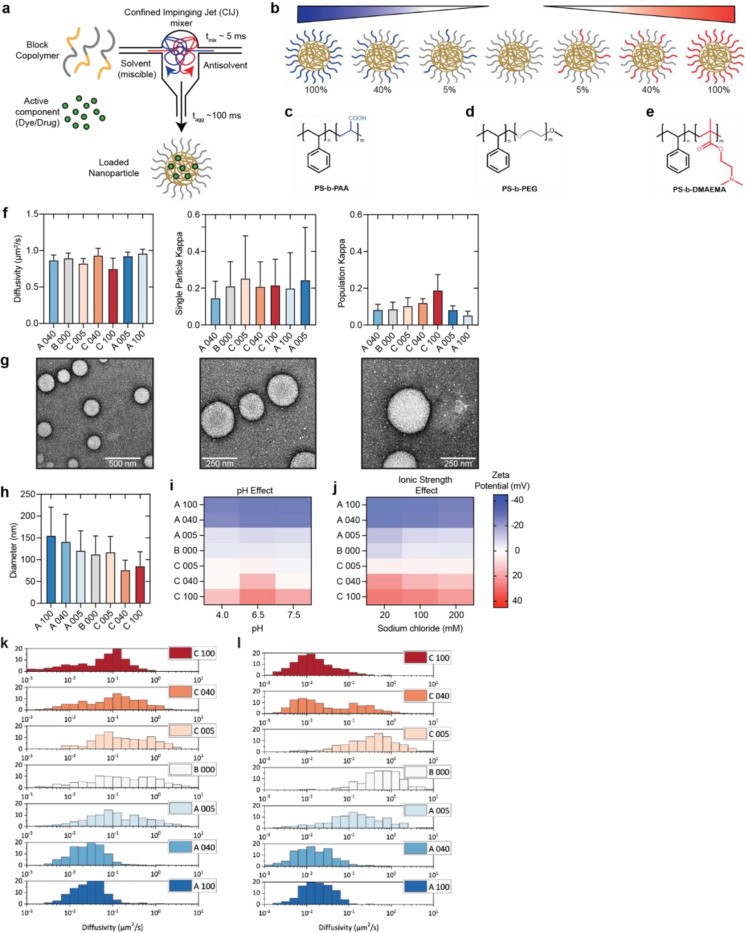
Mucus layers, viscoelastic gels abundant in anionic mucin glycoproteins, obstruct therapeutic delivery across all mucosal surfaces. We found that strongly positively charged nanoparticles (NPs) rapidly adsorb a mucin protein corona in mucus, impeding cell binding and uptake. To overcome this, we developed mucus-evading, cell-adhesive (MECS) NPs with variable surface charge using Flash NanoPrecipitation, by blending a neutral poly(ethylene glycol) (PEG) corona for mucus transport with a small amount, 5 wt%, of polycationic dimethylaminoethyl methacrylate (PDMAEMA) for increased cell targeting. In vitro experiments confirmed rapid mucus penetration and binding to epithelial cells by MECS NPs, suggesting a breakthrough in mucosal drug delivery.


Conflict of interest statement

Competing interests CAS, BS, BKW, KR and RKP are co-inventors on a patent disclosure MBHB 24-0253-US-PRO.

Figures

Figure 1.
Figure 1.. NP transport across the mucus barrier.
(a) Schematic representation of mucosal tissue and NPs for drug delivery across the mesh-like mucus network. (b) CryoSEM image of a reconstituted 1 wt% MUC2 gel. (c) Two-dimensional pore analysis of cryoSEM images of 1 wt% MUC2 gels. Counts represent the number of pores with the corresponding radius. (d) SPT-determined diffusivity of polystyrene NPs in a 1:1 water:glycerol mixture (anionic particles in blue, cationic particles in red). (e–f) Diffusivity of positively (red) and negatively (blue) charged NPs in 1 wt% (e) MUC2 and (f) MUC5AC gels.
Figure 2.
Figure 2.. Formation of a mucin corona on cationic NPs.
(a–b) CryoSEM images of 1 μm (a) anionic and (b) cationic particles in 1 wt% MUC2 gels. (c) Zeta potential measurements of particles pre- and post-incubation with MUC2. (d) Schematic representation of anionic NPs diffusing in mucin gel. (e) Schematic representation of cationic NPs with a mucin corona diffusing in a mucin gel. (f–h) Confocal images depicting the influence of the mucin corona on NP adhesion to epithelial cells: (f) cationic NPs adhering to HeLa cell monolayers, (g) anionic NPs incubated with HeLa cell monolayers, and (h) cationic NPs pretreated with MUC2 prior to incubation with a HeLa cell monolayer (green: particles, blue: nuclei [DAPI], red: actin [phalloidin]). (i) Quantification of microscopy images shown in f–h.
Figure 3.
Figure 3.. Production of MECS NPs by Flash NanoPrecipitation.
(a) Schematic representation of the Flash NanoPrecipitation procedure used to produce an MECS NP library. (b) Representation of NPs tested for mucosal transport and cell targeting. (c–e) Diagrams of (c) negatively charged PAA, (d) polystyrene-block-PEG, and (e) positively charged PDMAEMA. (f) SPT-determined diffusivity of NPs produced by Flash NanoPrecipitation in a 1:1 water:glycerol mixture (left), with individual particle kappa values (middle) and kappa values averaged over the entire population of tracked particles (right). (g) Transmission electron microscopy images of B000 particles, showing polymers brushed about the particle surfaces. (h) Dynamic light scattering size measurement of each NP. (i–j) Zeta potential measurements of each particle in solutions of different (i) pH and (j) ionic strength. (k–l) Histogram showing the diffusivity of every tracked particle in 1 wt% (k) MUC2 or (l) MUC5AC.
Figure 4.
Figure 4.. Superior epithelial cell targeting of MECS NPs.
(a–c) Confocal microscopy images of HeLa cells overlaid with 100 μL of 1 wt% MUC2 and A005 (a), B000 (b), or C005 (c). (d) Flow cytometry quantification of HeLa cells coated with MUC2 and incubated with NPs. (e) Schematic representation of the mesofluidic intestinal chip (right) and an image of the mucosal interface within the system (left). (f–h) Quantification of confocal images of organoid systems incubated with equivalent amounts of A005, B000, and C005 showing the (f) total remaining fluorescence, (g) NP green fluorescence overlapped with nuclei (DAPI), and (h) NP green fluorescence overlapped with mucus (WGA-Texas Red). (i–k) Volumetric analysis of (i) A005, (j) B000, and (k) C005 particles incubated with gut-on-a-chip organoid. Z-stack confocal images were converted into volumes for each fluorescent channel (blue: Hoechst; red: WGA-Texas Red; green: NPs). Hostasol Yellow 3G.

Similar articles

References

    1. Vargason A. M., Anselmo A. C. & Mitragotri S. The evolution of commercial drug delivery technologies. Nat. Biomed. Eng. 5, 951–967 (2021). - PubMed
    1. Mitchell M. J. et al. Engineering precision nanoparticles for drug delivery. Nat. Rev. Drug Discov. 20, 101–124 (2021). - PMC - PubMed
    1. Wagner C. E., Wheeler K. M. & Ribbeck K. Mucins and Their Role in Shaping the Functions of Mucus Barriers. Annu. Rev. Cell Dev. Biol. 34, 189–215 (2018). - PMC - PubMed
    1. Johansson M. E. V. & Hansson G. C. Immunological aspects of intestinal mucus and mucins. Nat. Rev. Immunol. 16, 639–649 (2016). - PMC - PubMed
    1. Lai S. K., Wang Y.-Y. & Hanes J. Mucus-penetrating nanoparticles for drug and gene delivery to mucosal tissues. Adv. Drug Deliv. Rev. 61, 158–171 (2009). - PMC - PubMed

Publication types

LinkOut - more resources