[Preprint]. 2024 Sep 17:2024.09.17.613539.
doi: 10.1101/2024.09.17.613539.

Decoding polyubiquitin regulation of KV7. 1 functional expression with engineered linkage-selective deubiquitinases

Affiliations

Decoding polyubiquitin regulation of KV7. 1 functional expression with engineered linkage-selective deubiquitinases

Sri Karthika Shanmugam et al. bioRxiv. .

Update in

Abstract

Protein posttranslational modification with distinct polyubiquitin linkage chains is a critical component of the 'ubiquitin code' that universally regulates protein expression and function to control biology. Functional consequences of diverse polyubiquitin linkages on proteins are mostly unknown, with progress hindered by a lack of methods to specifically tune polyubiquitin linkages on individual proteins in live cells. Here, we bridge this gap by exploiting deubiquitinases (DUBs) with preferences for hydrolyzing different polyubiquitin linkages: OTUD1 - K63; OTUD4 - K48; Cezanne - K11; TRABID - K29/K33; and USP21 - non-specific. We developed a suite of engineered deubiquitinases (enDUBs) comprised of DUB catalytic domains fused to a GFP-targeted nanobody and used them to investigate polyubiquitin linkage regulation of an ion channel, YFP-KCNQ1. Mass spectrometry of YFP-KCNQ1 expressed in HEK293 cells indicated channel polyubiquitination with K48 (72%) and K63 (24%) linkages being dominant. NEDD4-2 and ITCH both decreased KCNQ1 functional expression but with distinctive polyubiquitination signatures. All enDUBs reduced KCNQ1 ubiquitination but yielded unique effects on channel expression, surface density, ionic currents, and subcellular localization. The pattern of outcomes indicates K11, K29/K33, and K63 chains mediate net KCNQ1-YFP intracellular retention, but achieved in different ways: K11 promotes ER retention/degradation, enhances endocytosis, and reduces recycling; K29/K33 promotes ER retention/degradation; K63 enhances endocytosis and reduces recycling. The pattern of enDUB effects on KCNQ1-YFP differed in cardiomyocytes, emphasizing ubiquitin code mutability. Surprisingly, enDUB-O4 decreased KCNQ1-YFP surface density suggesting a role for K48 in forward trafficking. Lastly, linkage-selective enDUBs displayed varying capabilities to rescue distinct trafficking-deficient long QT syndrome type 1 mutations. The results reveal distinct polyubiquitin chains control different aspects of KCNQ1 functional expression, demonstrate ubiquitin code plasticity, and introduce linkage-selective enDUBs as a potent tool to help demystify the polyubiquitin code.

Keywords: KCNQ1; deubiquitinase; ion channel; ion channel trafficking; long QT syndrome; polyubiquitin; protein degradation; ubiquitin; ubiquitin code; ubiquitin linkage.


Conflict of interest statement

Declaration of interests S.A.K. and H.M.C. are inventors on a patent held by Columbia University for “Compositions and methods for using engineered deubiquitinases for probing ubiquitin-dependent cellular processes.” H.M.C. is a scientific co-founder and on the SAB of two startups, Stablix, Inc. and Flux Therapeutics, pursuing targeted protein stabilization therapeutics. S.A.K. is a co-founder and employee of Stablix, Inc.

Figures

Figure 1:
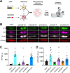
Figure 1:. KCNQ1-YFP subcellular distribution and polyubiquitination in HEK293 cells and design of linkage-selective enDUBs.
(A) Representative confocal images of HEK293 cells showing co-localization of expressed KCNQ1-YFP (green) and immunolabelled subcellular organelles [endoplasmic reticulum (ER), Golgi, early endosome (EE), late endosome (LE) and lysosome (lyso)] (red). (B) Co-localization of Q1-YFP with subcellular markers assessed by Pearson’s co-localization coefficient. (n>8 cells for each subcellular organelle imaged; N=2. (C) Schematic of BBS-KCNQ1-YFP with BTX-647 bound to an extracellular bungarotoxin-binding site (BBS). (D) Representative flow cytometry cumulative distribution function (CDF) plots showing total (YFP fluorescence) and surface (BTX-647) channels in cells expressing BBS-KCNQ1-YFP in untreated (black) and MG132 treated (red) conditions. (E) Representative Coomassie-stained SDS PAGE of pulled down KCNQ1-YFP. Black boxes indicate KCNQ1 monomeric and dimeric protein bands excised for mass spectrometry. (F) Representative ms2 spectra traces for ubiquitin peptides with di-glycine modification of K48 (top) and K63 (bottom), respectively. (G) Fractional distribution of Ub linkages on pulled down KCNQ1-YFP as assessed by mass spectrometry of associated di-glycine modified lysine residues on associated ubiquitin peptides. (H) Schematic of linkage-selective engineered deubiquitinases (enDUBs). (I) Top, KCNQ1-YFP pulldowns probed with anti-KCNQ1 in cells expressing KCNQ1-YFP with nano (control) or the indicated enDUBs. The four bands represent KCNQ1-YFP monomeric, dimeric, trimeric and tetrameric species. Bottom, same blot stripped and probed with anti-ubiquitin. (J) Relative KCNQ1-YFP ubiquitination computed as the ratio of anti-ubiquitin/anti-KCNQ1 signal intensities (mean ± SEM); N=3, #p<0.0001, *** p<0.001 and ** p<0.01, one-way ANOVA and Dunnett’s multiple comparisons test.
Figure 2:
Figure 2:. Differential impact of distinct enDUBs on KCNQ1 steady-state expression, subcellular localization, and function.
(A) Experimental design schematic; BBS-KCNQ1-YFP is co-expressed with either GFP/YFP targeting nanobody (nano, control) or an enDUB in HEK293 cells. (B) Flow cytometry representative CDF plots showing total (YFP) fluorescence in cells expressing BBS-Q1-YFP and either nano alone (black) or each of the enDUBs (enDUB-O1, cyan; enDUB-O4, green; enDUB-Cz, magenta; enDUB-Tr, orange; enDUB-U21, blue). (C) Quantification of flow cytometry experiments for total KCNQ1-YFP expression analyzed from YFP- and CFP- positive cells (n>5,000 cells per experiment, N=10 independent experiments; #p<0.0001, *p<0.05; one-way ANOVA with Dunnett’s multiple comparisons). Data were normalized to values from the nano control group (dotted line). (D) Representative CDF plots showing surface fluorescence (BTX-647) in cells expressing BBS-KCNQ1-YFP and either nano alone (black) or an enDUB (enDUB-O1, cyane; enDUB-O4, green; enDUB-Cz, magenta; enDUB-Tr, orange; enDUB-U21, blue). (E) Quantification of flow cytometry experiments for KCNQ1-YFP surface expression analyzed from YFP- and CFP- positive cells (n>5,000 cells per experiment, N=10 independent experiments; #p<0.0001, **p<0.01; one-way ANOVA with Dunnett’s multiple comparisons). Data were normalized to values from the nano control group (dotted line). (F-J) Co-localization of KCNQ1-YFP with subcellular markers assessed by Pearson’s co-localization coefficient (N=2, n>8 for each subcellular organelle; #p<0.0001, *p<0.05, one-way ANOVA and Dunnett’s multiple comparisons test). (K) Exemplar KCNQ1 + KCNE1 current traces from whole-cell patch clamp measurements in CHO cells. (L) Population I-V curves for nano control (black, n=25), enDUB-O1 (cyan, n=9), enDUB-O4 (green, n=9), enDUB-Cz (magenta, n=16), enDUB-Tr (orange, n=16) and enDUB-U21 (blue, n=8). #p<0.0001, two-way ANOVA, with Tukey’s multiple comparisons. Here and throughout, data was pooled from n cells tested across three or more independent experimental days.
Figure 3:
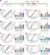
Figure 3:. Distinctive impact of different enDUBs on dynamic KCNQ1 delivery to and removal from the plasma membrane.
(A) Schematic of optical pulse-chase assay to measure BBS-KCNQ1-YFP forward trafficking. (B) Representative flow cytometry BTX-647 fluorescence histograms showing time evolution of surface channel increase in cells expressing BBS-KCNQ1-YFP with nano (black, top) or enDUB-Tr (orange, bottom). (C) Time evolution of channel forward to the surface in cells expressing BBS-KCNQ1-YFP with either nano or an enDUB (enDUB-O1, cyan; enDUB-O4, green; enDUB-Cez, magenta; enDUB-Tr, orange; enDUB-U21, blue; n>10,000 cells; N=3 for each data point; p>0.05, nano vs enDUB-O1; p<0.0001, nano vs enDUB-O4; p<0.01, nano vs enDUB-Cz; p<0.0001, nano vs enDUB-Tr; p<0.01, nano vs enDUB-U21; two-way ANOVA, with Tukey’s multiple comparisons). Data are normalized to the max value of nano control (black). Smooth curves are fits of an exponential growth function to the data: y= Ae1/τ +y0. (D) Schematic of endocytosis pulse-chase assay. (E) Representative flow cytometry BTX-647 fluorescence histograms showing time evolution of surface channel decrease in cells expressing BBS-KCNQ1-YFP with nano (black, top) or enDUB-O1 (turquoise, bottom). (F) Time evolution of channel removal from the surface in cells expressing BBS-KCNQ1-YFP with either nano or an enDUB (enDUB-O1, cyan; enDUB-O4, green; enDUB-Cz, magenta; enDUB-Tr, orange; enDUB-U21, blue; n>10,000 cells; N=3 for each data point; p<0.0001, nano vs enDUB-O1, p<0.05, nano vs enDUB-O4, p<0.05, nano vs enDUB-Cz, p>0.05, nano vs enDUB-Tr, p>0.05, nano vs enDUB-U21, two-way ANOVA with Tukey’s multiple comparisons). Smooth curves are fits of an exponential growth function to the data: y= Ae−1/τ +y0.
Figure 4:
Figure 4:. KCNQ1 functional expression is downregulated by the E3 ligases NEDD4–2 and ITCH with distinct polyubiquitin signatures.
(A) Representative flow cytometry CDF plots showing surface channel (BTX-647) fluorescence in cells expressing BBS-KCNQ1-YFP and nano (black) with NEDD4L (red) or ITCH (blue). (B) Mean BBS-KCNQ1-YFP surface expression (BTX-647 fluorescence) analyzed from YFP- positive cells (n>5,000 cells per experiment, N=3–4; #p<0.0001, one-way ANOVA and Dunnett’s multiple comparisons test). Data were normalized to values from the nano control group (dotted line). (C) Exemplar KCNQ1 current traces from whole-cell patch clamp measurements in CHO cells. (D) Population I-V curves for KCNQ1 + KCNE1 + nano (control; black, n=10), KCNQ1+ NEDD4–2 (red, n=9), and KCNQ1+ITCH (blue, n=11) (#p<0.0001, two-way ANOVA, with Tukey’s multiple comparisons). (E) Quantification of ubiquitin that was pulled down with KCNQ1-YFP expressed with nano (control), NEDD4–2 or ITCH, as assessed by mass spectrometry. Data were normalized to the control group. (F) Mass spectrometric evaluation of ubiquitin lysine residues that are di-gly modified (n=4, N=2). (G) Fractional distribution of Ub linkages as assessed by mass spectrometric analysis of immunoprecipitated and gel excised bands of KCNQ1-YFP co-expressed with NEDD4L or ITCH. (H) Cartoon of KCNQ1 with di-gly-modified lysine residues identified by mass spectrometry shown in red. Representative ms2 spectra trace of KCNQ1 peptide with di-gly modification of K598. (I) Quantification of di-gly-modified lysine residues on KCNQ1 peptides (N=2 independent experiments).
Figure 5:
Figure 5:. Differential impact of distinct linkage-selective enDUBs on reversing KCNQ1 functional downregulation by NEDD4–2.
(A) Experimental design schematic; BBS-KCNQ1-YFP is co-expressed with NEDD4–2 and either nano (control) or an enDUB in HEK293 cells. (B) Left, Western blot of KCNQ1-YFP pulldowns from cells expressing KCNQ1-YFP + NEDD4–2 and either nano or the indicated enDUB, probed with anti-KCNQ1 antibody. Right, same blot stripped and probed with anti-ubiquitin. (C) Relative KCNQ1 ubiquitination computed by the ratio of anti-ubiquitin/anti-KCNQ1 signal intensities; (n=3); *p<0.05, one-way ANOVA with Dunnett’s multiple comparisons. (D) Flow cytometry representative CDF plots showing total channel expression (YFP fluorescence) in cells expressing BBS-KCNQ1-YFP + NEDD4–2 and either nano or an enDUB. (E) Mean KCNQ1 expression analyzed from YFP- and CFP- positive cells. Data were normalized to values from the nano control group (dotted line) (n>5,000 cells per experiment; N=3; ***p<0.001 and **p<0.01, one-way ANOVA with Dunnett’s multiple comparisons). (F) Representative CDF plots showing surface channels (BTX-647 fluorescence) in cells expressing BBS-KCNQ1-YFP + NEDD4–2 and either nano or an enDUB. (G) Mean BBS-KCNQ1-YFP surface expression analyzed from YFP- and CFP- positive cells. Data were normalized to values from the nano control group (dotted line) (n>5,000 cells per experiment; N=3; ***p<0.001 and #p<0.0001, one-way ANOVA with Dunnett’s multiple comparisons). (H) Exemplar KCNQ1 + KCNE1 currents from whole-cell patch clamp measurements in CHO cells. (I) Population I-V curves for nano (control; red, n=18), enDUB-O1 (turquoise, n=9), enDUB-O4 (green, n=9), enDUB-Cz (pink, n=10), enDUB-Tr (orange, n=10) and enDUB-U21 (blue, n=8). #p<0.0001, two-way ANOVA, with Tukey’s multiple comparisons.
Figure 6:
Figure 6:. Differential impact of distinct linkage-selective enDUBs on reversing KCNQ1 functional downregulation by ITCH.
(A) Experimental design schematic; BBS-KCNQ1-YFP is co-expressed with ITCH and either nano (control) or an enDUB in HEK293 cells. (B) Left, Western blot of KCNQ1-YFP pulldowns from cells expressing KCNQ1-YFP + ITCH and either nano or the indicated enDUB, probed with anti-KCNQ1 antibody. Right, same blot stripped and probed with anti-ubiquitin. (C) Relative KCNQ1 ubiquitination computed by the ratio of anti-ubiquitin/anti-KCNQ1 signal intensities (n=3; **p<0.01 and *p<0.05, one-way ANOVA with Dunnett’s multiple comparisons). (D) Flow cytometry representative CDF plots showing total channel expression (YFP fluorescence) in cells expressing BBS-KCNQ1-YFP + ITCH and either nano or an enDUB. (E) Mean KCNQ1 expression analyzed from YFP- and CFP- positive cells (n>5,000 cells per experiment; N=3; #p<0.0001, ***p<0.001 and *p<0.05, one-way ANOVA with Dunnett’s multiple comparisons). Data were normalized to values from the nano control group (dotted line). (F) Representative CDF plots showing surface channels (BTX-647 fluorescence) in cells expressing BBS-KCNQ1-YFP + NEDD4–2 and either nano or an enDUB. (G) Mean BBS-KCNQ1-YFP surface expression analyzed from YFP- and CFP- positive cells (n>5,000 cells per experiments; N=3; #p<0.0001, **p<0.01 and *p<0.05, one-way ANOVA with Dunnett’s multiple comparisons). Data were normalized to values from the nano control group (dotted line). (H) Exemplar KCNQ1 + KCNE1 currents from whole-cell patch clamp measurements in CHO cells. (I) Population I-V curves for nano (control; red, n=18), enDUB-O1 (turquoise, n=9), enDUB-O4 (green, n=8), enDUB-Cez (pink, n=10), enDUB-Tr (orange, n=10) and enDUB-U21 (blue, n=8). #p< 0.0001, two-way ANOVA with Tukey’s multiple comparisons.
Figure 7:
Figure 7:. Differential impact of linkage-selective enDUBs on KCNQ1 functional expression in cardiomyocytes.
(A) Schematic of the experimental design. (B) Representative confocal images of adult guinea pig ventricular cardiomyocytes expressing BBS-KCNQ1-YFP with either nano-IRES-mCherry or an enDUB-IRES-mCherry, showing YFP (top row), BTX-647 (second row), YFP and BTX-647 overlay (third row), and mCherry (bottom) fluorescence signals. Scale bar, 20 μm. (C) Quantification of YFP fluorescence intensity (total channel expression) measured from confocal images (N=3 isolations; mean and SEM; #p< 0.0001 and **p<0.01, one-way ANOVA and Dunnett’s multiple comparisons test). (D) Quantification of the surface BTX-647 fluorescence intensity (surface channels) measured from confocal images (N=3 isolations; mean and SEM; #p<0.0001, *p<0.05 and **p<0.01, one-way ANOVA and Dunnett’s multiple comparisons test).
Figure 8:
Figure 8:. KCNQ1 disease mutants alter signature pattern of responsiveness to distinct enDUBs.
(A) Scheme of LQT1 mutations mapped on a cartoon of the Q1 C-terminus. (B) Left, representative flow cytometry CDF plots of YFP fluorescence (channel total expression) in cells expressing BBS-KCNQ1[R366W]-YFP and either nano or an enDUBs. Right, Mean channel expression analyzed from YFP- and CFP- positive cells (n>5,000 cells per experiment, N=3). Data were normalized to the values from WT BBS-KCNQ1-YFP + nano control group (dotted line, black). Red dotted line corresponds to the values of mutant BBS-KCNQ1-YFP + nano. (C) Left, representative CDF plots showing BTX-647 fluorescence (surface channels) in cells expressing BBS-KCNQ1[R366W]-YFP and either nano or an enDUB. Right, mean channel expression analyzed from YFP- and CFP- positive cells (n > 5,000 cells per experiment, N=3). Data were normalized to values from WT BBS-Q1-YFP+nano control group (black dotted line). (D, E) Data for BBS-KCNQ1[R539W]-YFP; same format as B and C, respectively. (F, G) Data for BBS-KCNQ1[T587M]-YFP; same format as B and C, respectively. (H, I) Data for BBS-KCNQ1[G589D]-YFP; same format as B and C, respectively. #p<0.0001, ***p<0.001, **p<0.01 and *p<0.05; one-way ANOVA and Dunnett’s multiple comparisons test relative to the mutant values.
Figure 9:
Figure 9:. Differential roles of distinct polyubiquitin linkages in regulating KCNQ1 expression and trafficking in HEK293 cells.
Cartoon showing proposed differential roles of distinct polyubiquitin chains in regulating KCNQ1 expression and trafficking among subcellular compartments in HEK 293 cells under basal conditions. This polyubiquitin code for KCNQ1 is not immutable and can be changed by both extrinsic (changing cellular conditions or cell types) and intrinsic (mutations in the substrate) factors.

Similar articles

References

    1. Abriel H., and Staub O. (2005). Ubiquitylation of ion channels. Physiology (Bethesda) 20, 398–407. - PubMed
    1. Aisenberg W.H., McCray B.A., Sullivan J.M., Diehl E., DeVine L.R., Alevy J., Bagnell A.M., Carr P., Donohue J.K., Goretzki B., et al. (2022). Multiubiquitination of TRPV4 reduces channel activity independent of surface localization. J Biol Chem 298, 101826. - PMC - PubMed
    1. Akutsu M., Dikic I., and Bremm A. (2016). Ubiquitin chain diversity at a glance. J Cell Sci 129, 875–880. - PubMed
    1. Aromolaran A.S., Subramanyam P., Chang D.D., Kobertz W.R., and Colecraft H.M. (2014). LQT1 mutations in KCNQ1 C-terminus assembly domain suppress IKs using different mechanisms. Cardiovasc Res 104, 501–511. - PMC - PubMed
    1. Bohnen M.S., Peng G., Robey S.H., Terrenoire C., Iyer V., Sampson K.J., and Kass R.S. (2017). Molecular Pathophysiology of Congenital Long QT Syndrome. Physiol Rev 97, 89–134. - PMC - PubMed

Publication types