[Preprint]. 2024 Sep 19:2024.09.17.613324.
doi: 10.1101/2024.09.17.613324.

The mechanism of peptidoglycan O-acetylation in Gram-negative bacteria typifies bacterial MBOAT-SGNH acyltransferases

Affiliations

The mechanism of peptidoglycan O-acetylation in Gram-negative bacteria typifies bacterial MBOAT-SGNH acyltransferases

Alexander C Anderson et al. bioRxiv. .

Update in

Abstract

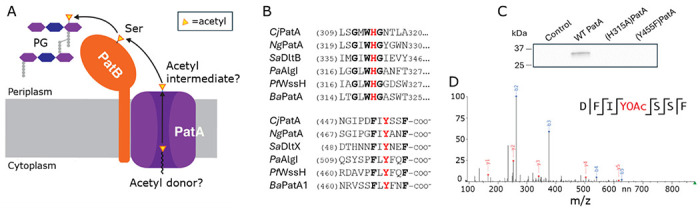
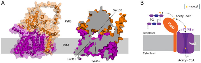
Bacterial cell envelope polymers are commonly modified with acyl groups that provide fitness advantages. Many polymer acylation pathways involve pairs of membrane-bound O-acyltransferase (MBOAT) and SGNH family proteins. As an example, the MBOAT protein PatA and the SGNH protein PatB are required in Gram-negative bacteria for peptidoglycan O-acetylation. The mechanism for how MBOAT-SGNH transferases move acyl groups from acyl-CoA donors made in the cytoplasm to extracellular polymers is unclear. Using the peptidoglycan O-acetyltransferase proteins PatAB, we explore the mechanism of MBOAT-SGNH pairs. We find that the MBOAT protein PatA catalyzes auto-acetylation of an invariant Tyr residue in its conserved C-terminal hexapeptide motif. We also show that PatB can use a synthetic hexapeptide containing an acetylated tyrosine to donate an acetyl group to a peptidoglycan mimetic. Finally, we report the structure of PatB, finding that it has structural features that shape its activity as an O-acetyltransferase and distinguish it from other SGNH esterases and hydrolases. Taken together, our results support a model for peptidoglycan acylation in which a tyrosine-containing peptide at the MBOAT's C-terminus shuttles an acyl group from the MBOAT active site to the SGNH active site, where it is transferred to peptidoglycan. This model likely applies to other systems containing MBOAT-SGNH pairs, such as those that O-acetylate alginate, cellulose, and secondary cell wall polysaccharides. The use of an acyl-tyrosine intermediate for MBOAT-SGNH acyl transfer is also shared with AT3-SGNH proteins, a second major group of acyltransferases that modify cell envelope polymers.

Keywords: Bacterial cell wall; O-acetylation; O-acetyltransferase; X-ray crystallography; peptidoglycan.


Conflict of interest statement

Competing Interest Statement: The authors declare no competing interest.

Figures

Figure 1.
Figure 1.. PatA forms a covalent acetyl-Tyr intermediate.
(A) Previous model for PatAB-catalyzed O-acetylation of PG. It was known that PatB transfers acetyl groups to PG via an acetyl-Ser intermediate, but the donor of acetyl to the pathway and how PatA acetylates this Ser on PatB was unknown. (B) Partial multiple sequence alignment of bacterial MBOAT proteins encoded with SGNH proteins. Both the His and Tyr residues essential for PatA activity are strictly conserved. Cj, C. jejuni; Sa, S. aureus; Pa, P. aeruginosa; Pf, P. fluorescens; B. anthracis. (C) SDS-PAGE autoradiography of PatA variants incubated in the presence of [14C]acetyl-CoA. (D) MS/MS sequencing analysis of O-acetylated peptide from tryptic digest of PatA incubated with acetyl-CoA.
Figure 2.
Figure 2.. PatB is a membrane protein and its N-terminal domain is required for membrane insertion but is dispensible for activity.
(A) Cartoon presentation of the AlphaFold2 model of full-length PatB toplology. Numbering in parentheses denotes residues associated with the domains. CM, cytoplasmic membrane. (B) Anti-His Western blot analysis of subcellular fractions of full length PatB from E. coli cells. (C) Acetylesterase activity of full-length CjPatB (black), CjPatBΔ31 (blue), and CjPatBΔ113 (red) on pNP-Ac as substrate. The enzyme variants (1 μM) in 50 mM sodium phosphate pH 7.0 at 37 °C with pNP-Ac at the concentrations indicated. Error bars denote standard deviation (n=3). (D) LC-MS analysis of reaction products of PatB variants acting as O-acetyltransferases. Chitopentaose (G5) and pNP-Ac served as acceptor and donor substrates, respectively, for (a) no enzyme control, (b) CjPatBΔ113, and (c) CjPatBΔ31.
Figure 3.
Figure 3.. The overall structure of PatB.
The native structure of NgPatBΔ100 (7TJB) is presented as a representative of all the structural models of the PatB SGNH domain. (A) Ribbon presentation of PatB depicting a central β/α fold typical of the SGNH hydrolase family. Two extended β-hairpin motifs at the C-terminal face of the central β-sheet are present, which is not a feature of any structurally resolved SGNH hydrolase member. A short, two-stranded β-sheet joins the N- and C-terminal loops of the SGNH domain at the opposite face to the β-hairpins. (B) Surface representation showing the catalytic triad and oxyanion hole residues (as sticks) arranged on the surface of NgPatB. The β-hairpin motifs extend the SGNH fold along the face that contains the active site to an approximate 60 planar surface. (C) B-factor putty model of PatB. The width and coloring of residues is based upon B-factor. The central SGNH fold contains lower (<30) B-factors, while the β-hairpin motifs display the highest B-factors (>50). (D) The surface electrostatic potential of PatB. Several positively charged Lys and Arg residues are positioned above the active site and across the β-hairpins. (E) The active site of NgPatBΔ100 depicting the positions of the Block I, II, and IV residues. An unusual Block II motif results in a family-atypical type I β-turn that composes the oxyanion hole, together with the Block III Asn residue. A hydrophobic “wall” is formed behind the active site. A sulfate ion is coordinated by Ser133, Ser161, Asn196 and His305.
Figure 4.
Figure 4.. O-Acetylated C-terminal peptide of PatA as acetyl donor for PatB.
(A) Esterase activity of NgPatBΔ100 was monitored at A278nm as the tyrosyl residue in the C-terminal peptide FIYANF of NgPatA was de-O-acetylated with the addition of the enzyme (arrow). (B) LC-MS analysis of the reaction products of the transferase reaction involving incubation of NgjPatBΔ100 with chitopentaose (G5) as acceptor in the (a) absence (negative control) and (b) presence of the O-acetylated peptide.
Figure 5.
Figure 5.. Models of the PatAB complex.
(A) AlphaFold Multimer model. The overall orientation of CjPatB is maintained, with the N-terminal region interacting with both the membrane and PatA (left). A cross-section (right) demonstrates that Tyr455 and His315 of PatA are positioned at the top of a solvent-accessible cavity where CoA presumably binds. Above the PatA active site is a solvent-accessible tunnel lined on top by the active site of PatB. (B) The working model for PG O-acetylation in Gram-negative bacteria. PatA transfers an acetyl group from a cytoplasmic CoA donor to a conserved periplasmic Tyr residue. PatB then transfers this acetyl group via a covalent Ser intermediate to PG.

Similar articles

References

    1. Silhavy T. J., Kahne D. & Walker S. The Bacterial Cell Envelope. Cold Spring Harb Perspect Biol 2, 1–16 (2010). - PMC - PubMed
    1. Vollmer W., Blanot D. & De Pedro M. A. Peptidoglycan structure and architecture. FEMS Microbiol. Rev. 32, 149–167 (2008). - PubMed
    1. Whitfield C. & Trent M. . S. Biosynthesis and export of bacterial lipopolysaccharides. Annu. Rev. Biochem. 83, 99–128 (2014). - PubMed
    1. Bera A., Herbert S., Jakob A., Vollmer W. & Götz F. Why are pathogenic staphylococci so lysozyme resistant? The peptidoglycan O-acetyltransferase OatA is the major determinant for lysozyme resistance of Staphylococcus aureus. Mol. Microbiol. 55, 778–787 (2005). - PubMed
    1. Kristian S. A. et al. d-Alanylation of teichoic acids promotes group A Streptococcus antimicrobial peptide resistance, neutrophil survival, and epithelial cell invasion. J. Bacteriol. 187, 6719–6725 (2005). - PMC - PubMed

Publication types