[Preprint]. 2024 Sep 22:2024.09.22.614367.
doi: 10.1101/2024.09.22.614367.

TDRD3 functions as a selective autophagy receptor with dual roles in autophagy and modulation of stress granule stability

Affiliations

TDRD3 functions as a selective autophagy receptor with dual roles in autophagy and modulation of stress granule stability

Matthew Deater et al. bioRxiv. .

Abstract

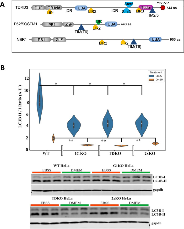
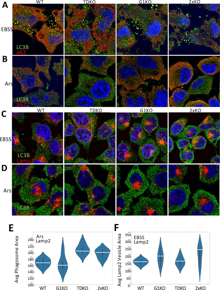
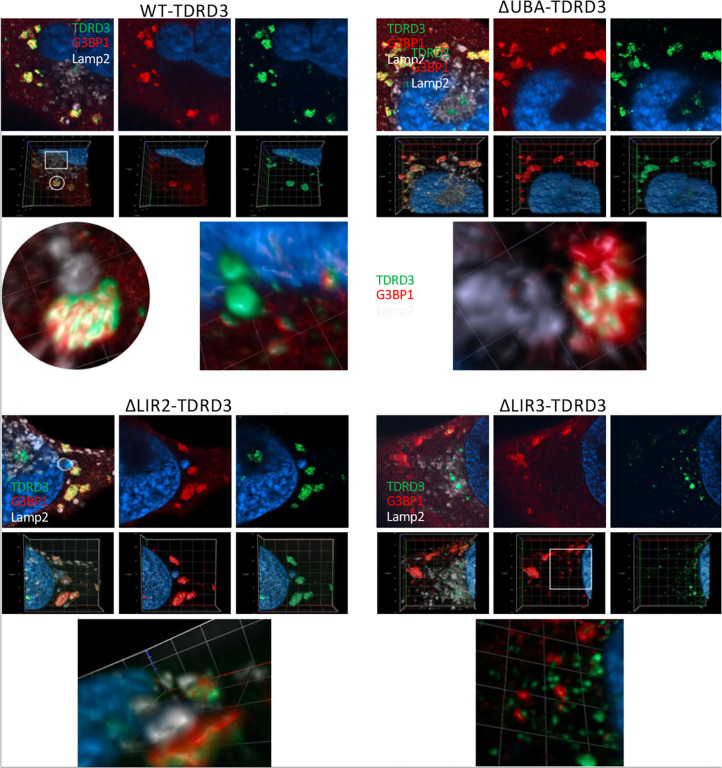
Tudor Domain Containing 3 (TDRD3) is a methylarginine-reader protein that functions as a scaffold in the nucleus facilitating transcription, however TDRD3 is also recruited to stress granules (SGs) during the Integrated Stress Response (ISR) although its function therein remains largely unknown. We previously showed that TDRD3 is a novel antiviral restriction factor that is cleaved by virus 2A protease, and plays complex modulatory roles in both interferon and inflammatory signaling during stress and enterovirus infections. Here we have found that TDRD3 contains structural motifs similar to known selective autophagy receptors such as p62/SQSTM1, sharing ubiquitin associated domains (UBA) and LC3 interacting regions (LIR) that anchor cargo destined for autophagosomes to activated LC3 protein coating autophagosome membranes. This is of interest since enteroviruses hijack autophagy machinery to facilitate formation of viral replication factories, virus assembly and egress from the infected cell. Here we explored possible roles of TDRD3 in autophagy, hypothesizing that TDRD3 may function as a specialized selective autophagy receptor. We found that KO of TDRD3 in HeLa cells significantly reduces starvation induced autophagy, while its reintroduction restores it in a dose-dependent manner. Autophagy receptors are degraded during autophagy and expression levels decrease during this time. We found that TDRD3 levels decrease to the same extent as the autophagy receptor p62/SQSTM1 during autophagy, indicating autophagy-targeted turnover in that role. Knockout of TDRD3 or G3BP1 did not make significant changes in overall cell localization of LC3B or p62/SQSTM1, but did result in greater concentration of Lamp2 phagosome marker for phagosomes and phagolysosomes. To test the potential roles of TDRD3 in autophagic processes, we created a series of deletion mutants of TDRD3 lacking either UBA domain or the various LIR motifs that are predicted to interact with LC3B. Microscopic examination of starved cells expressing these variants of TDRD3 showed ΔLIR-TDRD3 had defects in colocalization with LC3B or Lamp2. Further, super resolution microscopy revealed ring structures with TDRD3 interfacing with p62/SQSTM1. In examination of arsenite induced stress granules we found recruitment of TDRD3 variants disrupted normally tight SG condensation, altered the decay rate of SGs upon release from stress and the kinetics of SG formation. We found evidence that the LIR3 motif on TDRD3 is involved in TDRD3 interaction with LC3B in coIP experiments, colocalization studies, and that this motif plays a key role in TDRD3 recruitment to SGs and SG resolution. Overall, these data support a functional role of TDRD3 in selective autophagy in a mode similar to p62/SQSTM1, with specific roles in SG stability and turnover. Enterovirus cleavage of TDRD3 likely affects both antiviral and autophagic responses that the virus controls for replication.


Conflict of interest statement

Disclosures: The authors declare no competing interests exist

Figures

Fig. 1.
Fig. 1.
A. Domain structure of TDRD3 and archetype autophagy receptors. TDRD3 domains include DUF(domain of unknown function), OB fold, Tudor domain and two large intrinsically disordered regions (IDR). TDRD3 has three functional domains and motifs shared with key autophagy receptors p62/SQSTM1 and NBR1; LIR motif (LC3A/B/C interaction motif), UBA ubiquitin binding domain and Traf interaction motifs (TIM). LIR binds AT8 family proteins during autophagy. B. Both TDRD3 and G3BP1 facilitate basal and starvation-induced autophagy. Wild-type HeLa cells or knockouts for G3BP1 (G1KO), TDRD3 (TDKO) or both G3BP1 and TDRD3 (2xKO) were plated in triplicate, grown in DMEM supplemented with 10% FBS until confluency. Some plates were starved by washing in PBS then incubation in Earle’s Balanced Salt Solution (EBSS) for 4 hrs. All cells were collected for analysis, quickly lysed in RIPA buffer, sonicated briefly and analyzed by western blot against LC3BI and LC3BII. LC3B ratios were determined by densitometry and statistics performed using T-tests between cell types of similar treatments. Representative Western blots are shown below. *=p ≤ 0.05, **=p ≤ 0.01, ***=p ≤ 0.001.
Fig. 2.
Fig. 2.
A. TDRD3 and p62/SQSTM1 display coordinate altered expression levels during autophagy induction and inhibition. Cells were untreated (DMEM), or starved by washing in PBS then incubation in Earle’s Balances Salt Solution (EBSS) for 4 hrs. Alternatively, cells starved with EBSS supplemented with either 20 mM NH4Cl or 50 μg MG132 per manufacturer’s instruction. to inhibit autophagic protein turnover (autophagic flux). Cells were analyzed by western blot for LC3BI/LC3BII. LC3B ratios were determined by densitometry and statistics performed using T-tests between cell types of similar treatments. *=p ≤ 0.05, **=p ≤ 0.01, ***=p ≤ 0.001. B. TDRD3 rescues autophagy during starvation. WT HeLa, TDRD3 KO cells or TDRD3 KO cells ectopically expressing TDRD3-eGFP introduced at indicated plasmid levels/well were starved in EBSS for 2 hrs with no inhibition (N=3). Cells were analyzed by Western blot using antibodies for TDRD3, GAPDH or LC3B. Experiment was repeated (n=6). LC3B-II to LC3B-I ratios were calculated as a measure of autophagic potential for each condition. C. TDRD3 co-immunoprecipitates with LC3B. TDKO HeLa cells were cotransfected at a 1:1 ratio with TDRD3 variants expression plasmids lacking GFP tags along with an LC3B-GFP-RFP fusion construct for 18hr. Cells were starved with EBSS for 4 hrs and coIP was performed using Chromotec GFP-Trap and precipitates analysed by Western blot.
Fig. 3.
Fig. 3.
Altered morphology of LC3B/p62/SQSTM1 autophagosome and Lamp2 phagolysosomes in TDRD3 or G3BP1 knockout cells. Cells were starved 4hrs with EBSS (A, C) or treated with arsenite for 1 hr (B, D) before processing for immunofluorescence with indicated antibodies. E. Quantitation of phagosome sizes during arsenite stress. F. Quantitation of phagosome size during starvation.
Fig. 4.
Fig. 4.
A. Colocalization of endogenous levels of TDRD3 variants with LC3B and p62/SQSTM1 during starvation. TDKO cells expressing low endogenous levels of TDRD3 variants were starved 4 hrs before fixation and processing for IFA microscopy by immunostaining for LC3B and p62/SQSTM1. Vignettes shown are taken from indicated areas in images. B,C. Starved cells processed by immunostaining for LC3B and p62/SQSTM1 were subjected to super-resolution microscopy.
Fig. 5.
Fig. 5.
Colocalization of TDRD3 variants with LC3B and p62/SQSTM1 during arsenite stress and recovery from stress. A. IFA microscopy of TDRD3 variants expressed at endogenous levels in cells treated for one hr with arsenite, immunostained for G3BP1 (A) or LC3B and p62/SQSTM1 (B). Arrows indicate regions with numerous small TDRD3 foci that do not colocalize with G3BP1. C. The number of cells observed with similar small TDRD3 foci at indicated times post-Arsenite recovery. D. The number of cells containing G3BP1-stained SGs vs TDRD3-stained SGs (contain TDRD3 and G3BP1) at indicated times post-arsenite recovery. E. Examples of common phenotypes of TDRD3 condensates undergoing disassembly from SGs during 45, 60 or 120 min recovery.
Fig. 6.
Fig. 6.
Kinetics of SG recruitment of TDRD3 deletion variants during arsenite induced SG assembly. Microscopy images were taken every 10 sec post-addition of arsenite for 60 sec and SG content determined by CellProfiler analysis. Images below graph are representatives from time 0, 30 min or 60 min. B. Super resolution microscopy of non-SG associated ring structures observed in some cells during Ars stress.
Fig. 7.
Fig. 7.
Super resolution microscopy of non-SG associated ring structures observed in some cells during Ars stress. Cells were expressing endogenous levels of the indicated TDRD3-eGFP variant. Top row for each indicated 2D micrograph, a 3D image of the same area is below that, followed by enlargements of indicated areas of 3D images.

Similar articles

Cited by

References

    1. Goulet I., Boisvenue S., Mokas S., Mazroui R. & Côté J. TDRD3, a novel Tudor domain-containing protein, localizes to cytoplasmic stress granules. Human Molecular Genetics 17, 3055–3074 (2008). - PMC - PubMed
    1. Yang Y. et al. TDRD3 is an effector molecule for arginine-methylated histone marks. Mol Cell 40, 1016–1023 (2010). - PMC - PubMed
    1. Linder B. et al. Tdrd3 is a novel stress granule-associated protein interacting with the Fragile-X syndrome protein FMRP. Human Molecular Genetics 17, 3236–3246 (2008). - PubMed
    1. Morettin A. et al. Tudor Domain Containing Protein 3 Promotes Tumorigenesis and Invasive Capacity of Breast Cancer Cells. Sci Rep 7, 5153 (2017). - PMC - PubMed
    1. Saha S. et al. The TDRD3-USP9X complex and MIB1 regulate TOP3B homeostasis and prevent deleterious TOP3B cleavage complexes. Nat. Commun. 14, 7524 (2023). - PMC - PubMed

Publication types