[Preprint]. 2024 Sep 20:2024.09.19.613645.
doi: 10.1101/2024.09.19.613645.

Integrating collecting systems in kidney organoids through fusion of distal nephron to ureteric bud

Affiliations

Integrating collecting systems in kidney organoids through fusion of distal nephron to ureteric bud

Min Shi et al. bioRxiv. .

Update in

Abstract

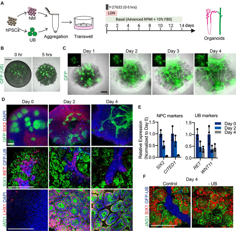
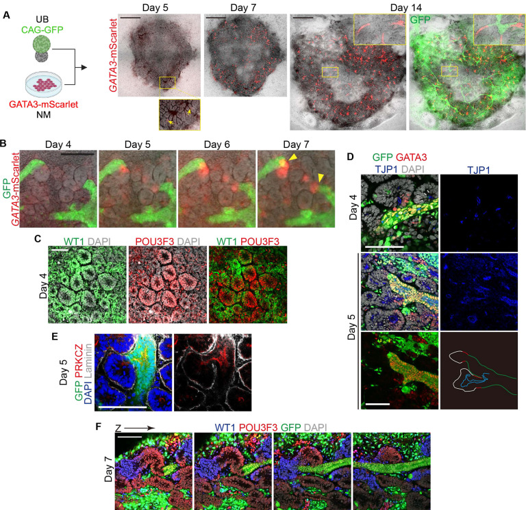
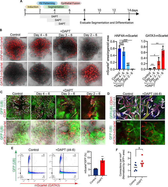
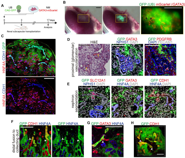
The kidney maintains homeostasis through an array of parallel nephrons, which all originate in development as isolated epithelial structures that later fuse through their distal poles to a system of collecting ducts (CD). This connection is required to generate functional nephrons by providing a pathway for excretion of metabolic waste and byproducts. Currently, methods for differentiating human pluripotent stem cells into kidney organoids generate nephrons that lack CDs and instead terminate as blind-ended tubules. Here we describe a developmentally inspired system that addresses this deficiency through assembly of induced nephrogenic mesenchyme with ureteric bud (UB) tissues, the embryonic building blocks of the kidney's collecting system. The UB progenitors grow and develop into a network of CDs within the organoid, and importantly, they functionally integrate with the nephrons through recapitulating fusion between the distal tubule and CD to create a continuous epithelial lumen. We further showed that proximal-distal nephron specification, fusion frequency, and maturation of the CD can be augmented through temporal manipulation of developmental signaling pathways. This work provides a platform for interrogating the principles and mechanisms underlying nephron-UB fusion and a framework for engineering unobstructed nephrons with patterned collecting systems, an important step toward the de novo generation of functional kidney tissue.


Conflict of interest statement

Declaration of Interests M.S., K.W.M., and J.V.B. are co-inventors on pending UB organoid patents, and M.S. and K.W.M. are coinventors on pending integrated organoid technologies described herein. The other authors have no competing financial interests to declare.

Figures

Figure 1.
Figure 1.. Assembly and dynamics of UB and NM progenitor cells in integrated kidney organoids.
A. Schematized protocol for integration of induced UB and NM into kidney organoids on transwell membranes. B. Within 5 hours, the dissociated NM condensed into a disk-shaped structure and surrounded the embedded GFP+ UB spheroids. C. In several days, the NM was induced into epithelialized renal vesicle-like structures, while the GFP+ UBs grew as elongating and sometimes bifurcating tubular epithelia that penetrated throughout the organoid. Insets show the isolated GFP channel. D. Whole-mount IF staining revealed progressive loss of NM progenitor markers SIX1/2 from days 0–4 with concomitant nearly uniform induction of renal vesicle markers LHX1 and JAG1 accompanying adoption of epithelial morphology. The GFP+ UBs exhibited similar reduction of the tip progenitor gene RET, although patchy expression was still observed on day 4. E. qPCR analyses confirmed loss of undifferentiated NPC markers SIX2 and CITED1 from day 0 to day 4 and reduction of UB tip progenitor markers RET and WNT11. n=3 organoid replicates per timepoint. F. NM progenitors displayed similar differentiation patterns when cultured in the absence of UB spheroids. Scale bars, 500 μm (B-C) and 200 μm (D, F).
Figure 2.
Figure 2.. Formation of collecting ducts in organoids that fuse to distal nephron tubules.
A. Following two weeks of culture, the GFP+ UBs generated extensive networks of CD tubules that were embedded amongst the nephron epithelia of the organoid. B. Numerous points of epithelial fusion (marked by red arrowheads) were observed between the GFP+ CDs and unlabelled nephron tubules. C. Gene expression analysis by qPCR at day 14 revealed lower levels of NPHS1 (**P = 0.0059) but otherwise comparable expression of nephron segment differentiation between mixed (Control) organoids and those without UB. Control organoids had significantly upregulated CD differentiation markers GATA3 (*P = 0.0236), CALB1 (*P = 0.036), AQP2 (*P = 0.0261), and SCNN1B (*P = 0.0214). n=3 independent organoid replicates per group; column and error bars represent mean and standard deviation, respectively. D. Expression of CALB1 in NM-only organoids marked the presumptive connecting segments that terminated as blind-ended tubules (yellow arrows), while in integrated organoids these segments fused to GFP+ CDs also expressing CALB1 (yellow arrowheads indicate epithelial fusion points). E. Nephron-CD anastomoses at day 14 exhibited uninterrupted apicobasal polarity with apparent continuity of the apical lumen across the junction. F. Organoids at day 14 contained an abundance of HNF4A+ proximal tubules and a relative scarcity of GATA3+ distal segments, as shown in micrographs of live organoids harboring fluorescent reporter alleles. The UBs are shown in green (GFP). G. IF staining and image quantification revealed that 96% of epithelial connections (as shown by yellow arrows) involved a GATA3-expressing nephron tubule, and fusions with HNF4A+ proximal tubules were not observed. Scale bars, 1,000 μm (A), 200 μm (B), 100 μm (D-E), and 500 μm (F-G).
Figure 3.
Figure 3.. Fusion to the UB follows nephron polarization and segmentation.
A. GATA3-mScarlet expression in the nephron lineage was first weakly detected as early as day 5 in small domains of renal vesicles (yellow arrows), and by day 7 it was strongly expressed in the presumptive distal segments of nascent nephrons. By day 14, the distal tubules were frequently fused to the GFP+ CDs. B. Daily imaging revealed the process by which the early GATA3+ segment interacts with and invades into nearby GFP+ UB epithelia. Yellow arrowheads indicate points of nephron fusion. C. At day 4, the renal vesicles exhibited polarization with coarse segregation of the proximal (WT1) and distal (POU3F3) domains. D. GATA3 expression in these early polarized vesicles was often associated with extension of the epithelium and its apical membrane (TJP1) toward the UB, and complete apical connections were observed by day 5. E. Loss of extracellular matrix (Laminin) was observed at the site of fusion while the apical membrane (PRKCZ) extended across the junction between the renal vesicle and UB. F. Optical sections through an organoid at day 7 revealed segmented and sometimes organized nephrons progressing from WT1+ presumptive glomerular structures through the distal epithelial fusion with the UB. Scale bars, 1,000 μm (A), 200 μm (B), and 100 μm (C-F).
Figure 4.
Figure 4.. NOTCH inhibition augments distal nephron specification and fusion competence.
A. Summary of early nephrogenesis events in organoids (day 0 = time of mixing NM and UB) and strategy for testing temporal inhibition of NOTCH signaling. B. Proximal (HNF4A) and distal (GATA3) nephron specification visualized and quantified in live organoids at day 8 via mScarlet fluorescent reporter activity. Prolonging exposure to the NOTCH inhibitor DAPT during the segmentation stage led to progressive reduction of proximal and increase in distal nephron formation. n=4 independent biological replicates per condition; ****P <0.0001, ***P = 0.0005, **P =0.0024 (control vs. day 4–6), **P = 0.0037 (control vs. day 2–6) and *P = 0.016. C. At day 14, nephron epithelia displayed disorganized morphology in organoids treated with 3–4 days of DAPT with widespread GATA3 expression and loss of HNF4A+ proximal tubules, whereas shorter treatment (days 4–6) led to increased abundance of the short GATA3+ distal segments compared to controls but with preserved overall morphology and maintenance of proximal tubular development. D. The organoids treated with DAPT from days 4–6 retained a comparable number of HNF4A+ proximal tubules and NPHS1+ podocytes, and they contained more GATA3+ distal segments that were fused to the UB-derived ducts. E. DAPT treatment induced 2.5-fold expansion in GATA3+ distal nephron cells by flow cytometry (n=3 independent biological replicates per condition; **P = 0.001) and (F) significantly increased frequency of nephron-UB fusion events (n=6 independent organoids per condition; *P = 0.045). Scale bars, 1,000 μm (B, C), and 200 μm (D). Column and error bars represent mean and standard deviation, respectively.
Figure 5.
Figure 5.. UB and NM progenitors are lineage-restricted and differentiate in parallel.
A. UMAP embedding of recombinant kidney organoids with supervised cluster annotation. B. DevKidCC assignment scoring for lineage classification (Tier 1) and nephron segmentation (Tiers 2/3). C. Cells in the dataset largely segregated by their stage of differentiation, days 3, 7, and 15. D. Expression of the lineage label GFP was specific to the two UB clusters, confirming the lineage fidelity of the early progenitors. These cells were extracted and re-analyzed to reveal 5 clusters representing the differentiation trajectory of the UB lineage. E. The UB cells at day 3 were enriched for tip/progenitor markers, while cells on day 15 exhibited a more mature CD signature. Day 7 cell exhibited an intermediate phenotype including expression of the stalk progenitor marker WNT9B. F. A CellChat analysis was performed to compare the putative signaling interactions that originate in the UB and are enriched in distal over proximal receiver probability. Pathways in blue show statistically significant (P < 0.05) increases in signaling probability. G. Bubble plot showing predicted increased receptivity of the early distal tubule for UB-derived WNT9B signaling, as well as other secreted factors including TGFB and NRG family members.
Figure 6.
Figure 6.. Fusion of UB and distal nephron following in vivo transplantation.
A. Overview of organoid transplantation experiments. B. Gross appearance and stereomicrographs of organoid tissue on the kidney surface following two weeks of in vivo growth. Short GATA3+ nephron tubules were seen throughout the graft, including many that were connected to larger GFP+ duct-like structures (black arrows). The general haziness of the GFP signal indicated robust growth of UB organoid-derived stromal cells. C. The engrafted tissue comprised numerous NM-derived proximal (HNF4A) and distal (CDH1) tubules and large UB organoid derived duct structures and interstitial cells. D. In vivo growth enabled vascularization and maturation of organoid glomerular structures that comprised an organized arrangement of podocytes (NPHS1), endothelial cells (PECAM1), and mesangial cells (PDGFRB, GATA3). Apparent perfusion of the glomerular tufts was indicated by the presence of red blood cells (yellow arrows). E. Segmented arrangement of nephrons in the graft was confirmed through serial sections highlighting the sequential progression of podocytes (NPHS1), proximal tubule (HNF4A), thick ascending limb (SLC12A1), and connecting tubule (GATA3). F-H. Fusion of nephron tubules to GFP+ UB-derived ducts was observed in the engrafted organoids (white arrowheads) in both sections (F-G) and wholemount staining (H), and it was restricted to the CDH1/GATA3+ distal segments. Scale bars, 1,500 μm (B), 500 μm (C), 100 μm (D-E), 200 μm (F-G), and 300 μm (H).
Figure 7.
Figure 7.. Induction of CD maturation in recombinant organoids.
A. The expression of AQP2 was induced in UB epithelia grown either in isolation or in recombinant organoids with NM when cultured in previously identified conditions to grow UB organoids (UB Medium), but not when grown in the minimal ‘Mix’ media. B. The combined organoids were transitioned at day 10 from ‘Mix’ medium to induce maturation. C-E. CD Medium (CDM) induced higher expression of AQP2 in the CDs and it was significantly further augmented by the addition of A83, U0126, and XAV, as shown through the reporter allele (C), protein staining (D), and qPCR (E). n=3 independent biological replicates per condition; column and error bars represent mean and standard deviation, respectively; P-values shown in figure. Scale bars, 500 μm (A, D), 1,000 μm (C) and 100 μm (F).

References

    1. Mugford J.W., Sipila P., McMahon J.A., and McMahon A.P. (2008). Osr1 expression demarcates a multi-potent population of intermediate mesoderm that undergoes progressive restriction to an Osr1-dependent nephron progenitor compartment within the mammalian kidney. Dev Biol 324, 88–98. 10.1016/j.ydbio.2008.09.010. - DOI - PMC - PubMed
    1. Taguchi A., Kaku Y., Ohmori T., Sharmin S., Ogawa M., Sasaki H., and Nishinakamura R. (2014). Redefining the in vivo origin of metanephric nephron progenitors enables generation of complex kidney structures from pluripotent stem cells. Cell Stem Cell 14, 53–67. 10.1016/j.stem.2013.11.010. - DOI - PubMed
    1. McMahon A.P. (2016). Development of the Mammalian Kidney. Curr Top Dev Biol 117, 31–64. 10.1016/bs.ctdb.2015.10.010. - DOI - PMC - PubMed
    1. Kao R.M., Vasilyev A., Miyawaki A., Drummond I.A., and McMahon A.P. (2012). Invasion of distal nephron precursors associates with tubular interconnection during nephrogenesis. J Am Soc Nephrol 23, 1682–1690. 10.1681/ASN.2012030283. - DOI - PMC - PubMed
    1. Georgas K., Rumballe B., Valerius M.T., Chiu H.S., Thiagarajan R.D., Lesieur E., Aronow B.J., Brunskill E.W., Combes A.N., Tang D., et al. (2009). Analysis of early nephron patterning reveals a role for distal RV proliferation in fusion to the ureteric tip via a cap mesenchyme-derived connecting segment. Dev Biol 332, 273–286. 10.1016/j.ydbio.2009.05.578. - DOI - PubMed

Publication types

LinkOut - more resources