[Preprint]. 2024 Sep 16:2024.09.16.613344.
doi: 10.1101/2024.09.16.613344.

RalB uncoupled exocyst mediates endothelial Weibel-Palade body exocytosis

Affiliations

RalB uncoupled exocyst mediates endothelial Weibel-Palade body exocytosis

Moua Yang et al. bioRxiv. .

Update in

Abstract

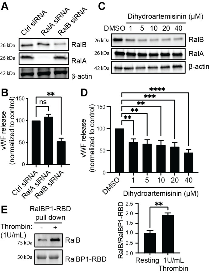
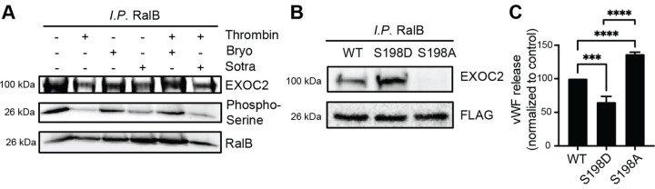
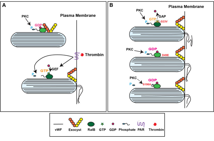
Ras-like (Ral) GTPases play essential regulatory roles in many cellular processes, including exocytosis. Cycling between GDP- and GTP-bound states, Ral GTPases function as molecular switches and regulate effectors, specifically the multi-subunit tethering complex exocyst. Here, we show that Ral isoform RalB controls regulated exocytosis of Weibel-Palade bodies (WPBs), the specialized endothelial secretory granules that store hemostatic protein von Willebrand factor. Remarkably, unlike typical small GTPase-effector interactions, RalB binds exocyst in its GDP-bound state in resting endothelium. Upon endothelial cell stimulation, exocyst is uncoupled from RalB-GTP resulting in WPB tethering and exocytosis. Furthermore, we report that PKC-dependent phosphorylation of the C-terminal hypervariable region (HVR) of RalB modulates its dynamic interaction with exocyst in endothelium. Exocyst preferentially interacts with phosphorylated RalB in resting endothelium. Dephosphorylation of RalB either by endothelial cell stimulation, or PKC inhibition, or expression of nonphosphorylatable mutant at a specific serine residue of RalB HVR, disengages exocyst and augments WPB exocytosis, resembling RalB exocyst-binding site mutant. In summary, it is the uncoupling of exocyst from RalB that mediates endothelial Weibel-Palade body exocytosis. Our data shows that Ral function may be more dynamically regulated by phosphorylation and may confer distinct functionality given high degree of homology and the shared set of effector protein between the two Ral isoforms.


Conflict of interest statement

Conflict of Interest The authors declare no financial conflict of interest.

Figures

Figure 1.
Figure 1.. RalB is switched ‘on’ upon thrombin stimulation and regulates endothelial Weibel-Palade body exocytosis.
(A) HUVECs depleted of RalA or RalB using specific siRNAs and compared with control siRNA. Western blot of total cell lysates showing RalA and RalB knockdown after 48 hours of transfection of siRNA, with β -actin as loading control. (B) Bar graphs showing total vWF antigen in media as estimated by ELISA 30 minutes after 1 U/ml thrombin stimulation 48 hours after siRNA transfection (**P<0.01; from 3 individual experiments each with duplicates). (C) HUVECs treated with increasing concentrations of dihydroartemisinin for 2 hours or DMSO. Western blot showing degradation of RalB but not RalA by dihydroartemisinin, with β-actin as loading control. (D) Bar graphs showing total vWF antigen in media as estimated by ELISA 30 minutes after 1 U/ml thrombin stimulation 2 hours after dihydroartemisinin treatment (**P<0.01, ***P<0.001; ****P<0.0001 from 3 individual experiments each with duplicates). (E) Total cell lysates of resting and thrombin-treated (1 U/ml for 2 minutes) HUVECs were incubated with RalBP1-RBD conjugated agarose slurry and eluates immunoblotted with anti-RalB antibody to determine total GTP-loaded RalB in each condition. Coomassie stain showing RalBP1-RBD as loading control (left). Bar graphs showing densitometric analysis (right). (**P<0.01; from 3 individual experiments).
Figure 2.
Figure 2.. RalB associates with mature WPBs.
(A-C) IF performed on HUVECs with anti-vWF and anti-RalB antibodies 48 hours after transfection with RalB siRNA compared with control siRNA. (A) Micrograph showing presence of RalB (red) in WPBs labeled with vWF (green) in control HUVECs. DAPI (blue) showing nuclei. Inset depicting magnified image of a single WPB. Scale bar 5 μm. (B) Micrograph showing unaffected distribution of WPBs (vWF; green) and RalB (red) in RalB-depleted HUVECs. Scale bar 5 μm. (C) Bar graph showing number of WPBs per cell 48 hours after transfection with RalB siRNA and compared with controls (ns=not significant; 30 random cells examined for each condition in 3 individual experiments). (D) IF performed with anti-vWF and anti-Rab27a antibodies 48 hours after transfection with RalB siRNA. Micrograph showing presence of Rab27A (red) in WPBs labeled with vWF (green). DAPI (blue) showing nuclei. Inset depicting magnified image of a WPB. Scale bar 5 μm.
Figure 3.
Figure 3.. RalB activation tethers WPBs to the plasma membrane.
(A) Expression of autoactivated RalB (RalB-G23V) in HUVECs augments vWF release as compared with RalB-WT. Bar graphs showing total vWF antigen in media estimated by ELISA from resting and thrombin stimulated HUVECs (1 U/ml thrombin for 30 minutes) (***P<0.001 or ****P<0.0001, from 3 individual experiments each with triplicates). (B) RalB-G23V and RalB-WT expressing HUVECs labeled with anti-vWF antibody and imaged with a confocal immunofluorescence microscope. 100 random cells counted, and cells expressing < 15 WPBs considered as empty cell. Bar graphs showing percent empty cells (**P<0.01; from 3 individual experiments). (C) CD63-EGFP expressing HUVECs transduced with lentiviral particles containing RalB-G23V or RalB-WT. WPBs appear as elongated ‘cigars’ (white arrowheads) distributed among round CD63-positive endolysosomes. WPBs appear tethered to the plasma membrane in RalB-G23V cells as compared with cytoplasmic distribution in RalB-WT cells. Scale bar 10 μm.
Figure 4.
Figure 4.. Exocyst interacts with GDP-loaded RalB.
(A) Confocal immunofluorescence micrographs showing presence of RalB (purple) and EXOC7 (red) with vWF (green) in mature WPBs. Insets depict magnified image of a mature WPB. DAPI (blue) showing nucleus. Scale bar 8 μm. (B) Transmission electron microscopy of HUVECs with double immunogold labeling with anti-EXOC4 (15 nm) and FLAG (10 nm) antibodies. Transmission electron micrograph showing both EXOC4 and RalB decorating mature electron-dense WPB ‘cigars’ (white asterisk). Scale bar 100 nm. (C) Western blots showing immunoprecipitation (IP) of resting and thrombin stimulated (1U/ml for 2 min) total HUVEC lysates with anti-RalB or anti-EXOC4 antibodies resolved by SDS-PAGE and immunoblotted with anti-RalB and anti-EXOC2 antibodies. Total HUVEC lysate (~5% of IP) used for input. (D) Total cell lysates of RalB-WT and RalB-G23V expressing HUVEC incubated with magnetic beads conjugated with anti-FLAG antibody and eluates immunoblotted with anti-FLAG and anti-EXOC2 antibodies. (E) Total cell lysates of resting and thrombin-treated (1 U/ml for 2 minutes) HUVECs incubated with EXOC2-RBD conjugated agarose slurry and eluates immunoblotted for with anti-RalB antibody. Ponceau S staining showing EXOC2-RBD as loading control.
Figure 5.
Figure 5.. PKC-dependent RalB phosphorylation modulates exocyst binding and WPB exocytosis.
(A) Thrombin stimulation dephosphorylates RalB and reduces EXOC2 binding. IP with anti-RalB or anti-EXOC4 antibodies of total cell lysates of resting and thrombin stimulated HUVECs (1U/ml for 2 min) with and without PKC activator bryostatin-1 (100nM for 10 min) or PKC inhibitor sotrostaurin (500nM for 1 hour), resolved by SDS-PAGE and immunoblotted with anti-phosphoserine, anti-RalB and anti-EXOC2 antibodies. Total HUVEC lysate (~5% of IP) used as input. (B) Expression of phosphomimetic RalB-S198D mutant increases EXOC2 binding, and phosphodeficient RalB-S198A mutant decreases EXOC2 binding in HUVECs. Total cell lysates of RalB-WT, RalB-S198D and RalB-S198A expressing cells incubated with magnetic beads conjugated with anti-FLAG antibody and eluates immunoblotted with anti-Flag and anti-EXOC2 antibodies. (C) Bar graphs showing total vWF antigen in media (at 30 minutes) collected from resting HUVECs expressing RalB-WT, RalB-S198D and RalB-S198A as estimated by ELISA (***P<0.001; ****P<0.0001, from 3 individual experiments each with triplicates).
Figure 6.
Figure 6.. RalB uncoupled exocyst triggers WPB exocytosis.
(A) Expression of RalB exocyst binding site mutation in HUVECs (RalB-WT D49E and RalB-G23V D49E). Total cell lysates of RalB-WT and RalB-G23V and their respective D49E mutants incubated with magnetic beads conjugated with anti-FLAG antibody and eluates immunoblotted with anti-FLAG and EXOC2 antibodies. (B) Bar graphs showing total vWF antigen in media from HUVECs expressing RalB-WT, RalB-WT D49E, RalB-G23V or RalB-G23V D49E mutants (*P<0.01; ns=non-significant; from 3 individual experiments each with triplicates). (C) RalB-WT and RalB-WT D49E expressing HUVECs labeled with anti-vWF antibody and imaged with a confocal immunofluorescence microscope. Micrograph showing WPBs labeled with vWF (red) decorating plasma membrane in RalB-WT D49E mutants as compared to cytoplasmic distribution in RalB-WT. DAPI (blue) depicting nuclei. Scale bar 5 μm.
Figure 7.
Figure 7.. Proposed model for RalB-exocyst dependent WPB tethering.
(A) In resting HUVECs, RalB is GDP-loaded as well as phosphorylated by protein kinase C in its C-terminal hypervariable region. Upon endothelial cell stimulation by thrombin, RalB becomes GTP-loaded catalyzed by GEF, as well as dephosphorylated by activation of phosphatases. Consequently, exocyst is uncoupled from dephosphorylated and GTP-bound RalB and tethers WPBs to the plasma membrane facilitating WPB exocytosis and vWF release. (B) RalB mutations which render RalB autoactivated (G23V; top), phosphodeficient (S198A; middle) or uncoupled from exocyst (D49E; bottom), trigger WPB tethering and vWF release in the absence of endothelial cells stimulation. (GEF, guanine nucleotide exchange factor; GAP, GTPase activating protein; P, phosphate; PKC, protein kinase C; PAR, protease activated receptor)

Similar articles

References

    1. Gerber SH, Sudhof TC. Molecular determinants of regulated exocytosis. Diabetes. Feb 2002;51 Suppl 1:S3–11. doi: 10.2337/diabetes.51.2007.s3 - DOI - PubMed
    1. Sharda A, Flaumenhaft R. The life cycle of platelet granules. F1000Res. 2018;7:236. doi: 10.12688/f1000research.13283.1 - DOI - PMC - PubMed
    1. Cassatella MA, Ostberg NK, Tamassia N, Soehnlein O. Biological Roles of Neutrophil-Derived Granule Proteins and Cytokines. Trends Immunol. Jul 2019;40(7):648–664. doi: 10.1016/j.it.2019.05.003 - DOI - PubMed
    1. Campbell JE, Newgard CB. Mechanisms controlling pancreatic islet cell function in insulin secretion. Nat Rev Mol Cell Biol. Feb 2021;22(2):142–158. doi: 10.1038/s41580-020-00317-7 - DOI - PMC - PubMed
    1. Springer TA. von Willebrand factor, Jedi knight of the bloodstream. Blood. Aug 28 2014;124(9):1412–25. doi: 10.1182/blood-2014-05-378638 - DOI - PMC - PubMed

Publication types

LinkOut - more resources