[Preprint]. 2024 Sep 20:2024.09.20.612365.
doi: 10.1101/2024.09.20.612365.

Preventing Inappropriate Signals Pre- and Post-Ligand Perception by a Toggle-Switch Mechanism of ERECTA

Affiliations

Preventing Inappropriate Signals Pre- and Post-Ligand Perception by a Toggle-Switch Mechanism of ERECTA

Liangliang Chen et al. bioRxiv. .

Update in

Abstract

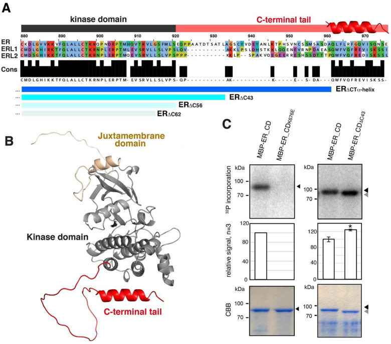
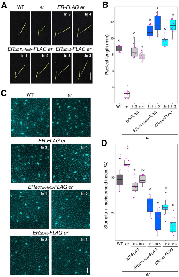
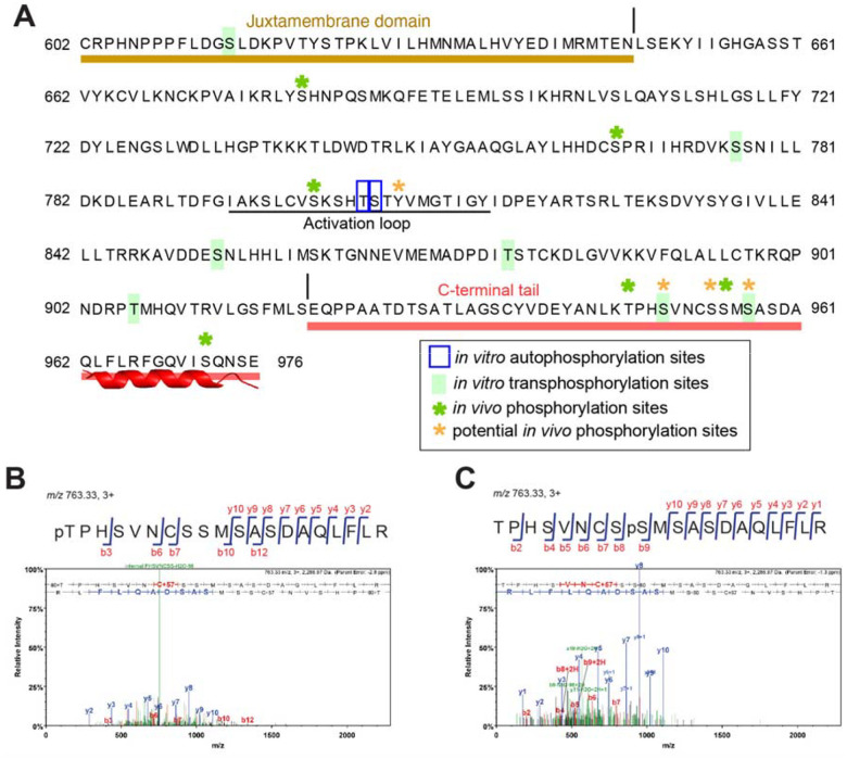
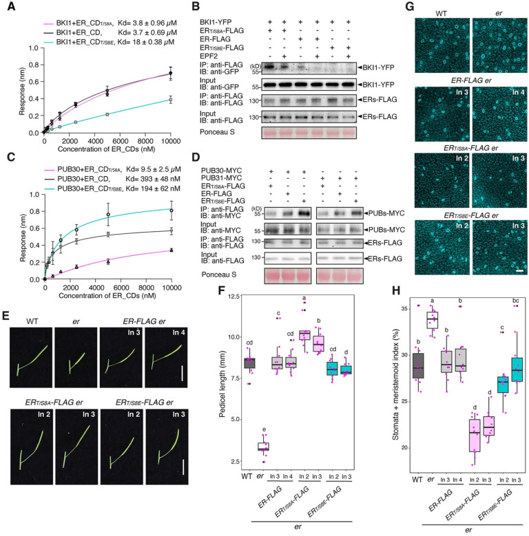
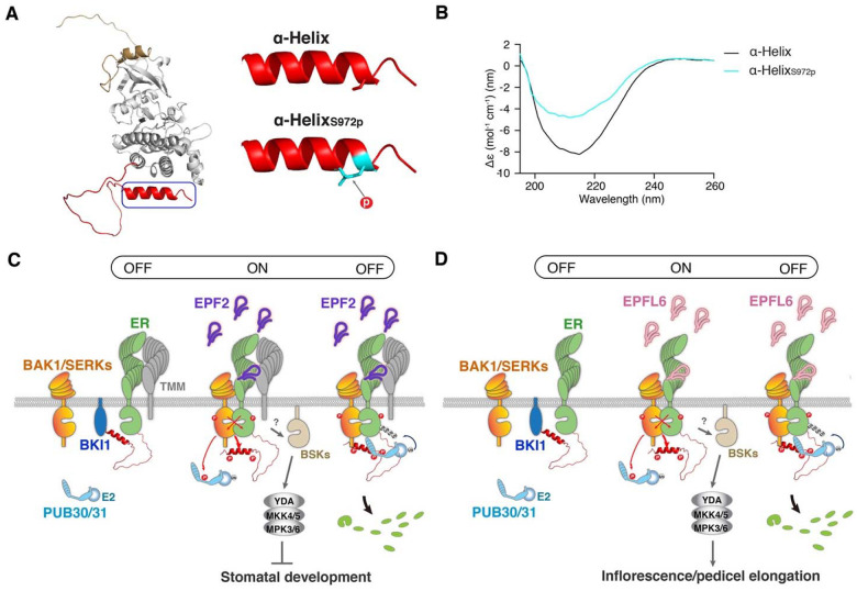
Dynamic control of signaling events requires swift regulation of receptors at an active state. By focusing on Arabidopsis ERECTA (ER) receptor kinase, which perceives peptide ligands to control multiple developmental processes, we report a mechanism preventing inappropriate receptor activity. The ER C-terminal tail (ER_CT) functions as an autoinhibitory domain: its removal confers higher kinase activity and hyperactivity during inflorescence and stomatal development. ER_CT is required for the binding of a receptor kinase inhibitor, BKI1, and two U-box E3 ligases PUB30 and PUB31 that inactivate activated ER. We further identify ER_CT as a phosphodomain transphosphorylated by the co-receptor BAK1. The phosphorylation impacts the tail structure, likely releasing from autoinhibition. The phosphonull version enhances BKI1 association, whereas the phosphomimetic version promotes PUB30/31 association. Thus, ER_CT acts as an off-on-off toggle switch, facilitating the release of BKI1 inhibition, enabling signal activation, and swiftly turning over the receptors afterwards. Our results elucidate a mechanism fine-tuning receptor signaling via a phosphoswitch module, keeping the receptor at a low basal state and ensuring the robust yet transient activation upon ligand perception.

Keywords: Arabidopsis; C-terminal Tail Domain; Phosphoregulation; Receptor Kinase; stomatal development.


Figures

Figure 1.
Figure 1.. The conserved C-terminal tail in ER-family LRR-RKs inhibits the kinase activity
(A) Alignment of the C-terminal tail domain (ER_CT) of ER and ERLs. Conserved residues (Cons) are filled in black. Kinase domain is highlighted in dark gray, and ER_CT is in red. The location of α-Helix is superimposed. Deletions used in this study are indicated in blue: ERΔCTα-Helix, ERΔC43, ERΔC56, and ERΔC62. (B) Structural modeling of the cytoplasmic domain of ER (ER_CD), with the Juxtamembrane domain in sand, the Kinase domain in gray, and the C-terminal tail domain in red. (C) In vitro phosphorylation assay of ER_CD. Left panels, control ER_CD and kinase-dead version (ER_CDK676E). Right panels, ER_CD and ER_CDΔC43. Top panels, autoradiography. Middle panels, mean and S.D. of densitometry plotted for the experiments performed 3 times. ** T-test p<0.05. Bottom panels, Coomassie Brilliant Blue staining (CBB) as a loading control.
Figure 2.
Figure 2.. Removal of the ER C-terminal tail domain leads to hyperactivity in promoting pedicel elongation and inhibiting stomatal development
(A) Representative pedicels and mature siliques of WT, er-105 (er), ER-FLAG er, ERΔCTα-Helix-FLAG er, and ERΔC43-FLAG er plants. For each transgenic constructs, two representative lines were subjected to analysis. Images were taken under the same magnification. Scale bar, 1 cm. (B) Morphometric analysis of pedicel length from each genotype. Six-week-old mature pedicels (n = 10) were measured. One-way ANOVA followed by Tukey’s HSD test was performed and classified their phenotypes into categories (e.g. a, b, c). (C) Representative confocal microscopy of cotyledon abaxial epidermal at 7 day-post-germination (DPG7) from wild type (WT), er-105 (er), ER-FLAG er, ERΔCTα-Helix-FLAG er, and ERΔC43-FLAG er. The same representative transgenic lines were observed. Images were taken under the same magnification. Scale bar = 50 μm. (D) Quantitative analysis. Stomata + meristemoid index (number of stomata and meristemoid per 100 epidermal cells) of the cotyledon abaxial epidermis from 7-day-old seedlings of respective genotypes (n = 10). One-way ANOVA followed by Tukey’s HSD test was performed and classified their phenotypes into categories (e.g. a, b, c).
Figure 3.
Figure 3.. The C-terminal tail is essential for the binding of ER with BKI1 and PUB30
(A) Quantitative analysis of interactions between BKI1 and ER_CD variants (ER_CD, ER_CDΔCTα-Helix, and ER_CDΔ43) using BLI. In vitro binding response curves for recombinantly purified GST-BKI1 and MBP-ER_CD variants at seven different concentrations (156.25, 312.5, 625, 1,250, 2,500, 5,000, and 10,000 nM) are shown. Kd values are indicated. Data are representative of three independent experiments. (B) Deletion of C-terminal tail decreases the association of ER with BKI1 in vivo. Proteins from double transgenic lines carrying ERpro::BKI1-YFP ERpro::ER-FLAG, ERpro::BKI1-YFP ERpro::ERΔCTα-Helix-FLAG, and ERpro::BKI1-YFP ERpro::ERΔC43-FLAG were immunoprecipitated with anti-FLAG beads (IP). The immunoblots (IB) were probed with anti-FLAG and anti-GFP antibodies, respectively. (C) Quantitative analysis of interactions between PUB30 and ER_CD variants (ER_CD, ER_CDΔCTα-Helix, and ER_CDΔC43) using BLI. In vitro binding response curves for recombinantly purified GST-PUB30 and MBP-ER_CD variants at seven different concentrations (156.25, 312.5, 625, 1,250, 2,500, 5,000, and 10,000 nM) are shown. Kd values are indicated. Data are representative of three independent experiments. (D) Deletion of C-terminal tail decreases the association of ER with PUB30 in vivo. Proteins from PUB30pro::PUB30-YFP; ERpro::ER-FLAG, PUB30pro::PUB30-YFP; ERpro:: ERΔCTα-Helix-FLAG, and PUB30pro::PUB30-YFP; ERpro:: ERΔC43-FLAG plants were immunoprecipitated with anti-GFP beads (IP), and the immunoblots (IB) were probed with anti-GFP and anti-FLAG antibodies, respectively. (E) Deletion of C-terminal tail decreases the ubiquitination of ER by PUB30 in vivo. Arabidopsis protoplasts were co-transfected with PUB30-MYC, FLAG-UBQ, together with ER-HA, ERΔCTα-Helix-HA, and ERΔC43-HA. Five micromolar EPFL6 was used for treatment for 1 hr. After immunoprecipitation using anti-FLAG beads, the ubiquitinated ER variants were probed with anti-HA antibody. The total ubiquitinated proteins were probed by anti-FLAG antibody and PUB30 proteins were probed by anti-MYC antibody. The inputs of ER were probed with anti-HA antibody. (F) Representative EPFL6 treatment destabilizes ER variants in Arabidopsis protoplasts co-expressing PUB30-MYC. Protoplasts expressing the indicated proteins were treated with 50 μM CHX and 5 μM EPFL6 for 3 hr. before total protein was examined with immunoblot. The experiment was repeated independently two times with similar results.
Figure 4.
Figure 4.. The C-terminal tail domain of ER is phosphorylated
(A) Phosphorylation sites in the C-terminal tail domain of ER. In vitro auto- and transphosphorylation sites identified by LC-MS/MS analysis are marked in blue and lime green, respectively. In vivo localized phosphorylation sites of ER found in this study are marked with green asterisks. Potential in vivo phosphorylation sites of ER are marked with orange asterisks. Juxtamembrane and C-terminal tail are highlighted in sand and red, respectively. Underline, the Activation Loop. (B, C) MS/MS spectra for selected in vivo phosphorylation sites of ER: Thr947 (B) and Ser955 (C).
Figure 5.
Figure 5.. Phosphorylation of the ER C-terminal tail evicts BKI1 and recruits PUB30/31
(A) Quantitative analysis of interactions between BKI1 and ER_CD variants (ER_CD, ER_CDT/S8A, and ER_CDT/S8E) using BLI. In vitro binding response curves for recombinantly purified GST-BKI1 and MBP-ER_CD variants at seven different concentrations (156.25, 312.5, 625, 1,250, 2,500, 5,000, and 10,000 nM) are shown. Kd values are indicated. Data are representative of three independent experiments. (B) Phosphorylation of C-terminal tail decreases the association of ER with BKI1 in vivo. Proteins from ERpro::BKI1-YFP; Col-0, ERpro::BKI1-YFP; ERpro::ER-FLAG, ERpro::BKI1-YFP; ERpro::ERT/S8A-FLAG, and ERpro::BKI1-YFP; ERpro::ERT/S8E-FLAG plants were immunoprecipitated with anti-FLAG beads (IP), and the immunoblots (IB) were probed with anti-FLAG and anti-GFP antibodies, respectively. (C) Quantitative analysis of interactions between PUB30 and ER_CD variants (ER_CD, ER_CDT/S8A, and ER_CDT/S8E) using BLI. In vitro binding response curves for recombinantly purified GST-PUB30 and MBP-ER_CD variants at seven different concentrations (156.25, 312.5, 625, 1,250, 2,500, 5,000, and 10,000 nM) are shown. Kd values are indicated. Data are representative of three independent experiments. (D) Phosphorylation of C-terminal tail promotes the association of ER with PUB30 and PUB31 in vivo. Arabidopsis protoplasts were co-transfected with PUB30-MYC or PUB31-MYC, together with ER-FLAG, ERT/S8A-FLAG, and ERT/S8E-FLAG. Five micromolar EPFL6 was used for treatment for 1 h. After immunoprecipitation using anti-FLAG beads, the immunoblots (IB) were probed with anti-FLAG and anti-MYC antibodies, respectively. (E) Representative pedicels and mature siliques of WT, er, ER-FLAG er, ERT/S8A-FLAG er, and ERT/S8E-FLAG er plants. Images were taken under the same magnification. Scale bar, 1 cm. (F) Morphometric analysis of pedicel length from each genotype. Six-week-old mature pedicels (n = 10) were measured. One-way ANOVA followed by Tukey’s HSD test was performed and classified their phenotypes into categories (a, b, c, and d). (G) Confocal microscopy of 7-day-old abaxial cotyledon epidermis of WT, er, ER-FLAG er, ERT/S8A-FLAG er, and ERT/S8E-FLAG er plants. Images were taken under the same magnification. Scale bar, 50 μm. (H) Quantitative analysis. Stomata + meristemoid index of the cotyledon abaxial epidermis from 7-day-old seedlings of respective genotypes (n = 10). One-way ANOVA followed by Tukey’s HSD test was performed and classified their phenotypes into categories (a, b, c, and d).
Figure 6.
Figure 6.. Mechanism preventing inappropriate signals pre- and post-activation of ER
(A) Structural modeling of ER_CD (left). Structural modeling of ER_CTα-Helix domain (right, top), with the Ser972 residue shown as sticks in red, and ER_CTα-HelixS972p (right, bottom), with the phosphorylated Ser972 residue shown as sticks in cyan. (B) CD spectra of ER_CTα-Helix (black) and ER_CTα-HelixS972p (cyan). (C, D) Regulation of stomatal development (C) and inflorescence/pedicel elongation (D) by ER_CT. (Left) BKI1 (blue) associates with ER (green) in the absence of ligand. (Middle) Upon perception of EPF2 (C, violet) or EPFL6 (D, pink), ER becomes activated. Both ER kinase domain and C-terminal tail are phosphorylated by its co-receptor BAK1/SERKs (orange) during the transphosphorylation events. The phosphorylation of C-terminal tail evicts BKI1. Activated BAK1/SERKs also phosphorylate PUB30/31 (cyan). The activated ER-BAK1/SERKs receptor complex transduces signals most likely via BSK (sand) before the activation of a MAPK cascade and subsequent inhibition of stomatal development (C) or promotion of inflorescence/pedicel elongation (D). TMM (gray) biases the signal activation for stomatal development (C). The identity of E2 ligase (cyan ball) is unclear.

References

    1. Shiu S.-H., Bleecker A. B., Plant Receptor-Like Kinase Gene Family: Diversity, Function, and Signaling. Science’s STKE 2001, re22–re22 (2001). - PubMed
    1. Morris E. R., Walker J. C., Receptor-like protein kinases: the keys to response. Current Opinion in Plant Biology 6, 339–342 (2003). - PubMed
    1. De Smet I., Voß U., Jürgens G., Beeckman T., Receptor-like kinases shape the plant. Nature Cell Biology 11, 1166 (2009). - PubMed
    1. Couto D., Zipfel C., Regulation of pattern recognition receptor signalling in plants. Nature Reviews Immunology 16, 537 (2016). - PubMed
    1. Tang D., Wang G., Zhou J.-M., Receptor kinases in plant-pathogen interactions: more than pattern recognition. The Plant Cell 29, 618–637 (2017). - PMC - PubMed

Publication types

LinkOut - more resources