[Preprint]. 2024 Sep 18:2024.09.18.613774.
doi: 10.1101/2024.09.18.613774.

A functional screen for ubiquitin regulation identifies an E3 ligase secreted by Pseudomonas aeruginosa

Affiliations

A functional screen for ubiquitin regulation identifies an E3 ligase secreted by Pseudomonas aeruginosa

Cameron G Roberts et al. bioRxiv. .

Abstract

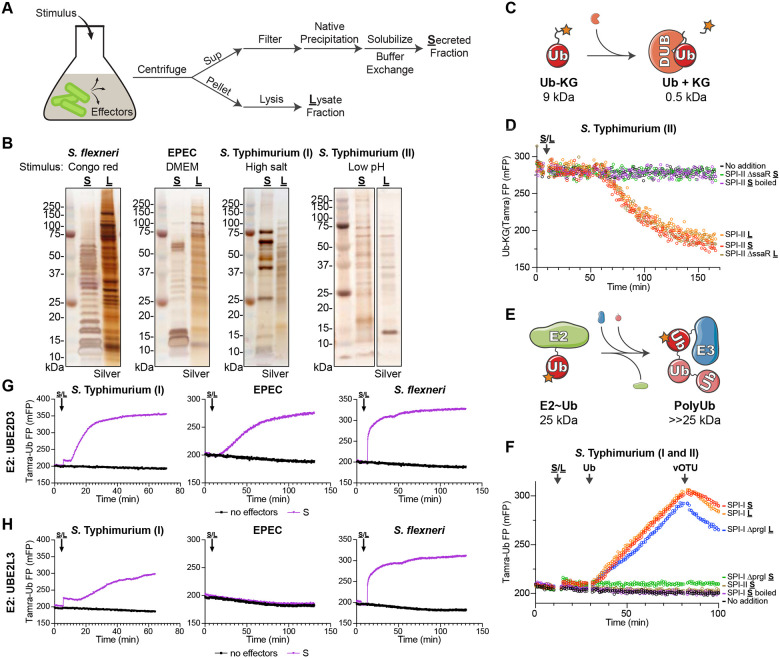
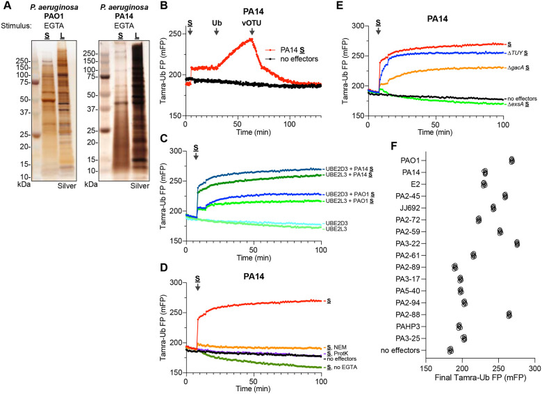
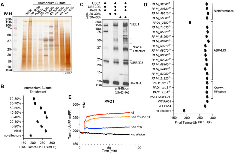
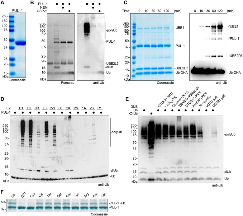
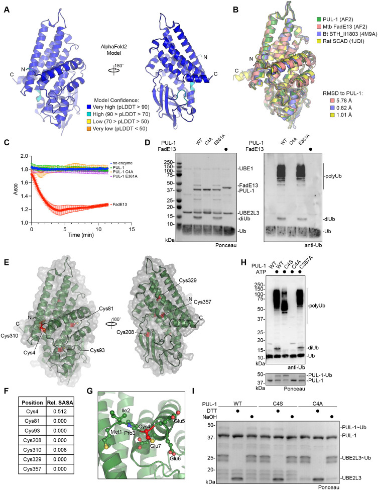
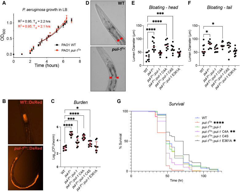
Ubiquitin signaling controls many aspects of eukaryotic biology, including targeted protein degradation and immune defense. Remarkably, invading bacterial pathogens have adapted secreted effector proteins that hijack host ubiquitination to gain control over host responses. These ubiquitin-targeted effectors can exhibit, for example, E3 ligase or deubiquitinase activities, often without any sequence or structural homology to eukaryotic ubiquitin regulators. Such convergence in function poses a challenge to the discovery of additional bacterial virulence factors that target ubiquitin. To overcome this, we have developed a workflow to harvest natively secreted bacterial effectors and functionally screen them for ubiquitin regulatory activities. After benchmarking this approach on diverse ligase and deubiquitinase activities from Salmonella Typhimurium, Enteropathogenic Escherichia coli, and Shigella flexneri, we applied it to the identification of a cryptic E3 ligase activity secreted by Pseudomonas aeruginosa. We identified an unreported P. aeruginosa E3 ligase, which we have termed Pseudomonas Ub ligase 1 (PUL-1), that resembles none of the other E3 ligases previously established in or outside of the eukaryotic system. Importantly, in an animal model of P. aeruginosa infection, PUL-1 ligase activity plays an important role in regulating virulence. Thus, our workflow for the functional identification of ubiquitin-targeted effector proteins carries promise for expanding our appreciation of how host ubiquitin regulation contributes to bacterial pathogenesis.

Keywords: Pseudomonas aeruginosa; Ubiquitin; bacterial effector; deubiquitinase; ubiquitin ligase.


Conflict of interest statement

COMPETING INTEREST STATEMENT The authors declare no competing interests.

Figures

Figure 1:
Figure 1:. A functional screen for ubiquitin regulation
A. Schematic for the strategy used to stimulate effector secretion in bacterial culture prior to harvesting the secreted and lysate fractions. B. Silver-stained SDS-PAGE analysis of secreted (S) and lysate (L) fractions prepared following stimulation as indicated. C. Schematic for the FP-based assay for detection of DUB activity. D. Representative FP traces monitoring the Ub-KG(Tamra) DUB substrate following addition of the indicated pools of S. Typhimurium protein. E. Schematic for the FP-based assay for detection of E3 ligase activity. F. Representative FP traces monitoring the Tamra-Ub ligase substrate following addition of the indicated pools of S. Typhimurium protein. At the indicated timepoints, additional unlabeled Ub or the DUB vOTU were added to stimulate product extension or deconjugation, respectively. G. Representative FP traces monitoring the Tamra-Ub ligase substrate following addition of the indicated bacterial secreted fractions to reactions containing the promiscuous E2 enzyme, UBE2D3. H. As in G), for reactions containing the Cys-specific E2 enzyme, UBE2L3.
Figure 2:
Figure 2:. Detection of E3 ligase activity secreted by P. aeruginosa
A. Silver-stained SDS-PAGE analysis of secreted (S) and lysate (L) fractions prepared following EGTA stimulation of P. aeruginosa PAO1 and PA14 strains. B. Representative FP traces monitoring the Tamra-Ub ligase substrate following addition of the PA14 secreted fraction. At the indicated timepoints, additional unlabeled Ub or the DUB vOTU were added to stimulate product extension or deconjugation, respectively. C. Representative FP traces monitoring the Tamra-Ub ligase substrate following addition of either the PA14 or PAO1 secreted fractions to reactions containing either the promiscuous E2 UBE2D3 or the Cys-specific E2 UBE2L3. D. Representative FP traces monitoring the Tamra-Ub ligase substrate following addition of the PA14 secreted fraction that was untreated, pre-treated with NEM or Proteinase K (ProtK), or generated without stimulation with EGTA. E. Representative FP traces monitoring the Tamra-Ub ligase substrate following addition of secreted fractions generated from the indicated PA14 mutant strains. F. Final FP values of the Tamra-Ub ligase substrate following addition of secreted fractions from the indicated P. aeruginosa clinical isolates.
Figure 3:
Figure 3:. Identification of a P. aeruginosa E3 ligase
A. Silver-stained SDS-PAGE analysis of the PA14 secreted fraction following stepwise ammonium sulfate fractionation. B. Final FP values of the Tamra-Ub ligase substrate following addition of the indicated ammonium sulfate fractions of PA14 secreted protein. C. Reactivity of the cascading Ub-DHA activity-based probe with the indicated ammonium sulfate fractions of PA14 secreted protein. Reactions were resolved by SDS-PAGE and visualized by western blot for the biotinylated Ub-DHA probe. Reacted proteins, including putative probe-reactive PA14 effectors, are labeled with an asterisk. D. Final FP values of the Tamra-Ub ligase substrate following addition of secreted fractions generated from the indicated P. aeruginosa mutant strains, which test ligase candidates from bioinformatic prediction, ABP-MS analysis, or known effectors. E. Representative FP traces monitoring the Tamra-Ub ligase substrate following addition of secreted fractions generated from PAO1 wild-type, the pul-1Tn mutant strain, or a pul-1Tn mutant strain complemented with pul-1.
Figure 4:
Figure 4:. Characterization of PUL-1 E3 ligase activity
A. Coomassie-stained SDS-PAGE analysis of recombinantly purified PUL-1. B. E3 ligase assays for recombinant PUL-1, including conditions with pre-treatment of NEM or post-treatment of the DUB USP21. Reactions were resolved by SDS-PAGE and visualized by Ponceau stain and anti-Ub western blot. C. Time course monitoring reactivity of the cascading Ub-DHA activity-based probe with recombinant PUL-1. Reactions were resolved by SDS-PAGE and visualized by Coomassie stain or anti-Ub western blot. Reacted proteins are labeled with an asterisk. D. E3 ligase assays for recombinant PUL-1 and the indicated panel of E2 enzymes. Reactions were resolved by SDS-PAGE and visualized by anti-Ub western blot. E. UbiCRest assay of PUL-1 ligase products. A PUL-1 ligase reaction was treated with the indicated panel of linkage-specific DUBs, a combination of all linkage-specific DUBs, or the indicated nonspecific DUBs. Reactions were resolved by SDS-PAGE and visualized by anti-Ub western blot. F. Amino acid reactivity analysis of the activated PUL-1~Ub thioester intermediate. PUL-1 was loaded with Ub, and discharge was monitored following addition of DTT or the indicated amino acids. Reactions were resolved by SDS-PAGE and visualized by Coomassie stain.
Figure 5:
Figure 5:. Structural analysis of the PUL-1 ligase fold
A. AlphaFold2 model of PUL-1, colored by pLDDT confidence scores, with N- and C-termini labeled. B. Structural overlay of the AlphaFold2 models of PUL-1, putative acyl-CoA dehydrogenases from M. tuberculosis (Mtb) and B. thailandensis (Bt, PDB: 4M9A), and the mitochondrial short-chain specific acyl-CoA dehydrogenase (SCAD, PDB: 1JQI) from rat. C-alpha root mean square deviation (RMSD) values are listed. C. Octanoyl-CoA dehydrogenase activity assay monitoring reduction of DCPIP at 600 nm wavelength. D. E3 ligase assays for recombinant PUL-1 and FadE13. Reactions were resolved by SDS-PAGE and visualized by Ponceau stain and anti-Ub western blot. E. AlphaFold2 model of PUL-1 (green), with surface representation (grey) and all Cys residues highlighted (red). F. Relative solvent-accessible surface area (SASA) of each Cys residue within the PUL-1 AlphaFold2 model. G. Detailed view of Cys4 within the PUL-1 AlphaFold2 model, with Cys4 (red) and neighboring residues shown as ball-and-stick. H. E3 ligase assays for wild-type or the indicated PUL-1 mutants. Reactions were resolved by SDS-PAGE and visualized by Ponceau stain and anti-Ub western blot. I. Chemical stability toward reducing agent (DTT) or basic pH (NaOH) of the activated UBE2L3~Ub and PUL-1~Ub intermediates. Enzymes were loaded with Ub, and discharge was monitored following the indicated chemical treatments. Reactions were resolved by SDS-PAGE and visualized by Ponceau stain.
Figure 6:
Figure 6:. PUL-1 ligase activity modulates P. aeruginosa virulence
A. Growth curves for wild-type (WT) and the PAO1 pul-1Tn mutant strain in LB, with doubling times (Td) indicated. B. Representative fluorescent images of C. elegans infected with WT and the PAO1 pul-1Tn mutant strain expressing DsRed. C. Bacterial burden measured as colony-forming units (CFU) per worm, following infection with the indicated PAO1 strains. Mean values and standard deviation are indicated in red. Significance was determined by one-way ANOVA with Dunnett’s multiple comparisons test. D. Representative images of C. elegans intestinal bloating near the head, following infection with WT or the PAO1 pul-1Tn mutant strain. The intestinal lumen diameter is indicated by red arrows. E. Quantification of C. elegans intestinal lumen diameter near the head following infection with the indicated PAO1 strains. Median values are shown in red. Significance was determined by one-way ANOVA with Dunnett’s multiple comparisons test. F. As in E), for intestinal lumen diameters near the tail. G. Survival curves for C. elegans infected with the indicated PAO1 strains. Significance was determined by the Log-rank (Mantel-Cox) test.

References

    1. Komander D., and Rape M. (2012). The ubiquitin code. Annu Rev Biochem 81, 203–229. 10.1146/annurev-biochem-060310-170328. - DOI - PubMed
    1. Swatek K.N., and Komander D. (2016). Ubiquitin modifications. Cell Res 26, 399–422. 10.1038/cr.2016.39. - DOI - PMC - PubMed
    1. Pao K.C., Wood N.T., Knebel A., Rafie K., Stanley M., Mabbitt P.D., Sundaramoorthy R., Hofmann K., van Aalten D.M.F., and Virdee S. (2018). Activity-based E3 ligase profiling uncovers an E3 ligase with esterification activity. Nature 556, 381–385. 10.1038/s41586-018-0026-1. - DOI - PubMed
    1. Otten E.G., Werner E., Crespillo-Casado A., Boyle K.B., Dharamdasani V., Pathe C., Santhanam B., and Randow F. (2021). Ubiquitylation of lipopolysaccharide by RNF213 during bacterial infection. Nature 594, 111–116. 10.1038/s41586-021-03566-4. - DOI - PMC - PubMed
    1. Metzger M.B., Pruneda J.N., Klevit R.E., and Weissman A.M. (2014). RING-type E3 ligases: master manipulators of E2 ubiquitin-conjugating enzymes and ubiquitination. Biochim Biophys Acta 1843, 47–60. 10.1016/j.bbamcr.2013.05.026. - DOI - PMC - PubMed

Publication types

LinkOut - more resources