[Preprint]. 2024 Sep 19:2024.09.17.613393.
doi: 10.1101/2024.09.17.613393.

RIPK1 is essential for Herpes Simplex Virus-triggered ZBP1-dependent necroptosis in human cells

Affiliations

RIPK1 is essential for Herpes Simplex Virus-triggered ZBP1-dependent necroptosis in human cells

Oluwamuyiwa T Amusan et al. bioRxiv. .

Abstract

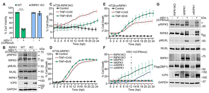
Necroptosis initiated by the host sensor Z-NA Binding Protein-1 (ZBP1) is essential for host defense against a growing number of viruses, including Herpes Simplex Virus-1 (HSV-1). Studies with HSV-1 and other necroptogenic stimuli in murine settings have suggested that ZBP1 triggers necroptosis by directly complexing with the kinase RIPK3. Whether this is also the case in human cells, or whether additional co-factors are needed for ZBP1-mediated necroptosis, is unclear. Here, we show that ZBP1-induced necroptosis in human cells requires RIPK1. We have found that RIPK1 is essential for forming a stable and functional ZBP1-RIPK3 complex in human cells, but is dispensable for the formation of the equivalent murine complex. The RIP Homology Interaction Motif (RHIM) in RIPK3 is responsible for this difference between the two species, because replacing the RHIM in human RIPK3 with the RHIM from murine RIPK3 is sufficient to overcome the requirement for RIPK1 in human cells. These observations describe a critical mechanistic difference between mice and humans in how ZBP1 engages in necroptosis, with important implications for treating human diseases.

Keywords: Cell Death; HSV-1; Necroptosis; RIPK1; RIPK3; ZBP1.


Conflict of interest statement

DECLARATION OF INTERESTS The authors declare no competing interests.

Figures

Figure 1.
Figure 1.. HSV-1(ICP6mut) infection triggers endogenous ZBP1-driven necroptosis in human cells.
(A) Genomic DNA sequences of ZBP1 locus in ZBP1 KO HT29 cells. ‘-’ indicates none of the nucleotides. (B) Immunoblotting analysis of endogenous expression of Wild-type (WT) or ZBP1 KO HT29 cells with or without IFN-β priming. (C) Cell death kinetics of WT or ZBP1 KO HT29 cells after IFN-β priming, followed by HSV-1(ICP6mut) infection. (D) Immunoblotting of WT or ZBP1 KO HT29 cells with or without IFN-β priming, followed by HSV-1(ICP6mut) infection. Cells were collected after 16hrs infection and subjected to immunoblotting analysis with pRIPK1, RIPK1, pRIPK3, RIPK3, p-MLKL, MLKL, ICP0, and GAPDH antibodies. (E) Cell death kinetics of primary human foreskin fibroblast (HFF) cells transfected with control or ZBP1 siRNAs using electroporation and primed with IFN-β (50ng/ml) for 24hrs, followed by HSV-1(ICP6mut) infection. The knockdown efficiency in HFFs was confirmed by immunoblotting assay in (F). Results are representative of at least two independent experiments. Error bars represent mean ± SD.
Figure 2.
Figure 2.. RIPK1 is essential for HSV-1(ICP6mut)-triggered endogenous ZBP1-driven necroptosis in human cells.
(A) Schematic illustration of the PROTAC-induced RIPK1 degradation system. (B) Cell death kinetics of HT29 cells pretreated with IFN-β (50 ng/mL) for 24 hours, followed by HSV-1(ICP6mut) infection, with or without Nec-1 (30 μM), GSK872 (5 μM), NSA (2 μM), R1-ICR-3 (100 nM), or zVAD (25 μM). (C) Immunoblotting of HT29 cells primed with IFN-β (50 ng/mL), followed by HSV-1(ICP6mut) infection, with or without Nec-1 (30 μM), GSK872 (5 μM), NSA (2μM), zVAD (25 μM), or R1-ICR-3 (100 nM). Cells were subjected to immunoblotting with pRIPK1, RIPK1, pRIPK3, RIPK3, pMLKL, MLKL, ICP0, and GAPDH antibodies at 16 hours post-infection (hpi). (D) Cell death kinetics of HS68 cells pretreated with IFN-β (50 ng/mL) for 24 hours, followed by HSV-1(ICP6mut) infection, with or without zVAD (25 μM), in combination with Nec-1 (30 (μM), GSK963 (5 μM), GSK872 (5 μM), or R1-ICR-3 (100 nM). (E) Immunoblotting of HS68 cells pretreated with IFN-β (50ng/ml) for 24 hrs, followed by HSV-1(ICP6mut) infection, with or without zVAD (25 μM), in combination with R1-ICR-3 (100 nM). Cells were collected at 16 hours post-infection and probed with pMLKL, MLKL, RIPK1, ZBP1, ICP0, and GAPDH antibodies. (F) Cell death kinetics of primary human foreskin fibroblast (HFF) cells pretreated with IFN-β (50 ng/mL) for 24 hours, followed by HSV-1(ICP6mut) infection, with or without zVAD (25 μM), in combination with Nec-1 (30 μM), GSK963 (5 μM), GSK872 (5 μM), or R1-ICR-3 (100 nM). (G) Immunoblotting of HFF cells pretreated with IFN-β (50ng/ml) for 24 hrs, followed by HSV-1(ICP6mut) infection, with or without zVAD (25 μM), in combination with R1-ICR-3 (100 nM). Cells were collected at 16 hpi and probed with pMLKL, MLKL, RIPK1, ZBP1, ICP0, and GAPDH antibodies. (H) Cell viability of U937 cells, followed by HSV-1(ICP6mut) infection, with or without zVAD (25 μM), in combination with R1-ICR-3 (100 nM) at 18 hpi. (I) Immunoblotting of U937 cells, followed by HSV-1(ICP6mut) infection, with or without zVAD (25 μM), in combination with R1-ICR-3 (100 nM). Samples were probed for p-MLKL, MLKL, RIPK1, ZBP1, ICP0, and GAPDH antibodies at 12 hpi. Results are representative of at least two independent experiments. Error bars represent mean ± SD.
Figure 3.
Figure 3.. RIPK1 binds to ZBP1 during HSV-1(ICP6mut) infection in human cells.
(A) Cell death kinetics of HT29 cells reconstituted Flag-hZBP1 (hereafter HT29-hZBP1), followed by HSV- 1(ICP6mut) infection, with or without Nec-1 (30 μM), GSK963 (5 μM), GSK872 (5 μM), NSA (2 μM), R1-ICR-3 (100 nM), or zVAD (25 μM). (B) Immunoblotting of HT29-hZBP1 cells, followed by HSV-1(ICP6mut) for 10 hrs with or without R1-ICR-3 (100 nM). Samples were probed with pRIPK1, RIPK1, pRIPK3, RIPK3, pMLKL, MLKL, Flag (ZBP1), ICP0, and GAPDH antibodies. (C) HT29-hZBP1 cells either mocked or infected with HSV-1(ICP6mut) for 10 hrs were harvested and subjected to immunoprecipitation with Anti-FLAG® M2 magnetic beads (Sigma-Aldrich). Total lysates (Input) and immunoprecipitates (IP) were subjected to SDS-PAGE and evaluated for RIPK1, RIPK3, and Flag (ZBP1). Input was also evaluated for viral protein ICP0 and GAPDH. (D) HT29 RIPK3 knockout (KO) cells reconstituted empty vector (EV), wild-type (WT) RIPK3, or RHIM-deleted (ΔRHIM) RIPK3 together with human ZBP1, were either mock or infected with HSV-1(ICP6mut) for 10 hrs. Co-immunoprecipitation was performed in these cells as described in C. (E) HT29-hZBP1 cells either mocked or infected with HSV-1(ICP6mut) for 10 hrs with or without R1-ICR-3 (100 nM), were harvested and subjected to immunoprecipitation as described in C. (F) HS68 cells pretreated with IFN-β (50ng/ml) for 24 hrs, followed by HSV-1(ICP6mut) infection in the presence of zVAD (25 μM), with or without R1-ICR-3 (100 nM) for 16 hrs, were harvested and subjected to immunoprecipitation as described in C. Results are representative of at least two independent experiments. Error bars represent mean ± SD.
Figure 4.
Figure 4.. Both human and mouse RIPK1 mediate ZBP1-driven necroptosis during HSV-1(ICP6mut) infection in human cells.
(A) Cell viability of HT29-hZBP1 or HT29-hZBP1(RIPK1 KO) cells infected with HSV-1(ICP6mut). Viability was determined at 18 hpi by CellTiter-Glo assay. (B) HT29-hZBP1 or HT29-hZBP1 (RIPK1 KO) cells infected with HSV-1(ICP6mut) were harvested at 10 hpi and subjected to immunoblotting with p-RIPK3, RIPK3, p-MLKL, MLKL, ICP0, and GAPDH antibodies. (C-E) Cell death kinetics of HT29-hZBP1 (RIPK1 KO) (C), HT29-hZBP1(hRIPK1) (D), and HT29-hZBP1(mRIPK1) (E) cells pretreated with Cumate (30μg/ml) for 3 hrs, followed by treatment of TNF+CHX or TNF+BV6. (F) Cell death kinetics of HT29-hZBP1 (RIPK1 KO), HT29-hZBP1(hRIPK1), and HT29-hZBP1(mRIPK1) cells pretreated with Cumate (30μg/ml) for 16 hrs, followed by HSV-1(ICP6mut) infection. (G) Immunoblotting of HT29-hZBP1 (RIPK1 KO), HT29-hZBP1 (hRIPK1), and HT29-hZBP1 (mRIPK1) cells pretreated with Cumate (30μg/ml) for 16hrs, followed by HSV-1(ICP6mut) infection. Cells were harvested at 10 hpi and subjected to immunoblotting with p-RIPK3, RIPK3, p-MLKL, MLKL, RIPK1, Flag (ZBP1), ICP0, and GAPDH antibodies. An unpaired t-test was used to test for statistical differences between indicated conditions in (A). *P < 0.1, **P < 0.0001, ***P < 0.001, ****P < 0.0001. Individual data points indicate three technical replicates. Results are representative of at least two independent experiments. Error bars represent mean ± SD.
Figure 5.
Figure 5.. Both Za2 and RHIM A domains of human ZBP1 are essential for RIPK1 binding and mediating necroptosis.
(A) Schematic of ZBP1 and its mutants. (B) Cell viability of HT29 cells reconstituted with indicated human ZBP1 constructs infected with HSV-1(ICP6mut). Viability was determined at 18 hpi by CellTiter-Glo assay. (C) HT29 cells reconstituted with indicated human ZBP1 constructs were either mocked or infected with HSV-1(ICP6mut) for 10 hrs. Co-immunoprecipitation was performed in these cells. (D) Cell viability of HT29 cells reconstituted with indicated mouse ZBP1 constructs infected with HSV-1(ICP6mut). Viability was determined at 18 hpi by CellTiter-Glo assay. (E) Cell viability of HT29-mZBP1 or HT29-mZBP1 (RIPK1 KO) cells infected with HSV-1(ICP6mut). Viability was determined at 18 hpi by CellTiter-Glo assay. (F) HT29-mZBP1 or HT29-mZBP1 (RIPK1 KO) cells infected with HSV-1(ICP6mut) were harvested at 10 hpi and subjected to immunoblotting with p-MLKL, MLKL, RIPK1, ICP0, and GAPDH antibodies. (G) HT29 cells reconstituted with indicated mouse ZBP1 constructs were mock-treated or infected with HSV-1(ICP6mut) for 10 hrs. Co-immunoprecipitation was performed in these cells. (H) Cell viability of MEF-hZBP1 transfected with scramble or RIPK1 siRNA for 48 hrs followed by HSV-1(ICP6mut) infection. Viability was determined at 18 hpi by CellTiter-Glo assay. (I) MEF-hZBP1 cells transfected with either scramble siRNA (Control) or RIPK1 siRNA for 48 hrs, followed by HSV-1(ICP6mut) infection. Cells were harvested at 10 hpi and subjected to IB with p-MLKL, MLKL, RIPK1, ICP0, and GAPDH antibodies. One-way ANOVA and Dunnett’s multiple comparisons tests were used to test for statistical differences between indicated conditions and EV groups in (B) and (D). An unpaired t-test was used to test for statistical differences between indicated conditions in (E) and (H). *P < 0.1, **P < 0.0001, ***P < 0.001, ****P < 0.0001. Individual data points indicate three technical replicates. Results are representative of at least two independent experiments. Error bars represent mean ± SD.
Figure 6.
Figure 6.. Species-Specific RIPK3 RHIM domain determines the requirement of RIPK1 in HSV-1(ICP6mut)-triggered ZBP1-dependent necroptosis.
(A) Alignment of RHIM sequences from human RIPK3 (hRIPK3) and mouse RIPK3 (mRIPK3). Several variable residues between hRIPK3 and mRIPK3 analyzed below were highlighted. The alignment was performed using ClustalW2 online software. (B) Structural comparison between mZBP1-mRIPK3 and hZBP1-hRIPK3 complexes. mZBP1 was colored in yellow, mRIPK3 in light green, hZBP1 in orange, and hRIPK3 in dark green. Hydrogen bonds were indicated by gray dotted lines. (C) Multiple sequence alignment of RIPK3 protein sequences from Primate and Rodent species. The conservative RHIM core amino acids were highlighted with a star (*). The variable residues in (A) were highlighted with a red box. (D) Phylogenetic tree of RIPK3 proteins by MEGA11. RIPK3 protein sequences from Primate and Rodent species were retrieved from GeneBank. The evolutionary history was inferred using the Maximum Likelihood method. All sequences from Primates were highlighted with Blue, and all sequences from Rodents were highlighted with Red. (E) Schematic of hRIPK3, mRIPK3, and chimeric hRIPK3 construct-hRIPK3mRIPK3_RHIM. (F) Cell death kinetics of HT29-hZBP1 (RIPK3 KO) cells reconstituted with hRIPK3, and hRIPK3mRIPK3_RHIM pretreated with DMSO or R1-ICR-3 (100 nM) for 5 hrs, followed by HSV-1(ICP6mut) infection. (G) Immunoblotting of HT29-hZBP1 (RIPK3 KO) cells reconstituted with hRIPK3, and hRIPK3mRIPK3_RHIM pretreated with DMSO or R1-ICR-3 (100 nM) for 5 hrs, followed by HSV-1(ICP6mut) infection and subjected to immunoblotting with p-MLKL, MLKL, p-RIPK3, RIPK3, RIPK1, Flag (ZBP1), ICP0, and GAPDH antibodies. Results are representative of at least two independent experiments. Error bars represent mean ± SD.
Figure 7.
Figure 7.. RIPK1 facilitates the formation of a ZBP1-RIPK3 amyloid structure in human cells.
(A) Thioflavin T (ThT) binding assay of endogenous ZBP1-containing complexes from HSV-1(ICP6mut) infected HT29-hZBP1 and HT29-hZBP1 (RIPK1 KO) cells. (B) HT29-hZBP1 (RIPK3 KO) cells transduced with human RIPK3 (hRIPK3) were either mock or HSV-1(ICP6mut) infection for 10 hrs, with or without R1-ICR-3 (100 nM). Cells were harvested and subjected to immunoprecipitation. ZBP1 complexes isolated by immunoprecipitation were lysed in regular lysis buffer or buffer containing 4M urea. (C) MEF-mZBP1 cells were either mock or HSV-1(ICP6mut) infection for 10 hrs, with or without R1-ICR-3 (1 μM). Cells were harvested and subjected to immunoprecipitation. ZBP1 complexes isolated by immunoprecipitation were lysed in regular lysis buffer or buffer containing 4M urea. (D) HT29-hZBP1 (RIPK3 KO) cells reconstituted with mouse RIPK3 (mRIPK3) were either mock or HSV-1(ICP6mut) infection for 10 hrs, with or without R1-ICR-3 (100 nM). Cells were harvested and subjected to immunoprecipitation. ZBP1 complexes isolated by immunoprecipitation were lysed in regular lysis buffer or buffer containing 4M urea. (E) Schematic model of the proposed ZBP1-driven necroptosis in both human and mouse cells during HSV-1 (ICP6mut) infection. Results are representative of at least two independent experiments. Error bars represent mean ± SD.

Similar articles

References

    1. Duprez L, Takahashi N, Van Hauwermeiren F, Vandendriessche B, Goossens V, Vanden Berghe T, et al. RIP kinase-dependent necrosis drives lethal systemic inflammatory response syndrome. Immunity. 2011;35(6):908–18. Epub 2011/12/27. doi: 10.1016/j.immuni.2011.09.020. - DOI - PubMed
    1. Kaiser WJ, Upton JW, Long AB, Livingston-Rosanoff D, Daley-Bauer LP, Hakem R, et al. RIP3 mediates the embryonic lethality of caspase-8-deficient mice. Nature. 2011;471(7338):368–72. Epub 20110302. doi: 10.1038/nature09857. - DOI - PMC - PubMed
    1. Welz PS, Wullaert A, Vlantis K, Kondylis V, Fernandez-Majada V, Ermolaeva M, et al. FADD prevents RIP3-mediated epithelial cell necrosis and chronic intestinal inflammation. Nature. 2011;477(7364):330–4. Epub 2011/08/02. doi: 10.1038/nature10273. - DOI - PubMed
    1. Cho YS, Challa S, Moquin D, Genga R, Ray TD, Guildford M, et al. Phosphorylation-driven assembly of the RIP1-RIP3 complex regulates programmed necrosis and virus-induced inflammation. Cell. 2009;137(6):1112–23. doi: 10.1016/j.cell.2009.05.037. - DOI - PMC - PubMed
    1. He S, Wang L, Miao L, Wang T, Du F, Zhao L, et al. Receptor interacting protein kinase-3 determines cellular necrotic response to TNF-alpha. Cell. 2009;137(6):1100–11. doi: 10.1016/j.cell.2009.05.021. - DOI - PubMed

Publication types