[Preprint]. 2024 Sep 19:2024.09.13.612942.
doi: 10.1101/2024.09.13.612942.

Chromosomal instability increases radiation sensitivity

Affiliations

Chromosomal instability increases radiation sensitivity

Pippa F Cosper et al. bioRxiv. .

Abstract

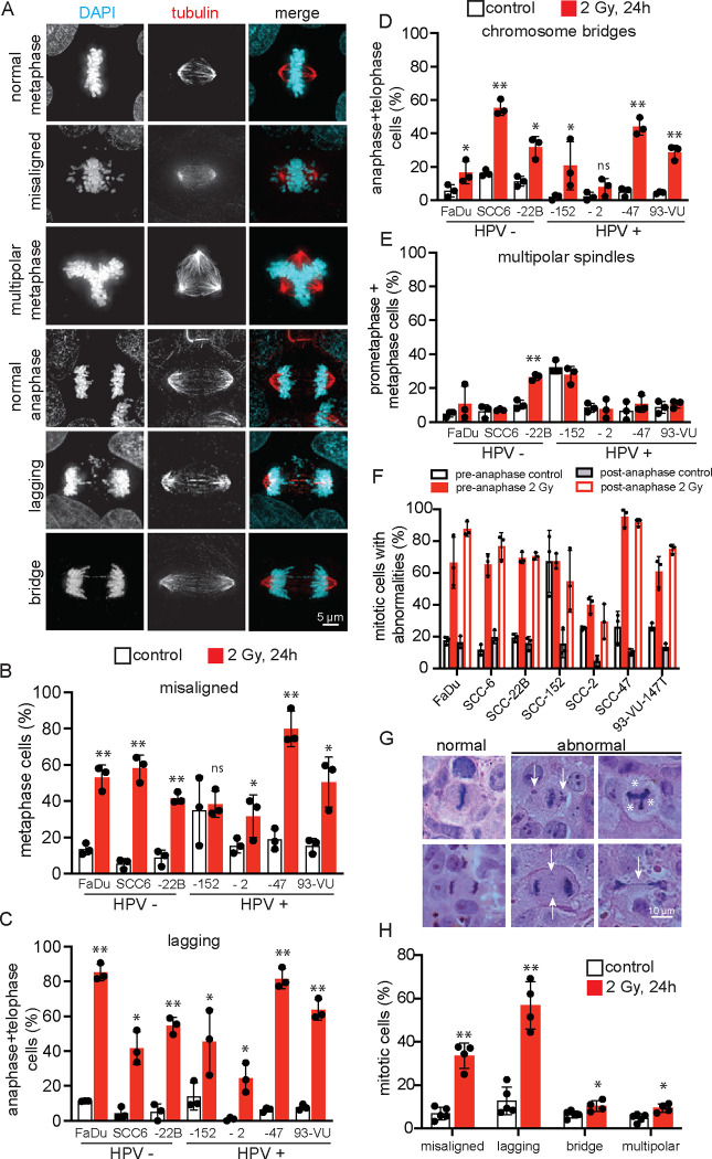
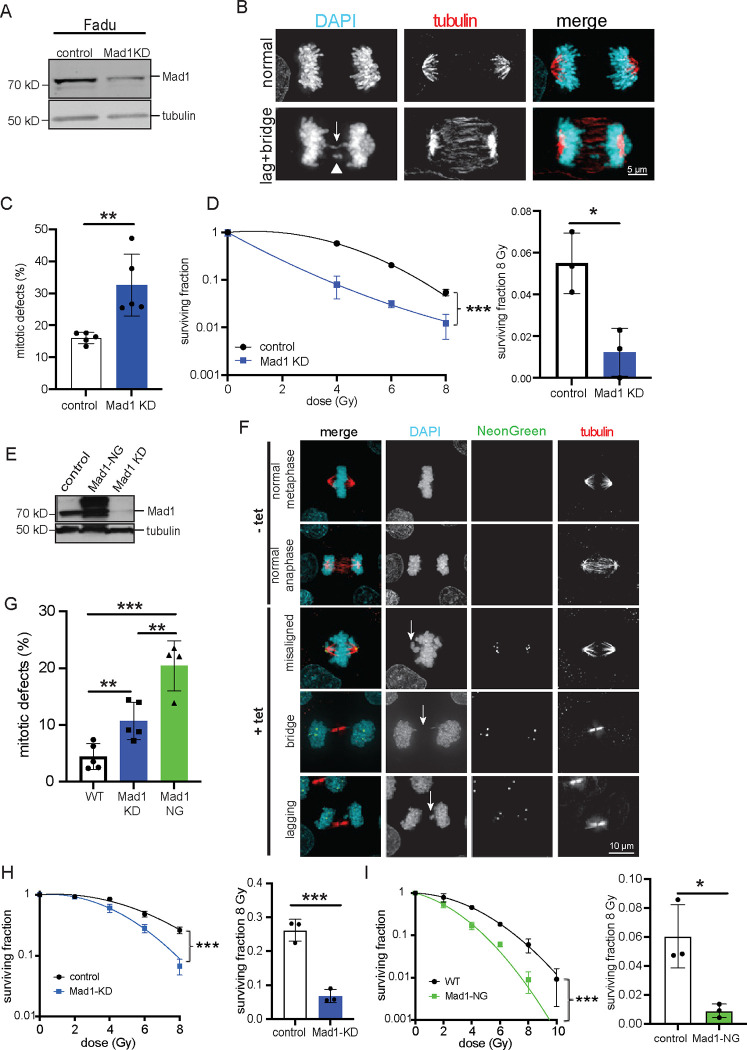
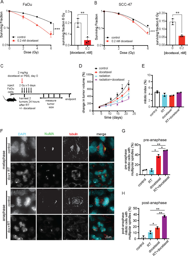
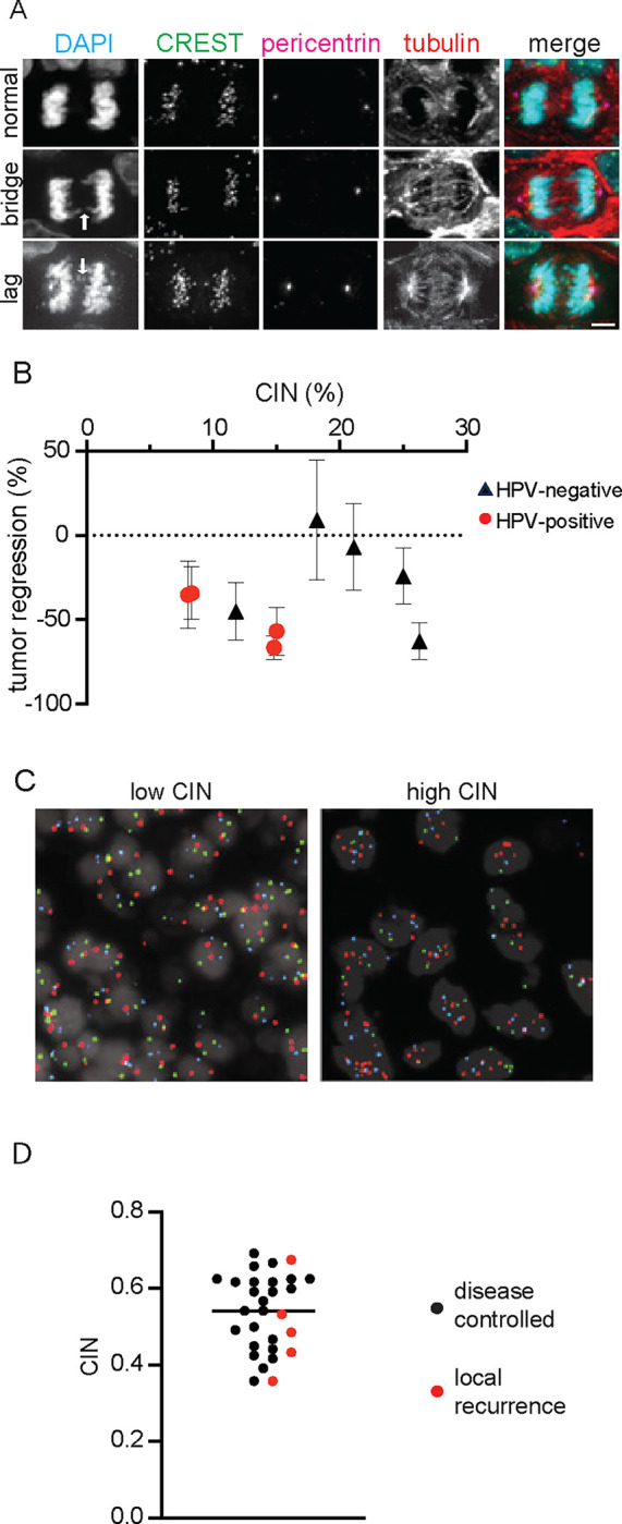
Continuous chromosome missegregation over successive mitotic divisions, known as chromosomal instability (CIN), is common in cancer. Increasing CIN above a maximally tolerated threshold leads to cell death due to loss of essential chromosomes. Here, we show in two tissue contexts that otherwise isogenic cancer cells with higher levels of CIN are more sensitive to ionizing radiation, which itself induces CIN. CIN also sensitizes HPV-positive and HPV-negative head and neck cancer patient derived xenograft (PDX) tumors to radiation. Moreover, laryngeal cancers with higher CIN prior to treatment show improved response to radiation therapy. In addition, we reveal a novel mechanism of radiosensitization by docetaxel, a microtubule stabilizing drug commonly used in combination with radiation. Docetaxel causes cell death by inducing CIN due to abnormal multipolar spindles rather than causing mitotic arrest, as previously assumed. Docetaxel-induced CIN, rather than mitotic arrest, is responsible for the enhanced radiation sensitivity observed in vitro and in vivo, challenging the mechanistic dogma of the last 40 years. These results implicate CIN as a potential biomarker and inducer of radiation response, which could provide valuable cancer therapeutic opportunities.

Statement of significance: Cancer cells and laryngeal tumors with higher chromosome missegregation rates are more sensitive to radiation therapy, supporting chromosomal instability as a promising biomarker of radiation response.

Keywords: Mad1; cervical cancer; docetaxel; head and neck cancer; mitosis.


Conflict of interest statement

Competing interests: The authors declare no competing interests.

Figures

Figure 1.
Figure 1.. Radiation induces multiple types of CIN in head and neck cancer cells.
(A) Representative images of types of chromosome segregation errors that lead to CIN in FaDu head and neck cancer cells 24 hours after 2 Gy of radiation. Normal metaphase and anaphase are shown for reference. (B-E) Quantification of different types of mitotic defects that cause CIN in HPV-negative (HPV−) and HPV-positive (HPV+) untreated control cells and 24 hours after 2 Gy of radiation. In (B), misaligned chromosomes include polar chromosomes. n≥50 cells in each phase of mitosis (prometaphase, metaphase, anaphase, and telophase) per condition in each of 3 biological replicates. (F) Quantification of the total CIN in each cell type before and 24 hours after 2 Gy of radiation. Misaligned chromosomes and multipolar spindles contribute to pre-anaphase CIN, while lagging and bridge chromosomes contribute to post-anaphase CIN. n=50 cells per condition in each of 3 biological replicates. (G) Representative H&E images of FaDu tumor xenografts 24 hours after 2 Gy radiation. Upper panel shows metaphase cells. From left to right: normal, misaligned chromosomes (arrows), multipolar spindle (*denotes inferred spindle pole). Lower panel shows anaphase cells. From left to right: normal, lagging chromosome (arrows), chromosome bridge (arrow). (H) Quantification of radiation-induced CIN in vivo. An average of 79 metaphases (range 36–132) and 35 anaphase/telophase (range 13–71) were counted per tumor. n=4 tumors in each condition. Error bars indicate SD. Statistical differences determined by 2-tailed t-test, * = p<0.05, ** = p<0.001.
Figure 2.
Figure 2.. CIN sensitizes to radiation in head and neck and cervical cancer cells.
(A-D) Mad1 knockdown (KD) induces CIN in FaDu HPV-negative head and neck cancer cells and sensitizes them to radiation. (A) Immunoblot showing efficient Mad1 knockdown. (B) Images of (top) normal anaphase and (bottom) anaphase cell with chromosome bridge (arrow) and lagging chromosome (arrowhead). (C) Quantification of mitotic defects (lagging and bridge chromosomes) in isogenic parental and Mad1 knockdown FaDu cells. n≥60 cells in metaphase and ≥70 cells in anaphase+telophase per condition in each biological replicate. (D) Clonogenic assays showing Mad1 knockdown FaDu cells with CIN have increased radiation sensitivity relative to isogenic parental cells. (E-I) Induction of CIN in HeLa HPV-positive cervical cancer cells sensitizes them to radiation. (E) Immunoblot showing expression of Mad1-mNeonGreen (NG) and Mad1 knockdown in HeLa cells. (F) Images showing examples of normal and abnormal mitotic figures in HeLa cells +/− tet inducible expression of Mad1-NG. Arrows indicate respective defect. (G) Quantification of mitotic errors due to Mad1 knockdown and expression of Mad1-NG, which both cause CIN. n≥70 cells in metaphase and ≥85 cells in anaphase or telophase per condition in each biological replicate. (H) Clonogenic assays showing that both Mad1 knockdown and Mad1-NG expression sensitize cells to radiation. The surviving fraction of cells after 8 Gy is shown to the right of each respective clonogenic curve. n=3 biological replicates each. Error bars indicate SD. Statistical differences determined by 2-tailed t-test, * = p<0.05, ** = p<0.01, *** = p<0.001.
Figure 3.
Figure 3.. Docetaxel causes cell death in head and neck cancer cells by inducing CIN on multipolar spindles without mitotic arrest.
(A) Left: Images of FaDu cells in metaphase showing normal bipolar spindle (top) or abnormal multipolar spindle (bottom). Right: Quantification of pre-anaphase multipolar spindles (in prometaphase and metaphase cells), which increase in a concentration dependent manner 24 hours after treatment with docetaxel. (B) Left: Images of FaDu cells in normal bipolar anaphase (top) and abnormal multipolar anaphase (bottom). Right: Quantification of multipolar spindles in post-anaphase (anaphase and telophase) cells 24 hours after docetaxel treatment. (A-B) n=100 cells per condition in each of 3 biological replicates. (C) Mitotic index after 24 hour treatment with the indicated concentrations of docetaxel showing that the concentrations used in A and B that induce multipolar spindles are too low to cause mitotic arrest. n≥500 cells in each of 3 biological replicates. (D) MTT assay showing concentrations of docetaxel that induce ≥20% multipolar spindles in post-anaphase cells impair proliferation. Y-axis values normalized to DMSO-treated cells at day 7. n=3 biological replicates. Error bars indicate SD. Statistical differences determined by 2-tailed t-test, * = p<0.05, ** = p<0.01, *** = p<0.001 versus DMSO.
Figure 4.
Figure 4.. Docetaxel sensitizes HPV-positive and HPV-negative head and neck cancer cells to radiation by inducing CIN on multipolar spindles without mitotic arrest.
(A-B) Clonogenic assays showing docetaxel concentrations that are too low to cause mitotic arrest (0.2 nM) sensitize HPV-negative FaDu (A) and HPV-positive SCC-47 (B) cells to radiation. The surviving fraction of cells after 6 Gy is shown to the right of each respective clonogenic curve. n=3 biological replicates. (C) Schematic of murine study. RT = radiation therapy. (D) Docetaxel sensitizes FaDu xenograft tumors to radiation. n=10 tumors per condition. Error bar, SEM. (E) Mitotic index in murine tumors is not elevated relative to PBS control by treatment with docetaxel, radiation, or docetaxel+radiation. n≥500 cells in each of 2 tumors per condition. (F-H) Docetaxel induces multipolar spindles in FaDu xenograft tumors. (F) Immunofluorescence images of normal bipolar and abnormal multipolar spindles in FaDu xenograft tumors. Spindle poles were identified by co-localization of NuMA and α-tubulin. (G-H) Quantification of multipolar spindles in xenograft tumor tissue in (G) pre-anaphase cells (prometaphase and metaphase) and (H) post-anaphase cells (in anaphase and telophase). n≥80 (range 83–187) pre-anaphase and ≥23 (range 23–57) post-anaphase cells in each of 2 biological replicates. Error bars indicate SD. Statistical differences determined by 2-tailed t-test * = p<0.05, ** = p<0.01.
Figure 5.
Figure 5.. CIN directly correlates with radiation response in head and neck cancer PDX tumors and laryngeal cancer patients.
(A) Images of normal anaphase and anaphase defects in head and neck cancer PDX tissues prior to irradiation. Arrows indicate the indicated defect. Scale bar, 5 μm. (B) PDX tumors with higher CIN are more sensitive to radiation. CIN is reported as the percentage of anaphase and telophase cells containing lagging chromosomes and/or chromosome bridges. Tumor regression is based on 12–14 tumors per PDX and treatment group treated with sham or 2 Gy daily for 5 days. Error bars represent SD. n≥19 cells in anaphase/telophase (range = 19–139, average = 47) per sample to quantify anaphase defects. (C) Interphase FISH images used to quantify CIN in laryngeal cancer. Two sections with centromeric probes (CEP) to chromosomes 3, 7, 9 (left, red, blue, green respectively) and 4, 10, 17 (right: green, blue, red, respectively) were used to quantify CIN for each sample. (D) Laryngeal tumors with CIN below the median (black bar, 0.54) had increased local recurrence rate (31%) compared to tumors with CIN above the median (6%).

References

    1. Taylor AM, Shih J, Ha G, Gao GF, Zhang X, Berger AC, et al. Genomic and Functional Approaches to Understanding Cancer Aneuploidy. Cancer Cell. 2018;33:676–689.e3. - PMC - PubMed
    1. Weaver BAA, Cleveland DW. Does aneuploidy cause cancer? Curr Opin Cell Biol. 2006;18:658–67. - PubMed
    1. Mitelman F.E., Johansson B. & Mertens F. 2017. Available from: (http://cgap.nci.nih.gov/Chromosomes/Mitelman
    1. Zasadil LM, Britigan EMC, Weaver BA. 2n or not 2n: Aneuploidy, polyploidy and chromosomal instability in primary and tumor cells. Semin Cell Dev Biol. 2013;24:370–9. - PMC - PubMed
    1. Sotillo R, Hernando E, Díaz-Rodríguez E, Teruya-Feldstein J, Cordón-Cardo C, Lowe SW, et al. Mad2 overexpression promotes aneuploidy and tumorigenesis in mice. Cancer Cell. 2007;11:9–23. - PMC - PubMed

Publication types