

Examining Penetration and Residual Depth in Modern Acrylic Foldable Intraocular Lenses: A Laboratory Study Using Differential Interference Contrast Microscopy to Compare Hydrophilic and Hydrophobic Materials
- PMID: 39345802
- PMCID: PMC11438304
- DOI: 10.7759/cureus.70383
Examining Penetration and Residual Depth in Modern Acrylic Foldable Intraocular Lenses: A Laboratory Study Using Differential Interference Contrast Microscopy to Compare Hydrophilic and Hydrophobic Materials
Abstract
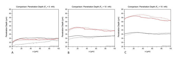
Introduction The material of modern intraocular lenses must meet the highest standards and fulfill various requirements. It is crucial that the material shows the best biocompatibility and should be flexible for an uncomplicated implantation process through small corneal incisions but also sufficiently rigid for good stability and centering in the capsular bag. In addition, the optic must remain clear for life and retain the best optical properties. Methods In this laboratory experiment, we performed scratch tests for the mechanical assessment of acrylic intraocular lenses. The aim was to determine differences in the behavior in regard to the manufacturing process and water content of hydrophilic and hydrophobic acrylic intraocular lenses. The scratch tests were performed using a Nano Scratch Tester. A conical indenter with a tip radius of 1 µm and a cone angle of 90° was selected to scratch the samples at three different constant loads of 5, 10, and 15 mN, respectively. The scratch length was set to 100 µm at a scratch speed of 200 µm/min. Hydrophilic and hydrophobic acrylic intraocular lenses (with different water content) were tested. Results The results showed that for sample A (hydrophilic acrylate), the penetration depth increases steadily with increasing force from 25-30 µm (5 mN) to 28-33 µm (10 mN) and 34-37 µm (15 mN). The penetration depths during the scratches seem to be load-dependent. In sample B (hydrophobic acrylate), the same forces lead to steadily increasing penetration depths: 25-30 µm (5 mN), 40-44 µm (10 mN), and 54-57 µm (15 mN). The evaluation of the residual depth showed much lower values for all samples. In the hydrophilic, softer samples (A), the residual depth was between 1 µm and 4 µm. In the hydrophobic, more solid, samples (B), the residual depth was more pronounced with values between 5 µm and 17 µm. The plastic influence and deformation zone seemed to be wider for the hydrophobic samples than for the hydrophilic samples. Conclusion The laboratory experiment confirms that modern, acrylic intraocular lenses are sensitive to scratches/touch, and penetration depths during scratching depend on the load. The remaining depths after the scratches are significantly lower and show a load dependence. The deforming zone was higher in the hydrophobic acrylates than in the hydrophilic acrylates. However, the results confirm that damage can occur with hydrophobic and hydrophilic acrylic materials, depending on the force applied. Therefore, careful handling during the preparation and implantation process is crucial to prevent permanent defects.
Keywords: damages in intraocular lenses; nanoindentation; penetration depth; residual depth; scratch test.
Copyright © 2024, Borkenstein et al.
Conflict of interest statement
Human subjects: All authors have confirmed that this study did not involve human participants or tissue. Animal subjects: All authors have confirmed that this study did not involve animal subjects or tissue. Conflicts of interest: In compliance with the ICMJE uniform disclosure form, all authors declare the following: Payment/services info: All authors have declared that no financial support was received from any organization for the submitted work. Financial relationships: All authors have declared that they have no financial relationships at present or within the previous three years with any organizations that might have an interest in the submitted work. Other relationships: All authors have declared that there are no other relationships or activities that could appear to have influenced the submitted work.
Figures


Similar articles
-
Nano-Indentation to Determine Mechanical Properties of Intraocular Lenses: Evaluating Penetration Depth, Material Stiffness, and Elastic Moduli.Ophthalmol Ther. 2023 Aug;12(4):2087-2101. doi: 10.1007/s40123-023-00728-7. Epub 2023 May 21. Ophthalmol Ther. 2023. PMID: 37211587 Free PMC article.
-
[Comparison of postoperative results after implantation of hydrophilic acrylic or hydrophobic acrylic intraocular lens: data of one-year prospective clinical study].Medicina (Kaunas). 2008;44(12):936-43. Medicina (Kaunas). 2008. PMID: 19142051 Lithuanian.
-
Scanning electron microscopic characteristics of manually loaded and preloaded foldable acrylic intraocular lenses.Eur J Ophthalmol. 2019 Jan;29(1):28-32. doi: 10.1177/1120672118762665. Epub 2018 Apr 5. Eur J Ophthalmol. 2019. PMID: 29619847
-
Biocompatibility of Intraocular Lenses.Turk J Ophthalmol. 2017 Aug;47(4):221-225. doi: 10.4274/tjo.10437. Epub 2017 Aug 15. Turk J Ophthalmol. 2017. PMID: 28845327 Free PMC article. Review.
-
[Influence of material on biocompatibility of intraocular lenses].Polim Med. 2007;37(1):35-45. Polim Med. 2007. PMID: 17703722 Review. Polish.
References
-
- Intraocular lens extraction using the cartridge pull-through technique. Fukuoka S, Kinoshita T, Morita S, Sakurai T. J Cataract Refract Surg. 2021;47:0–4. - PubMed
-
- Trisection technique: a 2-snip approach to intraocular lens explantation. Por YM, Chee SP. J Cataract Refract Surg. 2007;33:1151–1154. - PubMed
-
- Intraocular lens explantation after cataract surgery: indications, results, and explantation techniques. Fernández-Buenaga R, Alió JL. Asia Pac J Ophthalmol (Phila) 2017;6:372–380. - PubMed
LinkOut - more resources
Full Text Sources