

Glomus tumor: A rare differential diagnosis for subungual lesions
- PMID: 39345850
- PMCID: PMC11437606
- DOI: 10.1016/j.radcr.2024.08.116
Glomus tumor: A rare differential diagnosis for subungual lesions
Abstract
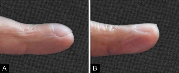
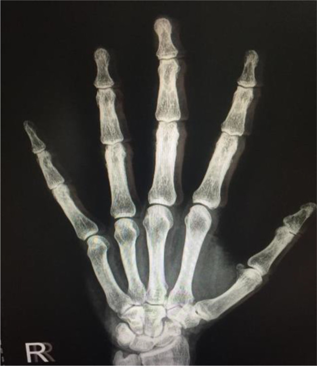
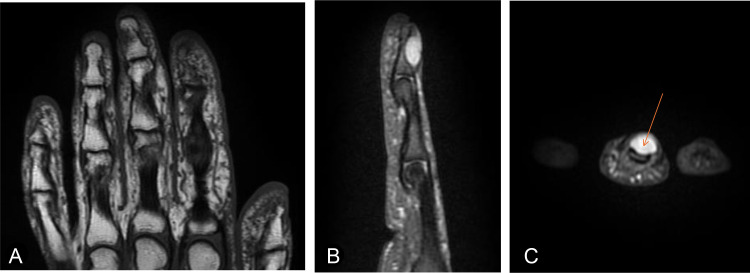
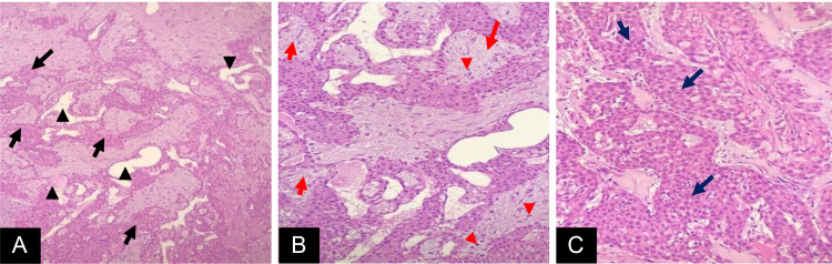
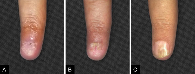
Glomus tumors are rare, benign vascular hamartomas of the glomus apparatus with unknown etiology. They can arise from anywhere in the body. However, up to 90% of them are located in the subungual region of the fingers, as in the case of our patient. These tumors typically present with the classic triad of pain, cold sensitivity, and point tenderness. Characteristic US and MRI findings aid the clinical diagnosis; nevertheless, a histopathologic examination is confirmatory. There is a well-documented mean delay in diagnosis of around 7 years, due to the rarity, benignity, small size, and lack of proper knowledge about the condition. However, we reported a case with a delay in diagnosis that reached 40 years, which is much longer than what is documented in the literature. A high index of suspicion is required for early diagnosis and management of glomus tumors to relieve the patient's long-term suffering and prevent possible secondary nail deformities. The curative treatment of glomus tumor is complete surgical excision, which is crucial to prevent recurrence and relieve the patient's symptoms.
Keywords: Finger sensitivity; Glomus tumor; Painful nail; Subungual lesion.
© 2024 The Authors. Published by Elsevier Inc. on behalf of University of Washington.
Figures
Similar articles
-
Subungual glomus tumors of the hand: Treated by transungual excision.Indian J Orthop. 2015 Jul-Aug;49(4):403-7. doi: 10.4103/0019-5413.159611. Indian J Orthop. 2015. PMID: 26229160 Free PMC article.
-
Digital Glomus Tumor - A Commonly Undiagnosed Cause of Finger Pain: Case Report.S D Med. 2024 Jan;77(1):37-41. S D Med. 2024. PMID: 38986147
-
Multiple glomus tumours in multidigit nail bed.Handchir Mikrochir Plast Chir. 2017 Oct;49(5):321-325. doi: 10.1055/s-0043-115115. Epub 2017 Oct 17. Handchir Mikrochir Plast Chir. 2017. PMID: 29041022 English.
-
Glomus tumours of the hand: Review of literature.J Clin Orthop Trauma. 2016 Oct-Dec;7(4):286-291. doi: 10.1016/j.jcot.2016.04.006. Epub 2016 Sep 1. J Clin Orthop Trauma. 2016. PMID: 27857505 Free PMC article. Review.
-
Glomus Tumor in the Left Submandibular Region: A Rare Case Report and Literature Review.Cancer Rep (Hoboken). 2025 Jan;8(1):e70113. doi: 10.1002/cnr2.70113. Cancer Rep (Hoboken). 2025. PMID: 39776317 Free PMC article. Review.
References
-
- Vernon, J, Forrester, M. (2022). Dermatologic manifestations of Glomus tumor clinical presentation, history, physical examination. https://emedicine.medscape.com/article/1083405-clinical?form=fpf. Accessed August 10, 2024.
Publication types
LinkOut - more resources
Full Text Sources
