

Multifunctional Bi2S3-Au nanoclusters for fluorescence/infrared thermal imaging guided photothermal therapy
- PMID: 39345871
- PMCID: PMC11437820
- DOI: 10.1016/j.ijpx.2024.100286
Multifunctional Bi2S3-Au nanoclusters for fluorescence/infrared thermal imaging guided photothermal therapy
Abstract
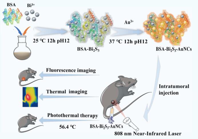
Nanotechnology has attracted extensive attention in the diagnosis and treatment of cancer. Therefore, the research aimed at developing new nanomaterials and exploring their applications in biomedicine has attracted more attention. In this study, Bi2S3-Au nanoclusters (Bi2S3-AuNCs) as fluorescence/infrared thermal imaging-guided photothermal therapy (PTT) was prepared for the first time. It was achieved in a facile and mild way by optimizing the amount of Bi3+ and Au3+ using bovine serum albumin (BSA) as reducer and stabilizer. The as-prepared Bi2S3-AuNCs with special morphology showed high stability, excellent biocompatibility and good photostability. Apart from these, it also can accumulate at tumor sites and exhibit considerable fluorescence/infrared thermal imaging-guided PTT. Bi2S3-AuNCs nanoparticles integrate imaging and therapeutic functions into an advanced application platform, which provides the possibility to build a novel nano-cancer diagnosis and treatment platform.
Keywords: Bi2S3-Au nanoclusters; Fluorescence imaging; Infrared thermal imaging; Multifunctional; Photothermal therapy.
© 2024 The Authors. Published by Elsevier B.V.
Conflict of interest statement
The authors declare that they have no known competing financial interests or personal relationships that could have appeared to influence the work reported in this paper.
Figures


Similar articles
-
Synthesis of gadolinium-based Bi2S3 nanoparticles as cancer theranostics for dual-modality computed tomography/magnetic resonance imaging-guided photothermal therapy.Nanotechnology. 2019 Feb 15;30(7):075101. doi: 10.1088/1361-6528/aaf442. Epub 2018 Nov 27. Nanotechnology. 2019. PMID: 30523911
-
Near-infrared-II-activatable sulfur-deficient plasmonic Bi2S3-x-Au heterostructures for photoacoustic imaging-guided ultrasound enhanced high performance phototherapy.J Colloid Interface Sci. 2023 Aug 15;644:437-453. doi: 10.1016/j.jcis.2023.04.108. Epub 2023 Apr 25. J Colloid Interface Sci. 2023. PMID: 37126893
-
Bistratal Au@Bi2S3 nanobones for excellent NIR-triggered/multimodal imaging-guided synergistic therapy for liver cancer.Bioact Mater. 2020 Sep 4;6(2):386-403. doi: 10.1016/j.bioactmat.2020.08.023. eCollection 2021 Feb. Bioact Mater. 2020. PMID: 32954056 Free PMC article.
-
Optical diagnostic imaging and therapy for thyroid cancer.Mater Today Bio. 2022 Sep 26;17:100441. doi: 10.1016/j.mtbio.2022.100441. eCollection 2022 Dec 15. Mater Today Bio. 2022. PMID: 36388462 Free PMC article. Review.
-
Gold nanoclusters as novel optical probes for in vitro and in vivo fluorescence imaging.Biophys Rev. 2012 Dec;4(4):313-322. doi: 10.1007/s12551-012-0076-9. Epub 2012 Apr 12. Biophys Rev. 2012. PMID: 28510207 Free PMC article. Review.
Cited by
-
Multifunctional gold nanoclusters as next-generation theranostic platforms for disease management.Int J Pharm X. 2025 Jul 21;10:100361. doi: 10.1016/j.ijpx.2025.100361. eCollection 2025 Dec. Int J Pharm X. 2025. PMID: 40777085 Free PMC article. Review.
References
-
- Alibakhshi A., Abarghooi Kahaki F., Ahangarzadeh S., Yaghoobi H., Yarian F., Arezumand R., Ranjbari J., Mokhtarzadeh A., de la Guardia M. Targeted cancer therapy through antibody fragments-decorated nanomedicines. J. Control. Release. 2017;268:323–334. doi: 10.1016/j.jconrel.2017.10.036. - DOI - PubMed
LinkOut - more resources
Full Text Sources
