. 2024 Sep 25:17:6743-6764.
doi: 10.2147/JIR.S477846. eCollection 2024.

Pan-Cancer Screening and Validation of CALU's Role in EMT Regulation and Tumor Microenvironment in Triple-Negative Breast Cancer

Affiliations

Pan-Cancer Screening and Validation of CALU's Role in EMT Regulation and Tumor Microenvironment in Triple-Negative Breast Cancer

Shi-Liang Chen et al. J Inflamm Res. .

Abstract

Purpose: Cancer-associated fibroblasts (CAFs) significantly contribute to tumor progression and the development of resistance to therapies across a range of malignancies, notably breast cancer. This study aims to elucidate the specific role and prognostic relevance of CALU across multiple cancer types.

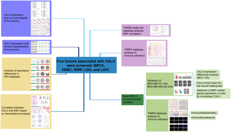
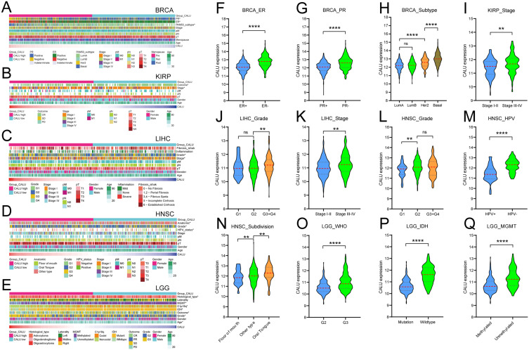
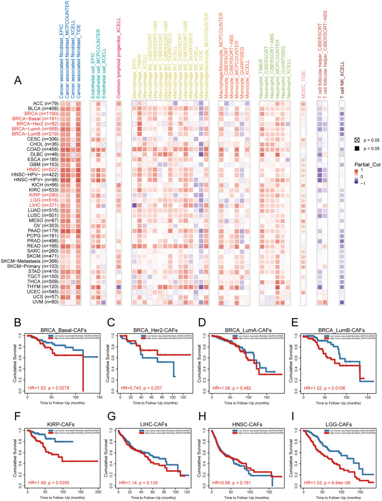
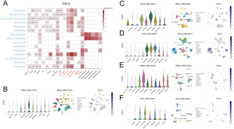
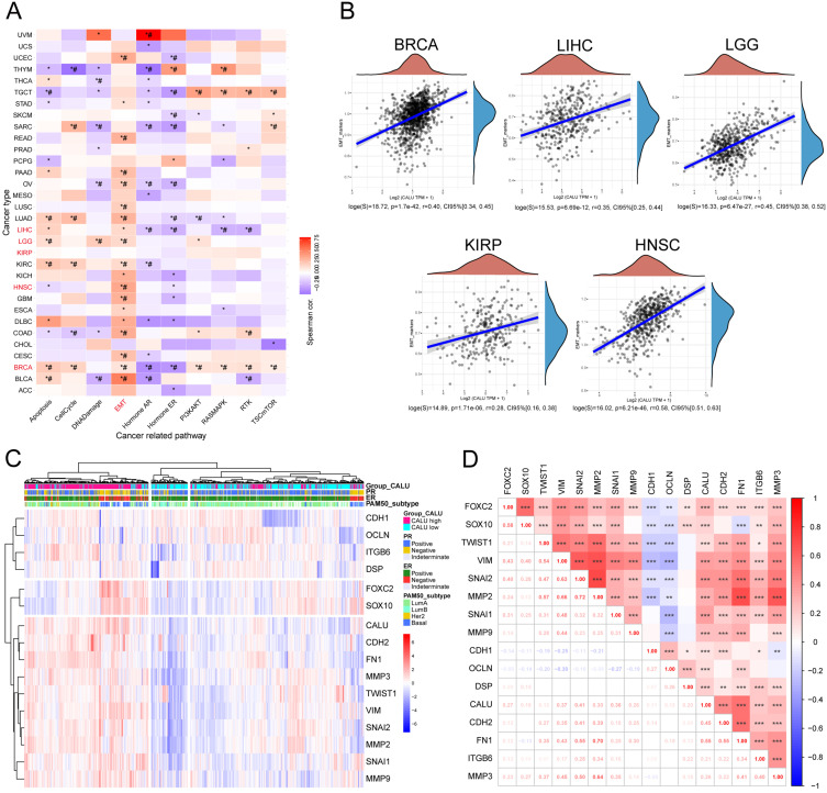
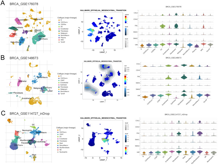
Patients and methods: The association between CALU expression and prognosis, along with clinical characteristics in BRCA, HNSC, KIRP, LGG, and LIHC, was analyzed using data from the TCGA, GTEx, and GEO databases. Transcriptomic analysis of TCGA BRCA project data provided insights into the interaction between CALU and epithelial-mesenchymal transition (EMT) marker genes. Using TIMER and TISCH databases, the correlation between CALU expression and tumor microenvironment infiltration was assessed, alongside an evaluation of CALU expression across various cell types. Furthermore, CALU's influence on TNBC BRCA cell lines was explored, and its expression in tumor tissues was confirmed through immunohistochemical analysis of clinical samples.

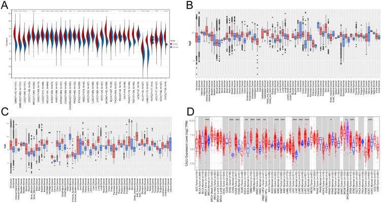
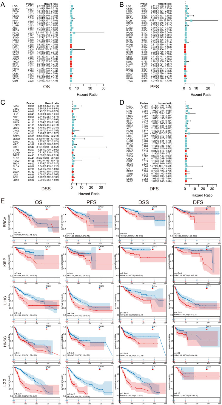
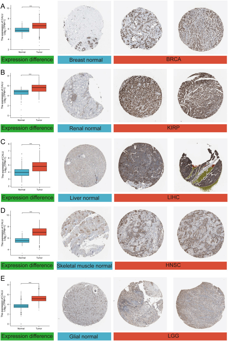
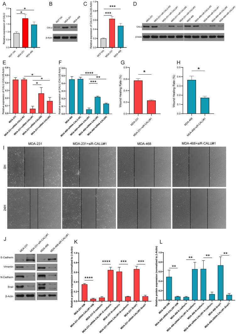
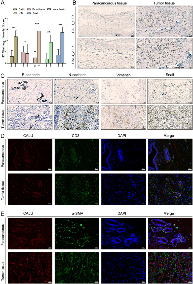
Results: This study revealed a consistent upregulation of CALU across several tumor types, including BRCA, KIRP, LIHC, HNSC, and LGG, with elevated CALU expression being associated with unfavorable prognoses. CALU expression was particularly enhanced in clinical contexts linked to poor outcomes. Genomic analysis identified copy number alterations as the principal factor driving CALU overexpression. Additionally, a positive correlation between CALU expression and CAF infiltration was observed, along with its involvement in the EMT process in both CAFs and malignant cells. In vitro experiments demonstrated that CALU is highly expressed in TNBC-BRCA cell lines, and knockdown of CALU effectively reversed EMT progression and inhibited cellular migration. Immunohistochemical analysis of clinical samples corroborated the elevated expression of CALU in tumors, along with alterations in EMT markers.

Conclusion: This comprehensive pan-cancer analysis underscores CALU's critical role in modulating the tumor microenvironment and facilitating cell migration via the EMT pathway, identifying it as a potential therapeutic target.

Keywords: CALU; breast cancer; cancer-associated fibroblasts; epithelial-mesenchymal transition; pan-cancer analysis; prognosis.


Conflict of interest statement

The authors declare no financial non-financial or competing interests.

Figures

Figure 1
Figure 1
The flowchart of our study.
Figure 2
Figure 2
CALU mRNA expression levels in different tumors. (A) TCGA+GTEx: CALU expression is elevated in most tumor tissues, except in UCEC, CESC, BLCA, THCA, UCS, PCPG, and KICH. (B) GENT2 (GPL570): CALU expression is increased in tumor tissues of the colon, brain, kidney, skin, lung, breast, ovary, liver, stomach, thyroid, head and neck, adrenal gland, bladder, prostate, blood, pancreas, tongue, teeth, and adipose. However, its expression is decreased in endometrium, uterus, esophagus, and bone tumors. (C) GENT2 (GPL96): Elevated CALU expression is observed in tumor tissues of the adrenal gland, blood, bone marrow, brain, breast, cervix, colon, esophagus, immune system, kidney, liver, lung, ovary, prostate, soft tissue, stomach, and testis. Decreased expression is noted in cartilage and heart tumors. (D) TIMER (TCGA): CALU expression is upregulated in BRCA, CHOL, COAD, ESCA, GBM, HNSC, KIRC, KIRP, LIHC, LUAD, LUSC, and STAD tumors, and downregulated in PRAD and UCEC. Notably, CALU expression is lower in HNSC HPV+ patients compared to HPV- patients, and higher in metastatic SKCM compared to non-metastatic SKCM. Red signifies the presence of neoplastic tissue, blue indicates the presence of healthy tissue, and purple denotes the existence of metastatic tissue. *p< 0.05; **p< 0.01; ***p< 0.001.
Figure 3
Figure 3
CALU Prognostic analysis of different tumors. (A) Overall survival (OS), (B) Progression-Free Survival (PFS), (C) Disease-Specific Survival (DSS), (D) Disease-Free Survival (DFS), and (E) Kaplan-Meier analysis of CALU in BRCA, KIRP, LIHC, HNSC, and LGG for different survival indicators.
Figure 4
Figure 4
CALU Immunohistochemistry data (HPA) and mRNA expression data (TCGA) in different tumors. (A) BRCA, (B) KIRP, (C) LIHC, (D) HNSC, and (E) LGG. ***p< 0.001.
Figure 5
Figure 5
Clinical characteristics analysis of CALU in different tumors. (A) BRCA, (B) KIRP, (C) LIHC, (D)HNSC, (E)LGG. (FQ) Analysis of CALU mRNA expression among distinct clinical characteristics in diverse tumor types. **p< 0.01; ****p< 0.0001.
Figure 6
Figure 6
Correlation between CALU expression and tumor microenvironment cells utilizing bulk RNA-seq. (A) The correlation between CALU mRNA expression and various stromal cells and immune cells was analyzed by timer database. The impact of cancer-associated fibroblast (CAFs) cells with varying degrees of infiltration on the prognosis of BRCA-Basal (B), BRCA-Her2(C), BRCA-LumA(D), BRCA-LumB(E), KIRP(F), LIHC(G), HNSC(H), and LGG(I) is being examined.
Figure 7
Figure 7
CALU is highly expressed in tumor stromal cells compared to other cells. (A) Heatmap of CALU expression levels by cell type in 15 single cell datasets including BRCA. (B) BRCA_GSE176078; (C) BRCA_GSE148673; (D) THCA_GSE148673; (E) HNSC_GSE103322; (F) LIHC_GSE125449.
Figure 8
Figure 8
CALU is closely related to the EMT process in BRCA. (A) Correlation analysis between CALU expression level and different signaling pathways in various cancers. (B) Correlation analysis between CALU and EMT scores in BRCA and other 4 tumors. (C and D) Heatmap and correlation analysis of CALU expression and EMT marker genes’ expression.*: p<0.05; #: FDR<0.05.
Figure 9
Figure 9
Visualization of EMT signatures in the t-SNE map of 3 scRNA-seq databases from BRCA. t-SNE plots of different cell types, EMT feature UMAP plots, and violin plots showing the expression of CALU and EMT markers, derived from the GSE176078 (A), GSE148673 (B), and GSE114727 (C) datasets, respectively.
Figure 10
Figure 10
Knockdown of CALU inhibits the EMT process in breast cancer cells. The expression of CALU in triple-negative breast cancer cell lines MDA-231 and MDA-468 was significantly higher than that in normal breast cell line MCF10A, both in mRNA(A) and protein (B and C). (DF) Detection of protein expression of CALU after transfection with different small interfering RNAs targeting CALU. (GI) Wound healing assay of MDA-231 and MDA-468 cell lines after transfection of siR-CALU#1. (JL) Detection of EMT marker gene expression in MDA-231 and MDA-468 cell lines before and after knockdown. *p< 0.05; **p< 0.01; ***p< 0.001; ****p< 0.0001.
Figure 11
Figure 11
Validation of CALU, EMT marker genes, CD3 and α-SMA in breast cancer samples. (A) IHC staining scores for CALU and EMT markers from 8 TNBC patients.(B and C) IHC staining results of CALU, E-cadherin, N-cadherin, Vimentin and Snail1 in TNBC tissues. (D) IF staining results of CALU and CD3 in breast cancer tissue. (E) IF staining results of CALU and α-SMA in breast cancer tissue.**p< 0.01; ***p< 0.001.

Similar articles

Cited by

References

    1. Siegel RL, Miller KD, Wagle NS, Jemal A. Cancer statistics, 2023. CA. 2023;73(1):17–48. doi:10.3322/caac.21763 - DOI - PubMed
    1. Siegel RL, Miller KD, Jemal A. Cancer statistics, 2019. CA. 2019;69(1):7–34. doi:10.3322/caac.21551 - DOI - PubMed
    1. Li YQ, Sun FZ, Li CX, et al. RARRES2 regulates lipid metabolic reprogramming to mediate the development of brain metastasis in triple negative breast cancer. Military Med Res. 2023;10(1):34. doi:10.1186/s40779-023-00470-y - DOI - PMC - PubMed
    1. Wang Y, Li Y, Jing Y, et al. Tubulin alpha-1b chain was identified as a prognosis and immune biomarker in pan-cancer combing with experimental validation in breast cancer. Sci Rep. 2024;14(1):8201. doi:10.1038/s41598-024-58982-z - DOI - PMC - PubMed
    1. Mao Y, Keller E, Garfield D, Shen K, Wang J. Stromal cells in tumor microenvironment and breast cancer. Cancer Metastasis Rev. 2013;32:303–315. doi:10.1007/s10555-012-9415-3 - DOI - PMC - PubMed

LinkOut - more resources