. 2024 Sep 24:17:6691-6706.
doi: 10.2147/JIR.S474329. eCollection 2024.

Qing-Luo-Yin Eased Adjuvant-Induced Arthritis by Inhibiting SIRT1-Controlled Visfatin Production in White Adipose Tissues

Affiliations

Qing-Luo-Yin Eased Adjuvant-Induced Arthritis by Inhibiting SIRT1-Controlled Visfatin Production in White Adipose Tissues

Dan-Dan Wang et al. J Inflamm Res. .

Abstract

Background: Nicotinamide adenine dinucleotide (NAD)-dependent deacetylase SIRT1 regulates both metabolism and immune functions. This study investigated if SIRT1 inhibitory property of herbal formula Qing-Luo-Yin (QLY) contributed to its anti-rheumatic effects.

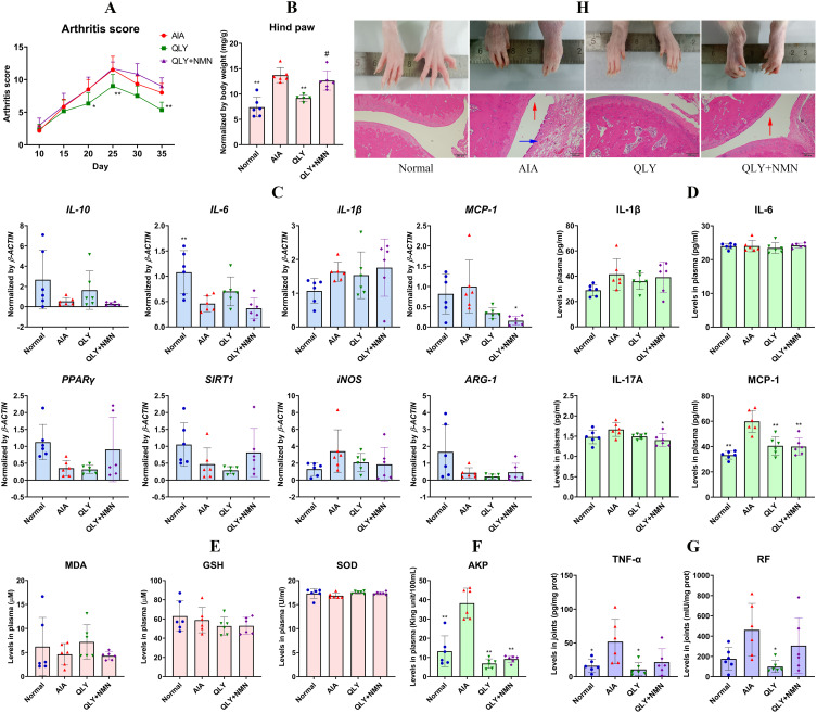
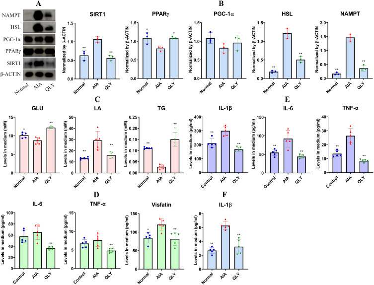
Methods: Adjuvant-induced arthritis (AIA) rats were treated by QLY and nicotinamide mononucleotide (NMN, a biosynthesis precursor of NAD) for 38 days. After sacrifice, blood, paws, liver and white adipose tissues (WAT) were collected. Pre-adipocytes were cultured by the rats' serum. The medium was used for monocytes culture. Some pre-adipocytes were treated by QLY-derived SIRT1 inhibitors. SIRT1 was silenced or overexpressed beforehand. The samples were subjected to kits-based quantification, polymerase-chain reaction, western-blot, immunofluorescence, and histology experiments.

Results: AIA rats experienced significant fat loss in liver and WAT. Expression of many SIRT1-related signals like PPARγ, PGC-1α, HSL, ATGL and CPT-1A were altered. QLY attenuated all these abnormalities and joint injuries. By pan-acetylation up-regulation, visfatin was obviously reduced in QLY-treated AIA rats' blood (from 191.8 to 127.0 pg/mL). NMN sustained SIRT1 activation by replenishing NAD, and weakened these effects. QLY-containing serum and the related compounds showed similar impacts on pre-adipocytes, resembling the changes in QLY-treated AIA rats' WAT. These treatments suppressed AIA serum-induced visfatin secretion (from 49.3 to 36.1 and 30.7 pg/mL). This effect was impaired by SIRT1 overexpression. The medium from the compounds-treated pre-adipocytes impaired NF-κB activation in AIA serum-cultured monocytes.

Conclusion: Besides fat depletion, SIRT1 up-regulation in rheumatic subjects' WAT promotes visfatin production, and exacerbates inflammation. SIRT1 inhibition in WAT is an anti-rheumatic way of QLY independent of immune regulation.

Keywords: PPARγ; Traditional Chinese Medicine; adipocyte; rheumatoid arthritis; visfatin.


Conflict of interest statement

The authors declare no conflict of interest.

Figures

Figure 1
Figure 1
QLY therapy improved arthritic manifestations of AIA rats. (A) AIA progression evaluated based on arthritis scores; (B) relative weight of left hind paw; (C) expression of some polarization-related genes in blood monocytes; (D) levels of some RA-related cytokines (IL-1β, IL-6, IL-17A and MCP-1) in rats’ blood; (E) levels of oxidative indicators (GSH, MDA and SOD) in rats’ blood; (F) AKP levels in rats’ blood; (G) levels of TNF-α and RF detected in rats’ joints; (H) morphological observation and H&E staining-based histological examination of left hind ankle joint; the red and blue arrows indicate cartilage erosion and synovial invasion, respectively. All the data are presented in mean±SD. Statistical significance: *p < 0.05 and **p < 0.01 compared with AIA models; #p < 0.05 compared with QLY-treated AIA rats.
Figure 2
Figure 2
QLY therapy affected SIRT1-related signals as well as metabolic status in AIA rats’ liver. (A) levels of metabolism indicators in rats’ blood; (B) levels of GLU and TG in rats’ livers; (C) Oil Red O staining-based histological examination of livers; (D) WB assay performed using liver samples; (E) the quantification results of assay (D). All the data are presented in mean±SD. Statistical significance: *p < 0.05 and **p < 0.01 compared with AIA models; #p < 0.05 and ##p < 0.01 compared with QLY-treated AIA rats.
Figure 3
Figure 3
QLY therapy reshaped both metabolism and secretion of WAT in AIA rats by regulating SIRT1-related signals. (A) morphological observation of representative perirenal fat pads; (B) relative weight of perirenal fat pads; (C) H&E staining-based histological examination of perirenal fat pads; (D) WB and IP assays performed using rats’ WAT samples; (E) the quantification results of assay (D); (F) levels of representative adipokines (leptin, adiponectin, and visfatin) in rats’ blood; (G and H) TG and visfatin detected in rats’ WAT; (I) expression of pan-acetylated proteins and visfatin in rats’ WAT. All the data are presented in mean±SD. Statistical significance: *p < 0.05 and **p < 0.01 compared with AIA models; #p < 0.05 compared with QLY-treated AIA rats.
Figure 4
Figure 4
QLY-containing serum affected immune status of monocytes indirectly via pre-adipocytes. (A) WB assays performed using the rat pre-adipocytes, which were cultured with the serums from healthy, AIA model or QLY-treated AIA rats; (B) the quantification results of assay (A); (C) levels of some key metabolites (GLU, TG and LA) in the medium from assay (A); (D) levels of IL-6, TNF-α and visfatin in the medium from assay (A); (E) levels of IL-1β, IL-6, and TNF-α secreted by the monocytes cultured by the mediums from assay (A); (F) levels of IL-1β secreted by the monocytes cultured by the supernatants of different rats’ WAT homogenates. All the data are presented in mean±SD. Statistical significance: *p < 0.05 and **p < 0.01 compared with AIA group.
Figure 5
Figure 5
QLY-related SIRT1 inhibitors exerted the similar effects on pre-adipocytes to QLY-containing serum. (A) WB assays performed on the rat pre-adipocytes, which were cultured by healthy, AIA model rats’ serums and then treated by the compounds or QLY-containing serum; (B) the quantification results of assay (A); (C) levels of some key metabolites (GLU, TG and LA) in the medium above; (D) levels of representative adipokines and cytokines (leptin, adiponectin, visfatin, IL-1β, IL-6 and MCP-1) in the medium; (E) levels of visfatin secreted by the 3T3-L1 cells, which received the same treatments as above; (F) expression of pan-acetylated proteins and visfatin in the cells from assay E. All the data are presented in mean±SD. Statistical significance: *p < 0.05 and **p < 0.01 compared with AIA group.
Figure 6
Figure 6
QLY-induced visfatin decrease in pre-adipocytes was attributed to SIRT1 inhibition. (A) WB assays performed using the pre-adipocytes cultured in vitro; SIRT1 in some of them were overexpressed, a portion of which were exposed to QLY-related SIRT1 inhibitors; (B) the quantification results of assay (A); (C) levels of some key metabolites (GLU, TG and LA) in the medium from assay (A); (D) levels of representative WAT-released substances (visfatin, IL-6 and MCP-1) in the medium; (E), WB assays performed using the pre-adipocytes cultured in vitro; SIRT1 in some of them were silenced, a portion of which exposed to QLY-related SIRT1 inhibitors; (F) the quantification results of assay (E); (G) levels of WAT-released substances in the medium from assay (E); (H) expression and localization of visfatin and p65 in the THP-1 cells stimulated by some mediums from assay (A); the yellow and green arrows indicate p65 distribution in the cytoplasm of normal cells and visfatin-induced p65 aggregation in the nuclear of AIA serum-cultured cells, respectively. All the data are presented in mean±SD. Statistical significance: *p < 0.05 and **p < 0.01 compared with AIA group; ##p < 0.01 compared with compounds group.

References

    1. McInnes IB, Schett G. The pathogenesis of rheumatoid arthritis. N Engl J Med. 2011;365(23):2205–2219. doi:10.1056/NEJMra1004965 - DOI - PubMed
    1. Ding Q, Hu W, Wang R, et al. Signaling pathways in rheumatoid arthritis: implications for targeted therapy. Signal Transduct Target Ther. 2023;8(1):68. - PMC - PubMed
    1. Roberts CA, Dickinson AK, Taams LS. The interplay between monocytes/macrophages and CD4+ T cell subsets in rheumatoid arthritis. Front Immunol. 2015;6:571. doi:10.3389/fimmu.2015.00571 - DOI - PMC - PubMed
    1. Davignon JL, Hayder M, Baron M, et al. Targeting monocytes/macrophages in the treatment of rheumatoid arthritis. Rheumatology. 2013;52(4):590–598. doi:10.1093/rheumatology/kes304 - DOI - PubMed
    1. Tu J, Huang W, Zhang W, Mei J, Zhu C. A tale of two immune cells in rheumatoid arthritis: the crosstalk between macrophages and T cells in the synovium. Front Immunol. 2021;12:655477. doi:10.3389/fimmu.2021.655477 - DOI - PMC - PubMed

LinkOut - more resources