. 2024 Aug 9;51(5):480-486.
doi: 10.1055/a-2337-2131. eCollection 2024 Sep.

A Lifeboat for Failed Nasal Reconstructions: The Supraclavicular-Submental Sandwich Flap

Affiliations

A Lifeboat for Failed Nasal Reconstructions: The Supraclavicular-Submental Sandwich Flap

Michel L H T Vaena et al. Arch Plast Surg. .

Abstract

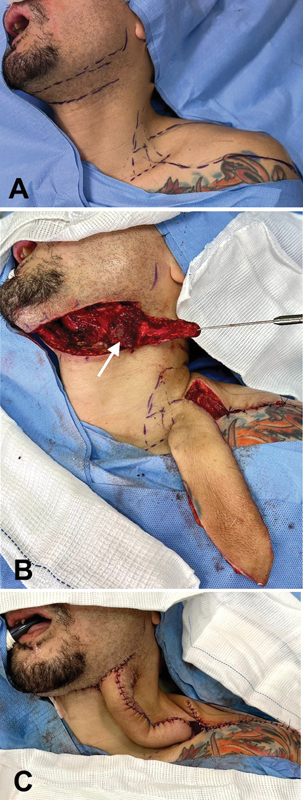
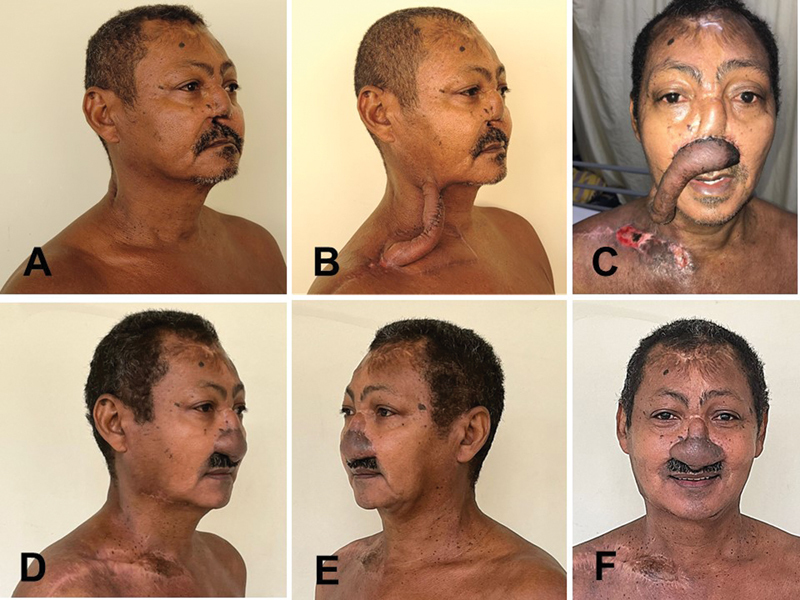
Many failures in total or subtotal nasal reconstruction result from an underestimation of the amount of skin required for an adequate result, especially for sufficient lining. Such planning errors usually lead to poor results, with exposure of structural grafts, infection, scar retraction, airway obstruction, and finally loss of projection and shape of the reconstructed nose. Reconstruction options for cases in which previous attempts have failed are always limited, as well as in cases of trauma or burns affecting the soft tissues of the forehead and face. In such complex situations, one may employ free flaps or tissue expansion, but such resources may not be always available. We describe a technique indicated for salvage surgeries in patients whose previous nasal reconstructions have failed, allowing a generous amount of tissue transfer for the nasal region. The technique combines the use of supraclavicular and submental flaps, with simple execution, not requiring microsurgical skills or devices such as tissue expanders. Done in three stages, the described technique provides enough skin for a total nasal reconstruction. The final result is obtained after subsequent refinements, and the total number of procedures is equivalent to when more sophisticated techniques are employed, such as tissue expansion or microsurgery.

Keywords: acquired; nose deformities; reconstructive surgical procedures; surgical flaps.


Conflict of interest statement

Conflict of Interest None declared.

Figures

Fig. 1
Fig. 1
A 35-year-old man with a total nasal defect. ( A ) The extensive scars on the forehead denote that both paramedian flaps (right and left) were used in the previous surgeries. ( B ) One month after the first stage, the left supraclavicular–submental sandwich (3SF) is healed and ready to be transferred. The length redundancy of the 3SF transfer ensures no restrictions to the patient's movements. ( C ) After the second stage, supraclavicular extremity was sectioned in the donor area and attached to the recipient's nasal area. This attachment is done above the piriform aperture, where neovascularization will occur. ( D ) Final aspect after insertion of structural grafts and subsequent refinements. Both left supraclavicular and submental scars are inconspicuous.
Fig. 2
Fig. 2
Schematic drawing of subplatysmal surgical anatomy. Above—The right submental flap is elevated together with the main portion of the right mylohyoid muscle (M). After blunt dissection of the right mylohyoid muscle (M), it can be mobilized laterally, exposing the deeper geniohyoid (G) and hyoglossus (H) muscles. The left mylohyoid muscle (M) and left anterior belly of the digastric muscle (D) remain in their anatomic positions. Below—The supraclavicular flap is raised from lateral do medial. The pedicle area is marked with an asterisk, whose anatomical landmarks can be easily identified preoperatively: the posterior edge of the sternocleidomastoid muscle (ECM), the clavicle (C), and the external jugular vein (EJV).
Fig. 3
Fig. 3
Schematic drawing of surgical stages. Above—First stage: ( A ) The skin paddle of the right submental flap is marked in green, and the right supraclavicular flap is marked in blue and purple. The area in purple corresponds to the skin that will cover the nose; therefore, it must have a minimum width of 7 to 8 cm. The pedicle area is marked with an asterisk. ( B ) Both flaps are elevated with their raw surfaces facing each other. ( C ) The flaps are sutured to each other, forming the “sandwich.” Note that the proximal redundant portion of the supraclavicular flap (blue area) has its raw edges sutured together forming a tube. No raw areas are left exposed. Below—Second stage: ( D ) After 1 month, the supraclavicular extremity is sectioned in the donor area. ( E ) The supraclavicular skin paddle (purple) is positioned in the recipient's nasal area. The supraclavicular donor area is closed primarily.
Fig. 4
Fig. 4
Surgical technique of the first stage in Case 1. ( A ) Marking for the dissection of supraclavicular and submental flaps. ( B ) The supraclavicular flap is raised from lateral to medial. The submental flap is dissected from medial to lateral leaving its skin portion connected to the submandibular area of the neck. The sectioned portion of the left mylohyoid muscle remains connected to the skin paddle, to avoid inadvertent injury to the submental perforating vessels (white arrow). ( C ) The raw sides of both flaps face one another, with the skin paddles being sutured to each other, forming a “sandwich.” Both donor areas (supraclavicular and submental) are closed primarily.
Fig. 5
Fig. 5
A 66-year-old man with a subtotal nasal defect. ( A ) Preoperative right oblique view. Previous reconstruction attempts have failed but left some frontal skin in the nasal radix and dorsum. ( B ) The right supraclavicular–submental sandwich flap (3SF) after the first stage. ( C ) Supraclavicular extremity demonstrating distal necrosis 1 week after its insertion in the nasal region. Note the length redundancy that allowed the flap to be detached, debrided, and readvanced. The wound in the right supraclavicular donor area (resulting from the second stage) can be observed. ( D ) Postoperative right oblique view. Supraclavicular and submental scars are unremarkable. ( E ) Postoperative left oblique view. ( F ) Postoperative frontal view. Notice the scarring on the forehead, demonstrating that both paramedian flaps (right and left) were used in the failed previous nasal reconstruction attempt.

References

    1. Whitaker I S, Karoo R O, Spyrou G, Fenton O M. The birth of plastic surgery: the story of nasal reconstruction from the Edwin Smith Papyrus to the twenty-first century. Plast Reconstr Surg. 2007;120(01):327–336. - PubMed
    1. Marck K W, Palyvoda R, Bamji A, van Wingerden J J. The tubed pedicle flap centennial: its concept, origin, rise and fall. Eur J Plast Surg. 2017;40:473–478.
    1. Lamberty B GH. The supra-clavicular axial patterned flap. Br J Plast Surg. 1979;32(03):207–212. - PubMed
    1. Pallua N, Magnus Noah E.The tunneled supraclavicular island flap: an optimized technique for head and neck reconstruction Plast Reconstr Surg 200010503842–851., discussion 852–854 - PubMed
    1. Patel U A, Bayles S W, Hayden R E. The submental flap: a modified technique for resident training. Laryngoscope. 2007;117(01):186–189. - PubMed

LinkOut - more resources