. 2024 Sep 24:12:e17904.
doi: 10.7717/peerj.17904. eCollection 2024.

A new species of alien land flatworm in the Southern United States

Affiliations

A new species of alien land flatworm in the Southern United States

Jean-Lou Justine et al. PeerJ. .

Abstract

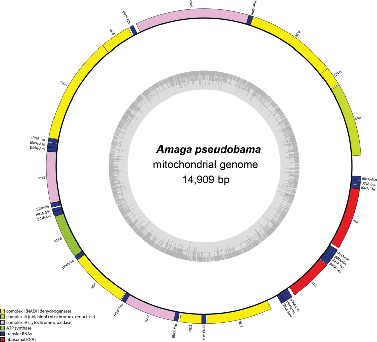
Specimens of a flat and dark brown land planarian were found in a plant nursery in North Carolina, USA in 2020. On the basis of examination of photographs of the live specimens only, the specimens were considered as belonging to Obama nungara, a species originally from South America, which has now invaded a large part of Europe. Unexpectedly, a molecular analysis revealed that the specimens did not belong to this species, neither to the genus Obama. We then undertook its histological study, which finally confirmed that the species is a member of the genus Amaga: the species is herein described as a new species, Amaga pseudobama n. sp. The species has been found in three locations in North Carolina and some infested plants were from Georgia. We reinvestigated specimens collected in Florida in 2015 and found that they also belong to this species. Citizen science observations suggest its presence in other states. Therefore, it is likely that A. pseudobama has already invaded a part of south-east USA and that the invasion took place more than ten years ago. The complete 14,909 bp long mitochondrial genome was obtained. The mitogenome is colinear with those of other Geoplanidae and it was possible to find and annotate a tRNA-Thr, which has been reported missing in several geoplanids. Amaga pseudobama shares with other Geoplaninae the presence of alternative start codons in three protein-coding genes of its mitogenome. The availability of this new genome helped us to improve our annotations of the ND3 gene, for which an ATT start codon is now suggested. Also, the sequence of the ATP6 gene raised questions concerning the use of genetic code 9 to translate the protein-coding genes of Geoplanidae, as the whole translated protein would not contain a single methionine residue when using this code. Two maximum likelihood phylogenies were obtained from genomic data. The first one was based on concatenated alignments of the partial 28S, Elongation Factor 1-alpha (EF1) and cox1 genes. The second was obtained from a concatenated alignment of the mitochondrial proteins. Both strictly discriminate A. pseudobama from O. nungara and instead associate it with Amaga expatria. We note that the nine species currently accepted within Amaga can be separated into two groups, one with extrabulbar prostatic apparatus, including the type species A. amagensis, and one with intrabulbar prostatic apparatus, including the new species A. pseudobama. This suggests that species of the latter group should be separated from Amaga and constitute a new genus. This finding again illustrates the possible emergence of new invasive species in regions naturally devoid of large land planarians, such as North America. Amaga pseudobama thus deserves to be monitored in the USA, although its superficial resemblance to O. nungara and Geoplana arkalabamensis will complicate the use of photographs obtained from citizen science. Our molecular information provides tools for this monitoring.

Keywords: Invasive alien species; Land flatworms; Mitogenome; New species; Platyhelminthes; Taxonomy; USA.


Conflict of interest statement

Jean-Lou Justine is an Academic Editor for PeerJ.

Figures

Figure 1
Figure 1. Amaga pseudobama n. sp., living specimen collected in Kingston, North Carolina in July 2020.
The specimen was photographed in a Petri dish on a white background. (A) Dorsal view from back; the head is at top. (B) Dorsal view; the head is on the right. (C) Ventral view; the pharynx (p) and gonopore (g), are indicated; the head is on the left. Unscaled. Photographs by Matthew A. Bertone.
Figure 2
Figure 2. Amaga pseudobama n. sp., general view of preserved holotype.
(A) Dorsal surface, (B) right side, and (C) ventral surface showing mouth (m) and gonopore (gp).
Figure 3
Figure 3. Amaga pseudobama n. sp., eyes of holotype.
(A) Eyes (arrowed) contouring the anterior tip, (B) eyes extending dorso-laterally, many of which are haloed (arrowed; a clear space surrounding the eye), (C) dorsum showing haloed eyes, and pigment surrounding the underlying testes (arrowed). Unscaled.
Figure 4
Figure 4. Amaga pseudobama n. sp., prepharyngeal region, histology.
Transverse section of the pre-pharyngeal region. In order to ensure adequate resolution of the histology, just over a half of the section is shown.
Figure 5
Figure 5. Amaga pseudobama n. sp., pharynx, histology.
(A) Pharynx, longitudinal sagittal section, and (B) more laterally, showing the folded pharyngeal lip.
Figure 6
Figure 6. Amaga pseudobama n. sp., anterior region, histology.
Anterior tip, longitudinal lateral section, showing mono- and bilobulated eyes, and ciliated pits.
Figure 7
Figure 7. Amaga pseudobama n. sp., reconstruction of reproductive organs.
Copulatory organs, composite reconstruction, longitudinal sagittal aspect. Some details of secretions and musculature have been omitted for clarity. The dashed line is the approximate area of transition between the male and female atria.
Figure 8
Figure 8. Amaga pseudobama n. sp., copulatory organs, histology.
Copulatory organs, longitudinal sagittal section. Secretion and musculature omitted from the composite reconstruction can be clearly seen.
Figure 9
Figure 9. Amaga pseudobama n. sp., testis, histology.
Testis, located above the supraintestinal transverse parenchymal muscles. Note the dark pigment that delineates the testes.
Figure 10
Figure 10. Amaga pseudobama n. sp., male organs, histology.
(A) The common sperm duct and seminal vesicle that arise laterally to the prostatic vesicle. The dashed line indicates the approximate junction of these vesicles; (B) details of the start of the opening of the slit in the prostatic vesicle, together with detail of the erythrophil secretions, and the cyanophil secretions of the male atrium.
Figure 11
Figure 11. Amaga pseudobama n. sp., female organs, histology.
Detail of the female atrium characterised by an epithelium with the necessary precursor secretions to form an egg-capsule: a tall nucleate columnar epithelium through which is discharged membrane bound chromophobe globules, fusiform erythrophil membrane bound packets, and amorphous lightly cyanophil secretions that form strands to which the chromophobe globules and erythrophil attach (arrowed). The dashed line is the approximate area of transition between the male and female atria.
Figure 12
Figure 12. Amaga pseudobama n. sp., female organs, histology.
(A) Right ovary and ovovitelline duct; (B) separate entry of the ovovitelline ducts from either side of the proximal end of the female glandular canal.
Figure 13
Figure 13. Map of the mitochondrial genome of Amaga pseudobama n. sp.
The mitogenome is 14,909 bp long and contains 12 protein coding genes, two ribosomal RNA genes and 22 transfer RNA genes. Canonical start codons for the genetic code 9 could not be found for ATP6, cox2 and ND3.
Figure 14
Figure 14. Three-gene phylogenetic tree.
The tree is based on concatenated alignments of the 28S, cox1 and EF1α genes. Maximum likelihood phylogenetic tree based on 72 taxa, using the TPM2uf+I+G4, GTR+I+G4 and GTR+G4 models of evolution for 28S, cox1 and EF1, respectively. The tree was computed for 1,000 ultrafast bootstrap replicates; ML bootstrap support values indicated at the nodes. The closest species to Amaga pseudobama n. sp. is Amaga expatria.
Figure 15
Figure 15. Maximum likelihood phylogenetic tree of concatenated mitochondrial proteins.
The tree is based on concatenated protein sequences extracted from 28 mitogenomes using the MTZOA+I+G4+F model and with 1000 ultrafast bootstrap replicates; ML bootstrap support values indicated at the nodes. The closest species to Amaga pseudobama n. sp. is Amaga expatria.

References

    1. Almeida AL, Álvarez-Presas M, Carbayo F. The discovery of new Chilean taxa revolutionizes the systematics of Geoplaninae Neotropical land planarians (Platyhelminthes: Tricladida) Zoological Journal of the Linnean Society. 2023;197:837–898. doi: 10.1093/zoolinnean/zlac072. - DOI
    1. Almeida AL, Francoy TM, Álvarez-Presas M, Carbayo F. Convergent evolution: a new subfamily for bipaliin-like Chilean land planarians (platyhelminthes) Zoologica Scripta. 2021;50:500–508. doi: 10.1111/zsc.12479. - DOI
    1. Bankevich A, Nurk S, Antipov D, Gurevich AA, Dvorkin M, Kulikov AS, Lesin VM, Nikolenko SI, Pham S, Prjibelski AD. SPAdes: a new genome assembly algorithm and its applications to single-cell sequencing. Journal of Computational Biology. 2012;19:455–477. doi: 10.1089/cmb.2012.0021. - DOI - PMC - PubMed
    1. Bernt M, Donath A, Jühling F, Externbrink F, Florentz C, Fritzsch G, Pütz J, Middendorf M, Stadler PF. MITOS: improved de novo metazoan mitochondrial genome annotation. Molecular Phylogenetics and Evolution. 2013;69:313–319. doi: 10.1016/j.ympev.2012.08.023. - DOI - PubMed
    1. Camacho C, Coulouris G, Avagyan V, Ma N, Papadopoulos J, Bealer K, Madden TL. BLAST+: architecture and applications. BMC Bioinformatics. 2009;10:421. doi: 10.1186/1471-2105-10-421. - DOI - PMC - PubMed

LinkOut - more resources