. 2024 Sep 25:12:e18107.
doi: 10.7717/peerj.18107. eCollection 2024.

Differences in cervical sagittal parameters and muscular function among subjects with different cervical spine alignments: a surface electromyography-based cross-sectional study

Affiliations

Differences in cervical sagittal parameters and muscular function among subjects with different cervical spine alignments: a surface electromyography-based cross-sectional study

Dian Wang et al. PeerJ. .

Abstract

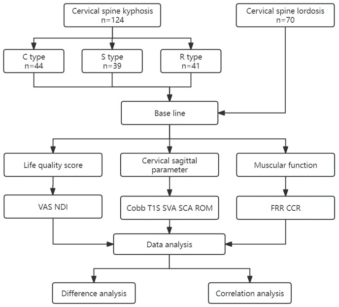
Background: We analyzed cervical sagittal parameters and muscular function in different cervical kyphosis types.

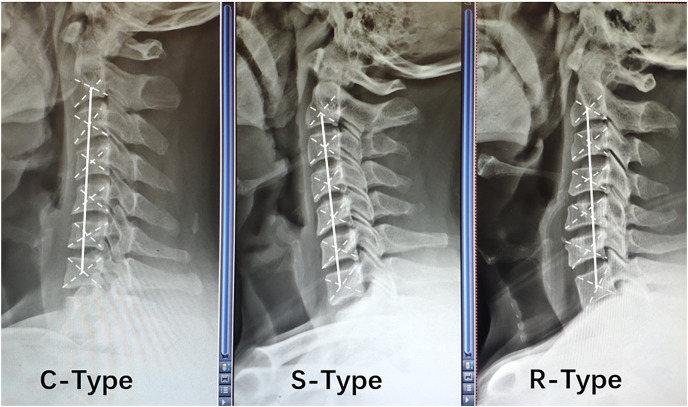
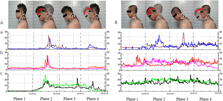
Methods: This cross-sectional study enrolled subjects with cervical spine lordosis (cervical curvature < -4°) or degenerative cervical kyphosis (cervical curvature > 4°), including C-, S-, and R-type kyphosis. We recorded patients' general information (gender, age, body mass index), visual analog scale (VAS) scores, and the Neck Disability Index (NDI). Cervical sagittal parameters including C2-C7 Cobb angle (Cobb), T1 slope (T1S), C2-C7 sagittal vertical axis (SVA), spino-cranial angle (SCA), range of motion (ROM), and muscular function (flexion-relaxation ratio (FRR) and co-contraction ratio (CCR) of neck/shoulder muscles on surface electromyography). Differences in cervical sagittal parameters and muscular function in subjects with different cervical spine alignments, and correlations between VAS scores, NDI, cervical sagittal parameters, and muscular function indices were statistically analyzed.

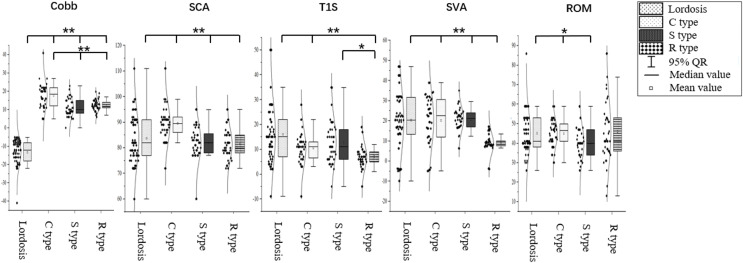
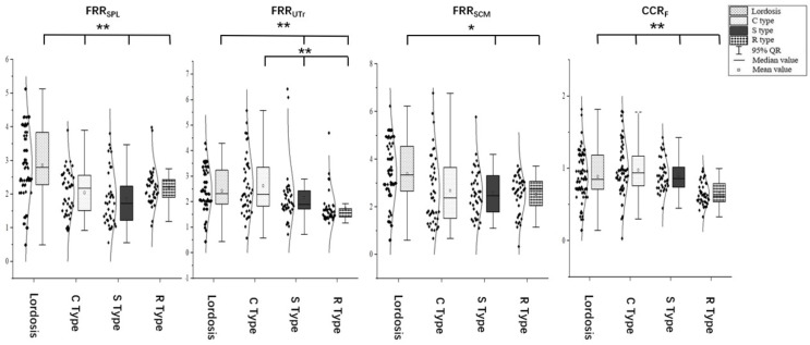
Results: The FRR of the splenius capitis (SPL), upper trapezius (UTr), and sternocleidomastoid (SCM) were higher in subjects with cervical lordosis than in subjects with cervical kyphosis. FRRSPL was higher in subjects with C-type kyphosis than in subjects with R- and S-type kyphosis (P < 0.05), and was correlated with VAS scores, Cobb angle, T1S, and SVA. FRRUTr was correlated with NDI, SCA, T1S, and SVA. FRRSCM was correlated with VAS scores and Cobb angle. CCR was correlated with SCA and SVA.

Conclusion: Cervical sagittal parameters differed among different cervical kyphosis types. FRRs and CCRs were significantly worse in R-type kyphosis than other kyphosis types. Cervical muscular functions were correlated with cervical sagittal parameters and morphological alignment.

Keywords: Cervical kyphosis; Cervical sagittal parameters; Flexion and relaxation ratio; Muscular function; Surface electromyography.


Conflict of interest statement

The authors declare that they have no competing interests.

Figures

Figure 1
Figure 1. Relative positions of the center of gravity of each vertebral body in different types of cervical spine kyphosis.
Figure 2
Figure 2. Study design.
VAS, visual analog scale; NDI, Neck Disability Index; Cobb, C2–C7 Cobb angle; T1S, T1 slope; SVA, C2–C7 sagittal vertical axis; SCA, spino-cranial angle; ROM, range of motion; FRR, flexion-relaxation ratio; CCR, co-contraction ratio.
Figure 3
Figure 3. Splenius capitis, upper trapezius, and sternocleidomastoid muscle surface anchor points.
(A) Splenius capitis body surface anchor points: line AB, the line between the apex of the auricle and the C7 spinous process; line AC, the posterior edge of the sternocleidomastoid muscle; line DE, the belly of the splenius capitis muscle; and an anchor point located outside point E. (B) Upper trapezius surface anchor points: line BG, line between the C7 spinous process and the acromion; the anchor point is at the midpoint of line BG. (C) Sternocleidomastoid body surface anchor points: line HI, line connecting the sternal notch and the mastoid bone; the fixation point was located at the junction of the middle and lower thirds of line HI.
Figure 4
Figure 4. Differences in cervical sagittal parameters in subjects with different morphological alignments of the cervical spine.
C2–C7 Cobb angle, Cobb; SCA, spino-cranial angle; T1S, T1 slope; SVA, C2–C7 sagittal vertical axis; ROM, range of motion. *P < 0.05; **P < 0.01.
Figure 5
Figure 5. Acquisition of neck and shoulder muscle signals via surface electromyography (sEMG; for the cervical extensor muscles as an example).
(A) Movement and muscle electrical signals of subjects with cervical lordosis. (B) Movement and muscle electrical signals of subjects with cervical kyphosis. a, Splenius capitis sEMG signals; b, upper trapezius sEMG signals; c, sternocleidomastoid sEMG signals. Phase 1, neutral position; phase 2, neutral position with gradual neck flexion to flexion position; phase 3, maintain the flexion position; phase 4, gradual extension from flexion position to neutral position.
Figure 6
Figure 6. Differences in muscular function in subjects with different cervical spinal alignments.
FRRSPL, flexion-relaxation ratio of the splenius capitis; FRRUTr, flexion-relaxation ratio of the upper trapezius; FRRSCM, flexion-relaxation ratio of the sternocleidomastoid; CCR, co-contraction ratio. *P < 0.05; **P < 0.01.

Similar articles

Cited by

References

    1. Abelin-Genevois K. Sagittal balance of the spine. Orthopaedics & Traumatology: Surgery & Research. 2021;107(1):102769. doi: 10.1016/j.otsr.2020.102769. - DOI - PubMed
    1. Ao S, Liu Y, Wang Y, Zhang H, Leng H. Cervical kyphosis in asymptomatic populations: incidence, risk factors, and its relationship with health-related quality of life. Journal of Orthopaedic Surgery and Research. 2019;14(1):681. doi: 10.1186/s13018-019-1351-2. - DOI - PMC - PubMed
    1. Been E, Shefi S, Soudack M. Cervical lordosis: the effect of age and gender. The Spine Journal. 2017;17(6):880–888. doi: 10.1016/j.spinee.2017.02.007. - DOI - PubMed
    1. Bokaee F, Rezasoltani A, Manshadi FD, Naimi SS, Baghban AA, Azimi H. Comparison of cervical muscle thickness between asymptomatic women with and without forward head posture. Brazilian Journal of Physical Therapy. 2017;21(3):206–211. doi: 10.1016/j.bjpt.2017.04.003. - DOI - PMC - PubMed
    1. Brumpt E, Aubry S, Vuillier F, Tatu L. Anatomo-sonographic identification of the longissimus capitis and splenius cervicis muscles: principles for possible application to ultrasound-guided botulinum toxin injections in cervical dystonia. Surgical and Radiologic Anatomy. 2021;43(6):909–915. doi: 10.1007/s00276-020-02646-w. - DOI - PMC - PubMed

LinkOut - more resources