. 2024 Sep 20;14(18):e5074.
doi: 10.21769/BioProtoc.5074.

A Full Good Manufacturing Practice-Compliant Protocol for Corneal Stromal Stem Cell Cultivation

Affiliations

A Full Good Manufacturing Practice-Compliant Protocol for Corneal Stromal Stem Cell Cultivation

Mithun Santra et al. Bio Protoc. .

Abstract

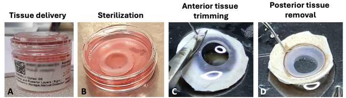
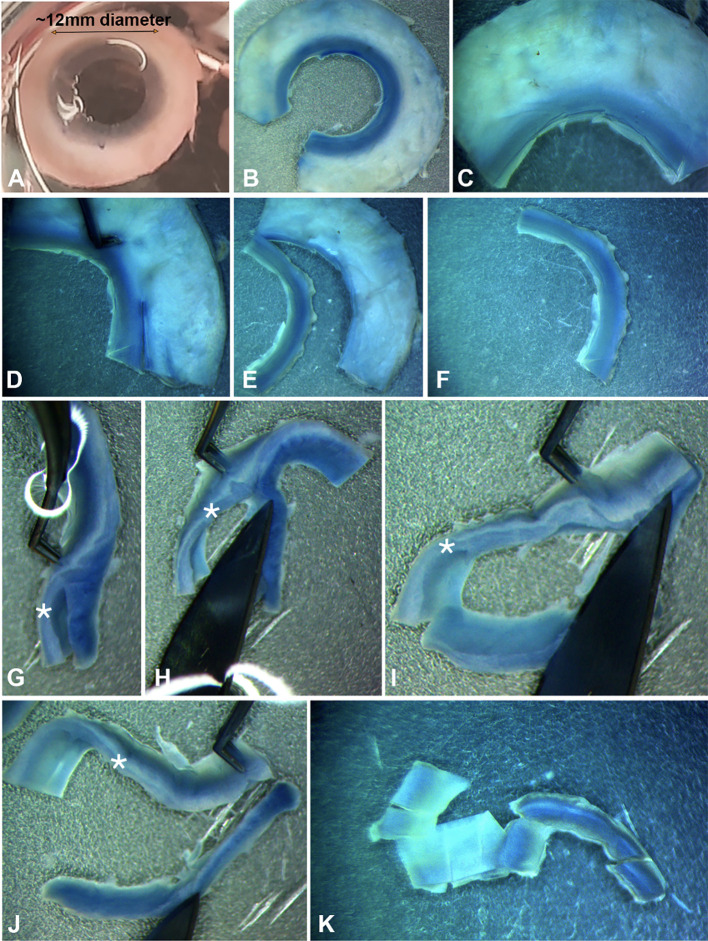
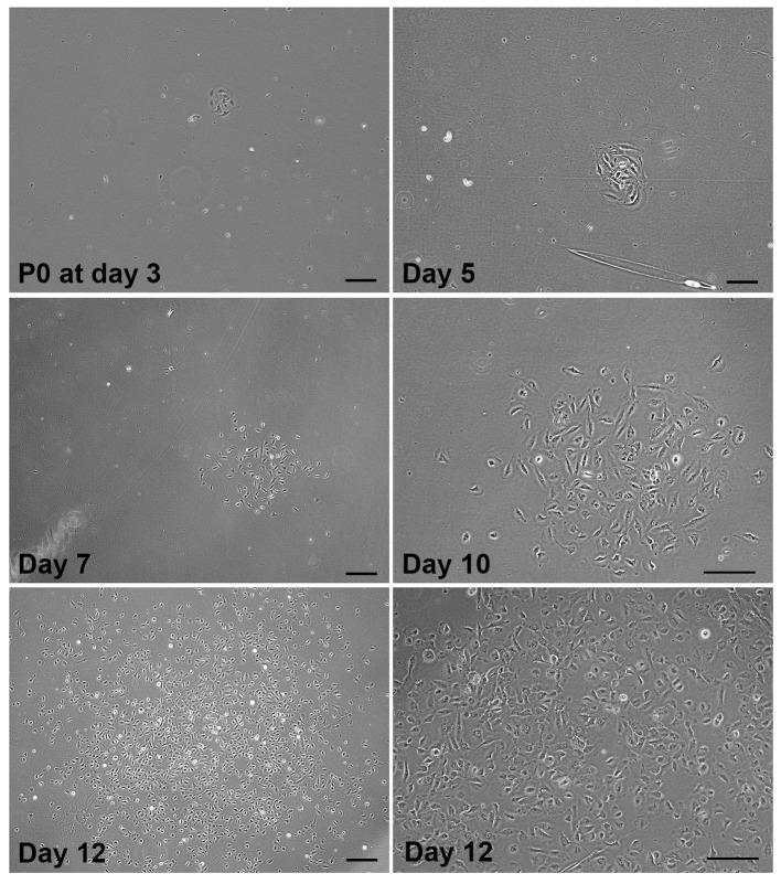
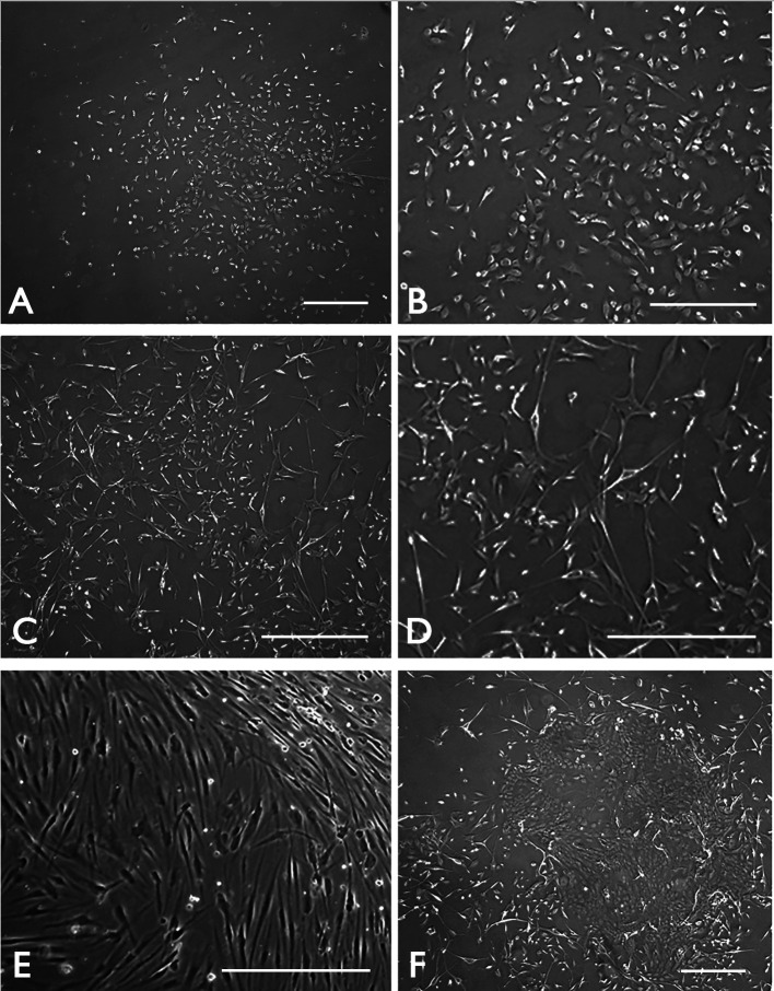
Corneal scarring, a significant cause of global blindness, results from various insults, including trauma, infections, and genetic disorders. The conventional treatment to replace scarred corneal tissues includes partial or full-thickness corneal transplantation using healthy donor corneas. However, only 1 in 70 individuals with treatable corneal scarring can undergo surgery, due to the limited supply of transplantable donor tissue. Our research focuses on cell-based strategies, specifically ex vivo-expanded corneal stromal stem cells (CSSCs), to address corneal scarring. Preclinical studies have demonstrated the efficacy of CSSC treatment in reducing corneal inflammation and fibrosis, inhibiting scar formation, and regenerating native stromal tissue. Mechanisms include CSSC differentiation into stromal keratocytes and the expression of regenerative cytokines. Here, we present a good manufacturing practice (GMP)-compliant protocol to isolate and expand human CSSCs. This method paves the way to produce clinical-grade CSSCs for transplantation and clinical trials. Key features • This protocol utilizes surgical skills to dissect human corneal tissues for CSSC isolation. • The yield and features of CSSCs rely on donor tissue quality (freshness) and have donor-to-donor variability. • Up to 0.5 billion CSSCs can be generated from a single cornea specimen, and cells at passage 3 are suitable for treatment uses.

Keywords: Anterior limbal stroma; Corneal opacities; Corneal stromal stem cells; Good manufacturing practices; Standard operating protocol.


Conflict of interest statement

Competing interestsThe authors declare that they have no competing interests.

Figures

Figure 1.
Figure 1.. Processing steps of donor corneal tissue.
(A) Donor corneal tissue stored in a primary container (sterile moist chamber) for delivery. (B) Tissue sterilization in DMEM with 2% antibiotics. (C) Removal of conjunctiva/tenons on the anterior side of donor cornea. (D) Clearing of all tissues on the posterior side.
Figure 2.
Figure 2.. Steps of isolating anterior limbal stroma.
(A) Whole adult cornea (~12 mm diameter) without the central region after removal by a circular surgical trephine. (B–C) Cut the corneal rim into quadrants. (D–F) Trimming of limbal strip with ~0.5 mm on scleral side and 1–1.5 mm on corneal side. (G–I) Making vertical cuts to separate anterior limbal stroma from the rest of tissue. (J) Separation of anterior limbal stroma. (K) Cuts of anterior limbal stromal strip into small blocks for efficient collagenase digestion. *denotes the anterior limbal stroma.
Figure 3.
Figure 3.. Primary good manufacturing practice (GMP)-corneal stromal stem cell (CSSC) cluster (TPF-23-29) at P0 and cell propagation.
Phase-contrast micrographs showing the cell cluster with 6–8 cells detected at day 3 post-seeding of primary CSSCs freshly isolated from anterior limbal stroma. The expansion of the same cluster at day 5, 7, and 10 showed an exponential increase in stem cell number. At day 12, the clonal expansion generated colonies with >500 cells. Magnified image at day 12 (bottom right) shows cells with moderate cell–cell contact at approximately 70% confluence. This stage is suitable for sub-passaging and cryopreservation. Scale bar, 50 μm.
Figure 4.
Figure 4.. Potential contaminating cell types in primary good manufacturing practice (GMP)-corneal stromal stem cell (CSSC) culture.
Phase-contrast micrographs showing (A) proper CSSC cluster with clonal expansion at P0 and (B) the growth of small-sized proliferating cells at P1. (C) Growth of fibroblast-like cells can occur in CSSC culture. (D) Under higher magnification, they display an heterogenous slender to stellate shape. (E) After sub-culture, the fibroblasts overgrow the CSSCs. (F) In case the limbal epithelium is not completely removed, epithelial colonies are formed due to the fast-growing limbal epithelial stem cells. Scale bar 120 μm.

Similar articles

References

    1. Mohan R. R., Kempuraj D., D'Souza S. and Ghosh A.(2022). Corneal stromal repair and regeneration. Prog Retin Eye Res. 91: 101090. - PMC - PubMed
    1. Chandran C., Santra M., Rubin E., Geary M. L. and Yam G. F.(2024). Regenerative Therapy for Corneal Scarring Disorders. Biomedicines. 12(3): 649. - PMC - PubMed
    1. Wilson S. E.(2020). Corneal wound healing. Exp Eye Res. 197: 108089. - PMC - PubMed
    1. Jeng B. H. and Ahmad S.(2021). In Pursuit of the Elimination of Corneal Blindness: Is Establishing Eye Banks and Training Surgeons Enough? Ophthalmology. 128(6): 813-815. - PubMed
    1. Wang E. Y., Kong X., Wolle M., Gasquet N., Ssekasanvu J., Mariotti S. P., Bourne R., Taylor H., Resnikoff S., West S., et al. .(2023). Global Trends in Blindness and Vision Impairment Resulting from Corneal Opacity 1984–2020. Ophthalmology. 130(8): 863-871. - PMC - PubMed

LinkOut - more resources