

RATTUS (Rat Thoracic Ultrasound): diagnosis of pneumothorax in pet rats
- PMID: 39346960
- PMCID: PMC11428198
- DOI: 10.3389/fvets.2024.1394291
RATTUS (Rat Thoracic Ultrasound): diagnosis of pneumothorax in pet rats
Abstract
Introduction: Rat thoracic ultrasound (RATTUS) is a non-invasive, easy-to-perform method for the evaluation of the pleural space and lungs in pet rats. The aim of the article is to present species-specific differences in the sonographic diagnosis of pneumothorax (PTX) in pet rats.
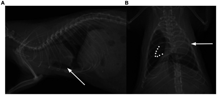
Methods: In total, 158 client-owned pet rats were examined during the period from July 2023 to January 2024. PTX was diagnosed in 20 of the examined rats (13.25%, the age of the animals ranged from 2 months to 32 months (19.08 ± 6.93 months; mean ± SD) and their body weight ranged from 97 g to 885 g (461.27 ± 138.97 g; mean ± SD). Radiographic confirmation of PTX was performed in all these 20 rats, in the control group radiography was used to confirm that PTX was not present.
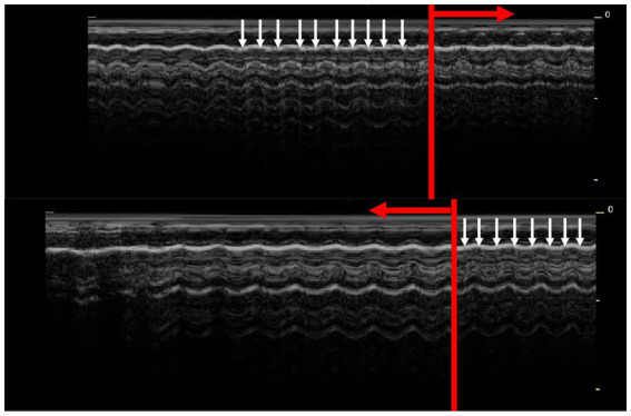
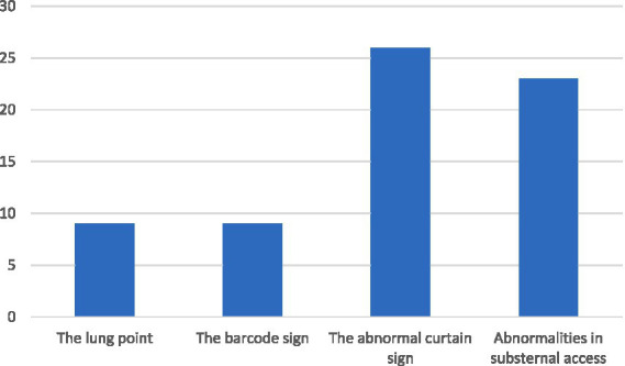
Results: The lung point and the barcode sign was found in 7/20 animals with sensitivity of 33.3% (95% CI, 0.16-0.59) and specificity of 100% (95% CI, 0.97-1.0). The abnormal curtain sign was found in 19/20 of animals with the sensitivity of 95% (95% CI, 0.73-0.99.7) and the specificity of 89% (95% CI, 0.82-0.93). The abnormalities in the substernal access were in 17/20 of animals with the sensitivity of 85% (95% CI, 0.61-0.96) and the specificity of 71% (95% CI, 0.62-0.78).
Discussion: In conclusion, RATTUS is a non-invasive method for the diagnosis of PTX in rats. Lung point and barcode sign are specific but not easily diagnosed signs. The curtain sign in RATTUS is not specific for PTX, as there are e.g. geriatric rats (rats older than 1,5 years) in which the abnormal curtain sign is visible without the presence of PTX. The presence of moderate to severe PTX can be assessed by the substernal approach based on the presence of cardiac displacement toward the collapsed lung lobe, and on evaluation of the lung inflation symmetry. This sign is not specific for PTX but in conjunction with other ultrasonic signs described makes the RATTUS a feasible tool for PTX diagnosis in rats.
Keywords: RATTUS; dyspnoea; pneumothorax; rat; respiratory disorders; thoracic disease; ultrasonography.
Copyright © 2024 Piskovská, Kraszewska, Hauptman, Chloupek, Linhart and Jekl.
Conflict of interest statement
The authors declare that the research was conducted in the absence of any commercial or financial relationships that could be construed as a potential conflict of interest.
Figures
Similar articles
-
The Rat Thoracic Ultrasound protocol: scanning technique and normal findings.Front Vet Sci. 2024 Feb 19;11:1286614. doi: 10.3389/fvets.2024.1286614. eCollection 2024. Front Vet Sci. 2024. PMID: 38440385 Free PMC article.
-
Abnormal Curtain Signs Identified With a Novel Lung Ultrasound Protocol in Six Dogs With Pneumothorax.Front Vet Sci. 2019 Aug 28;6:291. doi: 10.3389/fvets.2019.00291. eCollection 2019. Front Vet Sci. 2019. PMID: 31555674 Free PMC article.
-
A prospective comparison of supine chest radiography and bedside ultrasound for the diagnosis of traumatic pneumothorax.Acad Emerg Med. 2005 Sep;12(9):844-9. doi: 10.1197/j.aem.2005.05.005. Acad Emerg Med. 2005. PMID: 16141018 Clinical Trial.
-
Lung Ultrasonography for Pneumothorax in Dogs and Cats.Vet Clin North Am Small Anim Pract. 2021 Nov;51(6):1153-1167. doi: 10.1016/j.cvsm.2021.07.003. Epub 2021 Sep 9. Vet Clin North Am Small Anim Pract. 2021. PMID: 34511293 Review.
-
Bedside ultrasonography for diagnosis of pneumothorax.Quant Imaging Med Surg. 2015 Aug;5(4):618-23. doi: 10.3978/j.issn.2223-4292.2015.05.04. Quant Imaging Med Surg. 2015. PMID: 26435925 Free PMC article. Review.
References
-
- Rivas de Andrés JJ, Jiménez López MF, Molins López-Rodó L, Pérez Trullén A, Torres Lanzas J. Spanish Society of Pulmonology and Thoracic Surgery. Normativa sobre el diagnóstico y tratamiento del pneumotórax espontáneo [guidelines for the diagnosis and treatment of spontaneous pneumothorax]. Arch Bronconeumol. (2008) 44:437–48. doi: 10.1016/s1579-2129(08)60077-4, PMID: - DOI - PubMed
LinkOut - more resources
Full Text Sources