

Optimizing spatial accuracy in electroencephalography reconstruction through diffuse optical tomography priors in the auditory cortex
- PMID: 39347003
- PMCID: PMC11427190
- DOI: 10.1364/BOE.531576
Optimizing spatial accuracy in electroencephalography reconstruction through diffuse optical tomography priors in the auditory cortex
Abstract
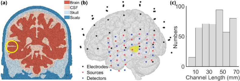
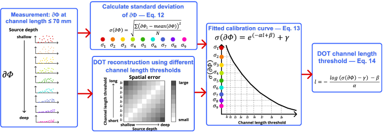
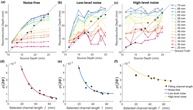
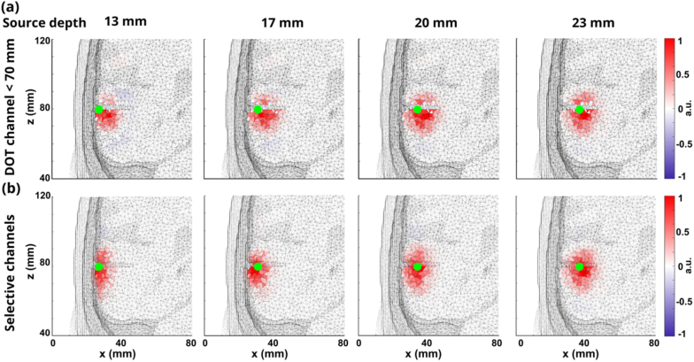
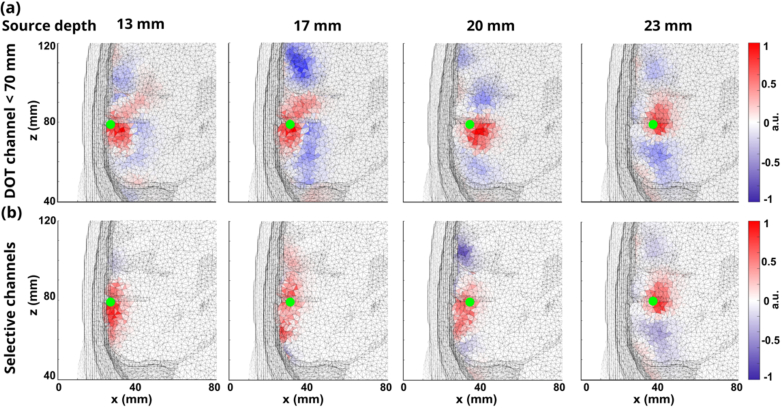
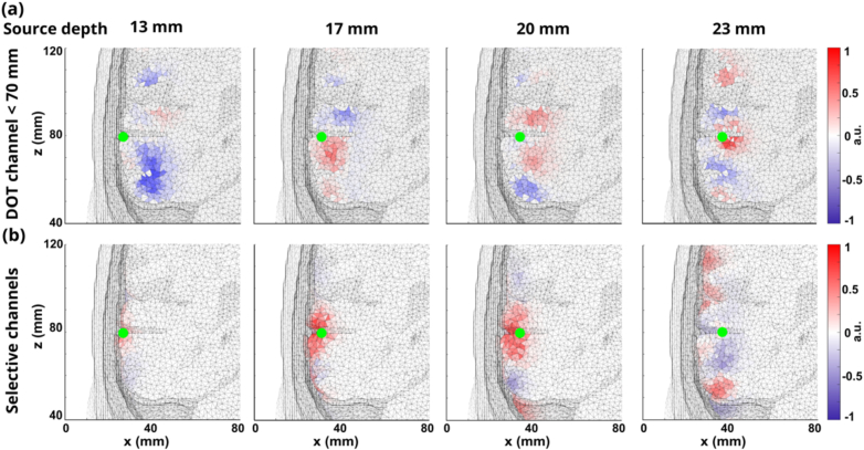
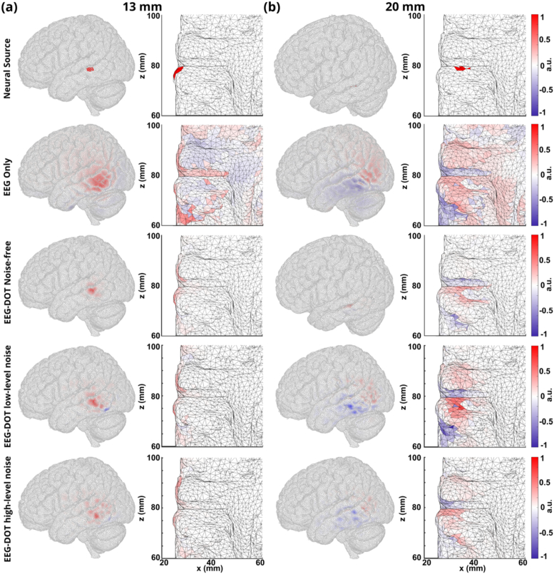
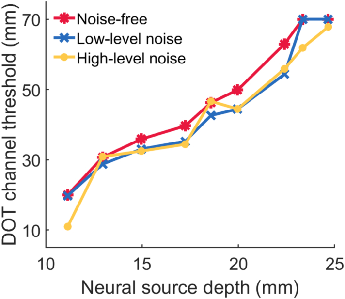
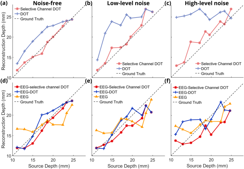
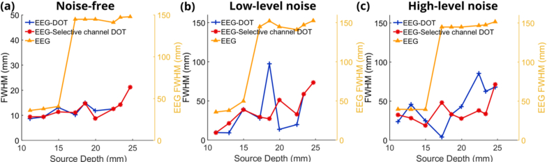
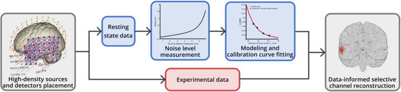
Diffuse optical tomography (DOT) enhances the localization accuracy of neural activity measured with electroencephalography (EEG) while preserving EEG's high temporal resolution. However, the spatial resolution of reconstructed activity diminishes for deeper neural sources. In this study, we analyzed DOT-enhanced EEG localization of neural sources modeled at depths ranging from 11-25 mm in simulations. Our findings reveal systematic biases in reconstructed depth related to DOT channel length. To address this, we developed a data-informed method for selecting DOT channels to improve the spatial accuracy of DOT-enhanced EEG reconstruction. Using our method, the average absolute reconstruction depth errors of DOT reconstruction across all depths are 0.9 ± 0.6 mm, 1.2 ± 0.9 mm, and 1.2 ± 1.1 mm under noiseless, low-level noise, and high-level noise conditions, respectively. In comparison, using fixed channel lengths resulted in errors of 2.6 ± 1.5 mm, 5.0 ± 2.6 mm, and 7.3 ± 4.5 mm under the same conditions. Consequently, our method improved the depth accuracy of DOT reconstructions and facilitated the use of more accurate spatial priors for EEG reconstructions, enhancing the overall precision of the technique.
© 2024 Optica Publishing Group.
Conflict of interest statement
No conflict of interest is declared.
Figures
References
-
- Grover P., Venkatesh P., “An information-theoretic view of EEG sensing,” Proc. IEEE 105(2), 367–384 (2017). 10.1109/JPROC.2016.2615179 - DOI
LinkOut - more resources
Full Text Sources