. 2024 Jul 9;15(8):4525-4539.
doi: 10.1364/BOE.523916. eCollection 2024 Aug 1.

In vivo bioluminescence tomography-guided system for pancreatic cancer radiotherapy research

Affiliations

In vivo bioluminescence tomography-guided system for pancreatic cancer radiotherapy research

Zijian Deng et al. Biomed Opt Express. .

Abstract

Recent development of radiotherapy (RT) has heightened the use of radiation in managing pancreatic cancer. Thus, there is a need to investigate pancreatic cancer in a pre-clinical setting to advance our understanding of the role of RT. Widely-used cone-beam CT (CBCT) imaging cannot provide sufficient soft tissue contrast to guide irradiation. The pancreas is also prone to motion. Large collimation is unavoidably used for irradiation, costing normal tissue toxicity. We innovated a bioluminescence tomography (BLT)-guided system to address these needs. We established an orthotopic pancreatic ductal adenocarcinoma (PDAC) mouse model to access BLT. Mice underwent multi-projection and multi-spectral bioluminescence imaging (BLI), followed by CBCT imaging in an animal irradiator for BLT reconstruction and radiation planning. With optimized absorption coefficients, BLT localized PDAC at 1.25 ± 0.19 mm accuracy. To account for BLT localization uncertainties, we expanded the BLT-reconstructed volume with margin to form planning target volume(PTVBLT) for radiation planning, covering 98.7 ± 2.2% of PDAC. The BLT-guided conformal plan can cover 100% of tumors with limited normal tissue involvement across both inter-animal and inter-fraction cases, superior to the 2D BLI-guided conventional plan. BLT offers unique opportunities to localize PDAC for conformal irradiation, minimize normal tissue involvement, and support reproducibility in RT studies.


Conflict of interest statement

The research group of Dr. Ken Kang-Hsin Wang and Xstrahl are supported by NIH academic-industrial partnership R37CA230341 in the development of BLT-guided system for pre-clinical radiation research.

Figures

Fig. 1.
Fig. 1.
illustrates the schematic of MuriGlo and the workflow of BLT-guided irradiation. (a) MuriGlo consists of an optical assembly, a thermostatic system, and a transportable mouse bed. The red dash line indicates the optical path from the imaging plane at the object through a 3-mirror system, a fixed mirror, a selected filter, and a camera lens to a CCD chip. The thermostatic system, including a heater, two circulation fans and a thermocouple at 3-mirror system, keeps the imaged object temperature at 37 °C to maintain stable intensity and consistent spectrum of bioluminescence emission throughout experiment. (b) The transportable mouse bed is composed of a bed base, a nose cone, eight fiducial markers and two anesthesia hoses. After optical image acquisition, the imaged animal is detached from MuriGlo along with the bed under anesthesia, and transferred to (c) a small animal irradiator for CBCT imaging and BLT reconstruction for BLT-guided irradiation.
Fig. 2.
Fig. 2.
shows the first open-field BLI (a) 1.5 and (b) 3 weeks after tumor implantation at (a1 and b1) 0 and (a2 and b2) 180° projections with corresponding SARRP CBCT images (a3 and b3).
Fig. 3.
Fig. 3.
The deviation between actual center of autoluminescent source implanted in pancreas and CoM of BLT-reconstructed source distribution corresponding to spectral-dependent μa (Table 1) is shown; three CoMs were projected at the same transverse slice in the CBCT image to visualize their deviations from the actual source.
Fig. 4.
Fig. 4.
BLT reconstruction for the mice bearing orthotopic PDAC; (a) the 0° and -90° projection BLIs at 630 nm mapped onto mesh surface after correcting time-resolved signal variations and source spectrum; data > 10% of maximum within these two projections were mapped and used for reconstruction. (b) Transverse view of CBCT image and (c) 3D rendering overlapped with aGTV (green contour) and GTVBLT (heat map in b and red contour in c) with 50% threshold; center of Ti wire (yellow dot), CoM of GTVBLT (black dot) and maximum point of BLI shown at 0° projection (red dot) were marked on animal surface. (d) shows the deviations between center of Ti wire and CoM of GTVBLT for the three mice at 1, 1.5, 2, 3, and 5 weeks after tumor implantation.
Fig. 5.
Fig. 5.
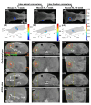
BLT-guided 6-arc conformal plan vs. BLI-guided conventional AP/PA plans for inter-animal comparison between Mouse (a) #1 and (b) #3 at 1 week after tumor implantation, and for inter-fraction comparison of Mouse #3 at (b) 1 and (c) 1.5 weeks after tumor implantation; for each data set (Fig. a, b, or c): (a-c1) show 0° projection BLI at 630 nm; (a-c2) are 0° and 90° projection BLIs at 630 nm mapped onto mesh surface; data > 10% of maximum (10% threshold) within these two projections were displayed. Transverse and coronal views of CBCT image overlapped with aGTV (green contour), GTVBLT (50% threshold, heat map), PTVBLT (red contour), and isodose distributions of BLT-guided conformal and BLI-guided AP/PA plans are shown in Fig. (a-c3 and a-c4) and (a-c5 and a-c6), respectively. GTVBLT CoM (black dot) and BLI maximum point (red dot) were projected onto the coronal view where the center of Ti wire was located, Fig. (a-c6).
Fig. 6.
Fig. 6.
The corresponding DVHs of BLT-guided 6-arc and BLI-guided AP/PA plans for the three data sets described in Fig. 5. The PTVBLT DVH is only for the BLT-guided plan and the aGTV and normal tissue (NT) DVHs are for both plans.

Similar articles

Cited by

References

    1. Sung H., Ferlay J., Siegel R. L., et al. , “Global cancer statistics 2020: Globocan estimates of incidence and mortality worldwide for 36 cancers in 185 countries,” Ca-Cancer J. Clin. 71(3), 209–249 (2021).10.3322/caac.21660 - DOI - PubMed
    1. Orth M., Metzger P., Gerum S., et al. , “Pancreatic ductal adenocarcinoma: Biological hallmarks, current status, and future perspectives of combined modality treatment approaches,” Radiat. Oncol. 14(1), 141 (2019).10.1186/s13014-019-1345-6 - DOI - PMC - PubMed
    1. . “Chemoimmunotherapy and radiation in pancreatic cancer (CRIT),” (ClinicalTrials.gov.).
    1. . “Pancreatic tumor cell vaccine (GVAX), low dose cyclophosphamide, fractionated stereotactic body radiation therapy (SBRT), and folfirinox chemotherapy in patients with resected adenocarcinoma of the pancreas,” (ClinicalTrials.gov.).
    1. . “Immune checkpoint inhibition (Tremelimumab and/or MEDI4736) in combination with radiation therapy in patients with unresectable pancreatic cancer,” (ClinicalTrials.gov.).

LinkOut - more resources