. 2024 Jul 29;15(8):4877-4890.
doi: 10.1364/BOE.531577. eCollection 2024 Aug 1.

High-speed reflectance confocal microscopy using speckle modulation

Affiliations

High-speed reflectance confocal microscopy using speckle modulation

Momoka Sugimura et al. Biomed Opt Express. .

Abstract

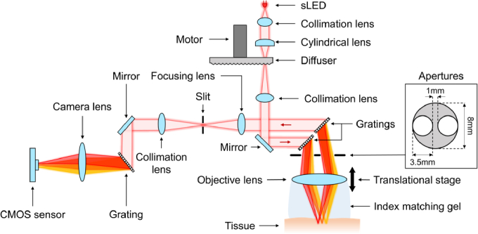
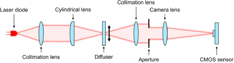
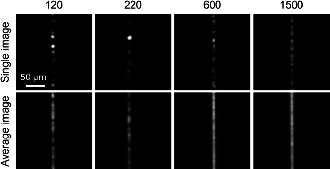
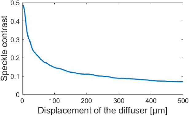
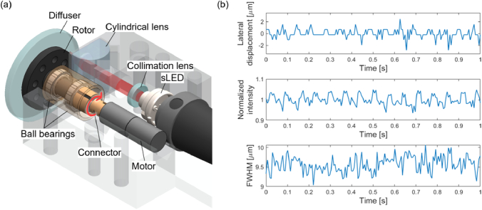
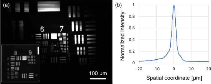
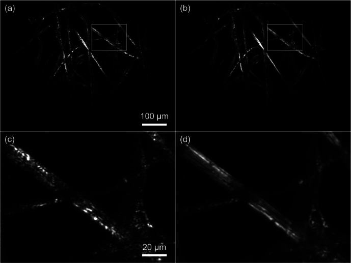
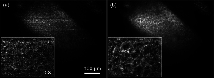
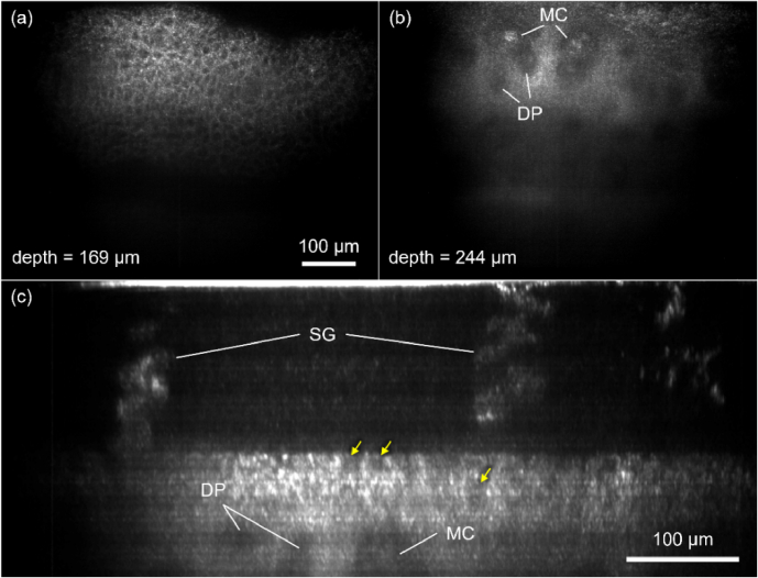
We developed a spectrally-encoded, line reflectance confocal microscope (RCM) that uses a rotating diffuser to rapidly modulate the illumination speckle pattern. The speckle modulation approach reduced speckle noise while imaging with a spatially coherent light source needed for high imaging speed and cellular resolution. The speckle-modulation RCM device achieved lateral and axial resolutions of 1.1 µm and 2.8 µm, respectively. With an imaging speed of 107 frames/sec, three-dimensional RCM imaging over 300-µm depth was completed within less than 1 second. RCM images of human fingers, forearms, and oral mucosa clearly visualized the characteristic cellular features without any noticeable speckle noise.


Conflict of interest statement

The University of Arizona has a technology-licensing agreement with ArgosMD on the reflectance confocal microscopy technology. DK has the rights to receive royalties as a result of this licensing agreement. DK serves as a scientific advisor to ArgosMD. Conflicts of interest resulting from this interest are being managed by the University of Arizona in accordance with its policies.

Figures

Fig. 1.
Fig. 1.
Schematic of the speckle-modulating RCM device.
Fig. 2.
Fig. 2.
Bench setup for diffuser evaluation.
Fig. 3.
Fig. 3.
Images of line illumination generated by diffusers with various grit numbers. Single images (top row) and aerage images (bottom row).
Fig. 4.
Fig. 4.
Measured speckle contrast as a function of displacement of the 1500 grit diffuser. The displacement on the horizontal axis is the distance over which the line illumination images were averaged.
Fig. 5.
Fig. 5.
SolidWorks design of the speckle-modulating illumination unit (a), and measured lateral displacement, normalized intensity, and FWHM of line illumination (b).
Fig. 6.
Fig. 6.
SolidWorks design (a) and photo (b) of the speckle modulating RCM device.
Fig. 7.
Fig. 7.
(a) RCM image of the USAF resolution target (inset – a magnified view around groups 8 and 9) and (b) axial response curve.
Fig. 8.
Fig. 8.
RCM image of a lens paper (a) without and (b) without speckle modulation, and their magnified views (c, d).
Fig. 9.
Fig. 9.
RCM images of a human finger in vivo without (a) and with (b) speckle modulation. insets –5x-magnified views.
Fig. 10.
Fig. 10.
En face RCM image of a human finger in vivo at different tissue layers: (a) basement membrane and (b) superficial dermis. (c) Cross-sectional image of a human finger in vivo. DP: derma papillae; MC: Meissner's corpuscles; SG: sweat glands; arrows – basal cells.
Fig. 11.
Fig. 11.
En face RCM images of a pigmented lesion on a human forearm in vivo at different tissue layers: (a) stratum corneum, (b) stratum granulosum, (c) dermato-epidermal junction, and (d) superficial dermis. arrows – keratinocytes; arrowheads – melanin-containing cells; DP: derma papillae; BV: blood vessel.
Fig. 12.
Fig. 12.
En face RCM image of a human oral mucosa in vivo. arrows – flowing blood cells; arrowheads – epithelial cell nuclei.

Similar articles

Cited by

References

    1. Busam K. J., Hester K., Charles C., et al. , “Detection of clinically amelanotic malignant melanoma and assessment of its margins by in vivo confocal scanning laser microscopy,” Arch. Dermatol. 137(7), 923–929 (2001).10.1016/j.jaad.2004.06.028 - DOI - PubMed
    1. Nori S., Rius-Díaz F., Cuevas J., et al. , “Sensitivity and specificity of reflectance-mode confocal microscopy for in vivo diagnosis of basal cell carcinoma: a multicenter study,” J. Am. Acad. Dermatol. 51(6), 923–930 (2004). - PubMed
    1. Astner S., Gonzalez E., Cheung A., et al. , “Pilot study on the sensitivity and specificity of in vivo reflectance confocal microscopy in the diagnosis of allergic contact dermatitis,” J. Am. Acad. Dermatol. 53(6), 986–992 (2005).10.1016/j.jaad.2005.08.026 - DOI - PubMed
    1. Pellacani G., Guitera P., Longo C., et al. , “The impact of in vivo reflectance confocal microscopy for the diagnostic accuracy of melanoma and equivocal melanocytic lesions,” J. Invest. Dermatol. 127(12), 2759–2765 (2007).10.1038/sj.jid.5700993 - DOI - PubMed
    1. Gerger A., Hofmann-Wellenhof R., Langsenlehner U., et al. , “In vivo confocal laser scanning microscopy of melanocytic skin tumours: diagnostic applicability using unselected tumour images,” Br. J. Dermatol. 158(2), 329–333 (2008).10.1111/j.1365-2133.2007.08389.x - DOI - PubMed

LinkOut - more resources