

Successful use of a 3D-printed surgical guide to facilitate transiliosacral fixation in a cat with bilateral sacroiliac luxation
- PMID: 39347013
- PMCID: PMC11437534
- DOI: 10.1177/20551169241273629
Successful use of a 3D-printed surgical guide to facilitate transiliosacral fixation in a cat with bilateral sacroiliac luxation
Abstract
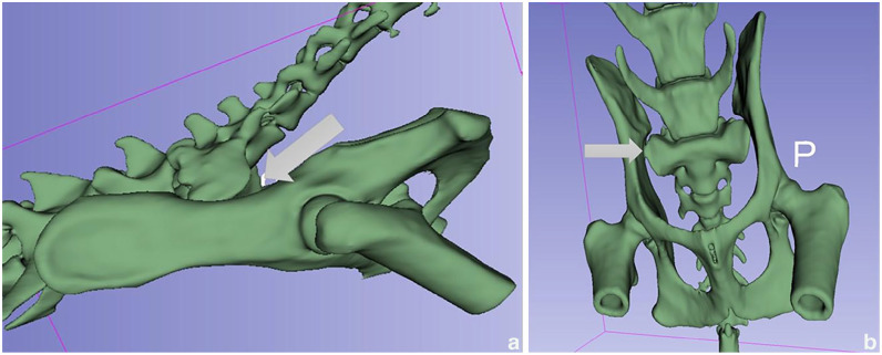
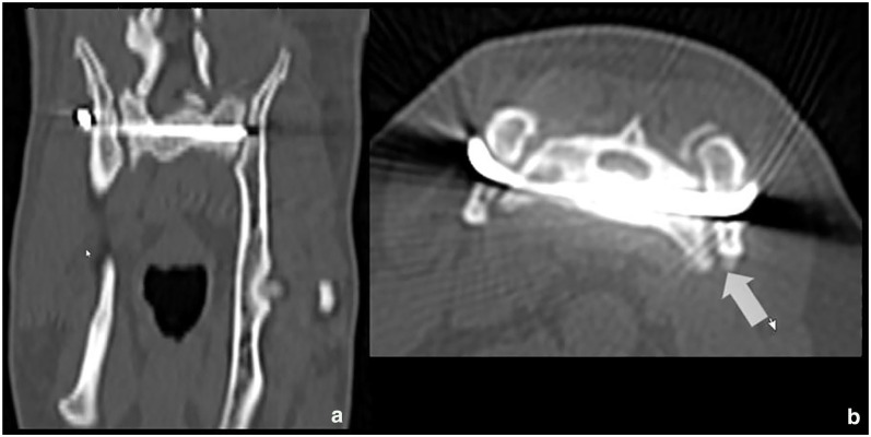
Case summary: A 7-year-old male castrated domestic shorthair cat was presented for treatment of a bilateral sacroiliac luxation (SIL). CT was performed and the data were extracted in a stereolithography (STL) file, after which a 3D-printed drill guide (3DPDG) was devised, using computer-aided design (CAD) software, and printed. Using an open surgical approach, the guide was used as an aid for drilling the sacrum. The ilial wings were drilled free-hand later and a transiliosacral pin (TP) was inserted to realign and stabilise the SIL. The cat exhibited an early return to normal limb function and a CT scan performed at the postoperative follow-up showed early signs of bone remodelling at the sacroiliac joint.
Relevance and novel information: To the authors' knowledge, this is the first report using a 3DPDG for implant placement in the feline sacrum without intraoperative imaging.
Keywords: 3D-printed drill guide; computer-aided design; sacroiliac fracture/luxation; stereolithography; transiliosacral pin.
© The Author(s) 2024.
Conflict of interest statement
The authors declared no potential conflicts of interest with respect to the research, authorship, and/or publication of this article.
Figures


References
-
- Bookbinder PF, Flanders JA. Characteristics of pelvic fracture in the cat. A 10-year retrospective study. Vet Comp Orthop Traumatol 1992; 5: 122–127.
-
- Bennett D. Orthopaedic disease affecting the pelvic region of the cat. J Small Anim Pract 1975; 16: 723–738. - PubMed
-
- Anderson A, Coughlan AR. Sacral fractures in dogs and cats: a classification scheme and review of 51 cases. J Small Anim Pract 1997; 38: 404–409. - PubMed
-
- Burger M, Forterre F, Brunnberg L. Surgical anatomy of the feline sacroiliac joint for lag screw fixation of sacroiliac fracture-luxation. Vet Comp Orthop Traumatol 2004; 17: 146–151.
Publication types
LinkOut - more resources
Full Text Sources
Miscellaneous