

Inhibitory impact of a mesoporous silica nanoparticle-based drug delivery system on Porphyromonas gingivalis-induced bone resorption
- PMID: 39347836
- PMCID: PMC11442573
- DOI: 10.1007/s10856-024-06827-6
Inhibitory impact of a mesoporous silica nanoparticle-based drug delivery system on Porphyromonas gingivalis-induced bone resorption
Abstract
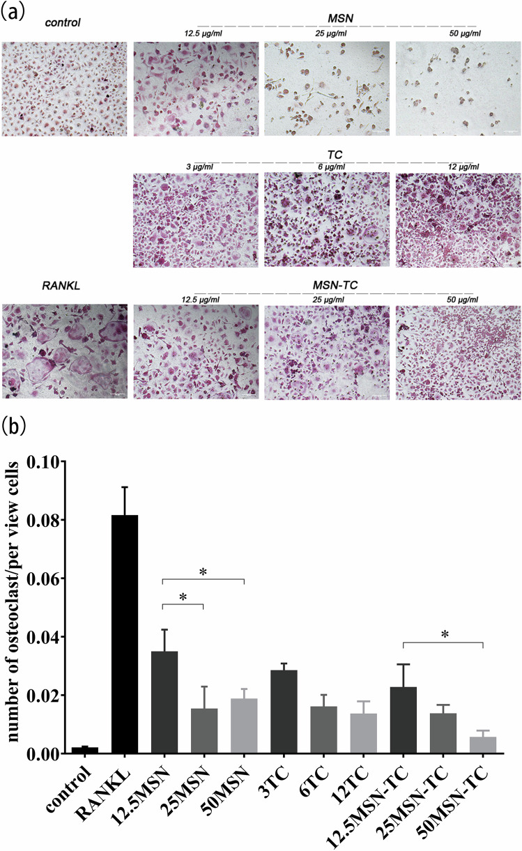
Controlling and reducing plaque formation plays a pivotal role in preventing and treating periodontal disease, often utilizing antibacterial drugs to enhance therapeutic outcomes. Mesoporous silica nanoparticles (MSN), an FDA-approved inorganic nanomaterial, possess robust physical and chemical properties, such as adjustable pore size and pore capacity, easy surface modification, and high biosafety. Numerous studies have exploited MSN to regulate drug release and facilitate targeted delivery. This study aimed to synthesize an MSN-tetracycline (MSN-TC) complex and investigate its inhibitory potential on Porphyromonas gingivalis (P. gingivalis)-induced bone resorption. The antibacterial efficacy of MSN-TC was evaluated through bacterial culture experiments. A P. gingivalis-induced bone resorption model was constructed by subcutaneously injecting P. gingivalis around the cranial bone of rats. Micro-computed tomography was employed to assess the inhibitory impact of MSN and MSN-TC on bone resorption. Furthermore, the influence of MSN and MSN-TC on osteoclast differentiation was examined in vitro. The MSN exhibited optimal pore size and particle dimensions for effective loading and gradual release of TC. MSN-TC demonstrated significant bacteriostatic activity against P. gingivalis. MSN-TC-treated rats showed significantly reduced cranial bone tissue destruction compared to MSN or TC-treated rats. Additionally, both MSN and MSN-TC exhibited inhibitory effects on the receptor activator of nuclear factor kappa-Β ligand-mediated osteoclast differentiation. The MSN-TC complex synthesized in this study demonstrated dual efficacy by exerting antibacterial effects on P. gingivalis and by resisting osteoclast differentiation, thereby mitigating bone resorption induced by P. gingivalis.
© 2024. The Author(s).
Conflict of interest statement
The authors declare no competing interests.
Figures


Similar articles
-
Micromolar sodium fluoride mediates anti-osteoclastogenesis in Porphyromonas gingivalis-induced alveolar bone loss.Int J Oral Sci. 2015 Dec 18;7(4):242-9. doi: 10.1038/ijos.2015.28. Int J Oral Sci. 2015. PMID: 26674426 Free PMC article.
-
Licorice isoliquiritigenin-encapsulated mesoporous silica nanoparticles for osteoclast inhibition and bone loss prevention.Theranostics. 2019 Jul 9;9(18):5183-5199. doi: 10.7150/thno.33376. eCollection 2019. Theranostics. 2019. PMID: 31410209 Free PMC article.
-
pH-Responsive Mesoporous Silica Nanoparticles Loaded with Naringin for Targeted Osteoclast Inhibition and Bone Regeneration.Int J Nanomedicine. 2024 Jun 24;19:6337-6358. doi: 10.2147/IJN.S456545. eCollection 2024. Int J Nanomedicine. 2024. PMID: 38946884 Free PMC article.
-
Polymer Nanoassembly as Delivery Systems and Anti-Bacterial Toolbox: From PGMAs to MSN@PGMAs.Chem Rec. 2018 Jan;18(1):45-54. doi: 10.1002/tcr.201700036. Epub 2017 Jul 4. Chem Rec. 2018. PMID: 28675576 Review.
-
Exploring the role of mesoporous silica nanoparticle in the development of novel drug delivery systems.Drug Deliv Transl Res. 2022 Jan;12(1):105-123. doi: 10.1007/s13346-021-00935-4. Epub 2021 Feb 18. Drug Deliv Transl Res. 2022. PMID: 33604837 Review.
References
-
- Kinane DF, Stathopoulou PG, Papapanou PN. Periodontal diseases. Nat Rev Dis Primers. 2017;3:17038. - PubMed
-
- Slots J. Periodontitis: facts, fallacies and the future. Periodontol 2000. 2017;75:7–23. - PubMed
-
- Pihlstrom BL, Michalowicz BS, Johnson NW. Periodontal diseases. Lancet. 2005;366:1809–20. - PubMed
-
- Herrera D, Sanz M, Jepsen S, Needleman I, Roldán S. A systematic review on the effect of systemic antimicrobials as an adjunct to scaling and root planing in periodontitis patients. J Clin Periodontol. 2002;29:136–62. - PubMed
MeSH terms
Substances
Grants and funding
LinkOut - more resources
Full Text Sources
Medical
Molecular Biology Databases
Research Materials
Miscellaneous
