Review
. 2024 Dec;26(6):912-925.
doi: 10.1007/s11307-024-01954-6. Epub 2024 Sep 30.

Neurotoxin-Derived Optical Probes for Elucidating Molecular and Developmental Biology of Neurons and Synaptic Connections : Toxin-Derived Optical Probes for Neuroimaging

Affiliations
Review

Neurotoxin-Derived Optical Probes for Elucidating Molecular and Developmental Biology of Neurons and Synaptic Connections : Toxin-Derived Optical Probes for Neuroimaging

Rohini Bijjam et al. Mol Imaging Biol. 2024 Dec.

Abstract

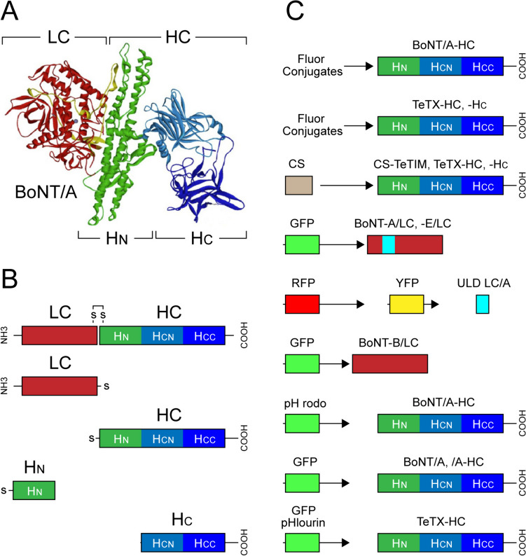
Botulinum neurotoxins (BoNTs) and tetanus toxin (TeTX) are the deadliest biological substances that cause botulism and tetanus, respectively. Their astonishing potency and capacity to enter neurons and interfere with neurotransmitter release at presynaptic terminals have attracted much interest in experimental neurobiology and clinical research. Fused with reporter proteins or labelled with fluorophores, BoNTs and TeTX and their non-toxic fragments also offer remarkable opportunities to visualize cellular processes and functions in neurons and synaptic connections. This study presents the state-of-the-art optical probes derived from BoNTs and TeTX and discusses their applications in molecular and synaptic biology and neurodevelopmental research. It reviews the principles of the design and production of probes, revisits their applications with advantages and limitations and considers prospects for future improvements. The versatile characteristics of discussed probes and reporters make them an integral part of the expanding toolkit for molecular neuroimaging, promoting the discovery process in neurobiology and translational neurosciences.

Keywords: Advanced biomaterials; Fluorescent probes; Fusion proteins; Molecular trafficking; Optical imaging; Retrograde transport; SNARE proteins.


Conflict of interest statement

Declarations. Conflict of Interest: The authors have no conflict of interest to report.

Figures

Fig. 1
Fig. 1
Structure and functional domains of neurotoxin-derived optical probes used for visualizing molecular, functional and developmental processes in neurons. (A) Crystal structure of botulinum neurotoxin type A (BoNT/A) with a colour-coded illustration of various sub-domains. The C-terminal binding domain (HC) of the heavy chain (HC) adopts a trefoil fold to bind the membrane surface of neurons, while the N-terminal jelly-roll motif of HN facilitates the translocation of the light chain (LC) protease from the vesicular lumen to the cytoplasm of neuron. The LC contains the conserved HExxH motif characteristic of zinc-dependent proteases, which targets and cleaves SNAP-25 SNARE protein. Adapted with permission from [26]. (B) Schematic of BoNT/A di-chain (linked via S–S bonds), which is conserved across all BoNT (A-G) serotypes and TeTX. Break down of S–S bond in acidic environment of synaptic vesicles lead to release of LC into the cytoplasm of neurons. (C) Graphical representations of optical probes derived from BoNTs and TeTX or their sub-domains conjugated with fluorophores, or fused with reporter proteins (e.g. GFP, GFP-phlourin, YFP and RFP). Further details of structure and function of probes with various subdomains and their applications are described throughout this study
Fig. 2
Fig. 2
Visualizing subcellular distribution and action sites of BoNT -LC. (A) GFP-LC/A and GFP-LC/E expression (left) and validation of proteolytic activity by cleaving SNAP25206 into SNAP25197 and SNAP25180, respectively (right). (B) SNAP25197 and BoNT/A-LC colocalize in the plasma membrane. Differentiated cells expressing GFP-LC/A were stained with antibodies to GFP (green) and SNAP25197 (red). White colour on merged imaging indicates areas of colocalization of the GFP-LC/A with substrate SNAP25. (C) Non-targeted GFP expressing PC12 cells stained with antibodies to GFP (green) and SNAP25206 (red). Note the absence of colocalization of the two proteins. (D) SNAP25180 and BoNT/E-LC are cytoplasmic proteins. GFP-LC/E showed cytoplasmic localization with nuclear exclusion. Cells displayed rounded morphology and absence of neurites even in differentiation media. PC12 cells were stained with antibodies to GFP (green) and SNAP25180 (red). Adapted with permission from [42]
Fig. 3
Fig. 3
Imaging endocytosis and molecular trafficking in neurons in vitro and in vivo using BoNT and TeTX-derived optical probes. (A) Visualizing motor nerve endings of mouse triangularis sterni by GFP-BoNT/A-HC (left) and Alexa647-α-BTX targeting acetylcholine receptors of NMJ (middle). Arrows indicate areas of intense labelling at the NMJ. Merged image on the right. Adapted with permission from [57]. (B) Imaging synaptic contacts in neuronal cultures by BoNT/A-Alexa-488 and presynaptic marker VAMP-2 (left). Cultured neurons were exposed to 40 nM BoNT/E-TR and BoNT/A-Alexa-488 (middle and right) reveal fluorescent puncta corresponding to presynaptic terminals (arrows). Adapted permission from [63]. (C) Top: imaging of the uptake and transport of TeTX-derived probes CS-TeTIM, CS-TeTX-HC and CS-TeTX-HC linked with biotin-4-fluorescein in spinal cord cultures. Bottom: fluorescence confocal images of CS-TeTX-HC in the NMJ of the rat tongue 8 h post-after injection. Dashed circles – random ROI in NMJ counter-stained by rhodamine α-BTX. Adapted with permission from [33]. (D) Retro-axonal labelling of motor neurons by Cy3-CS-TeTIM injected in m. gastrocnemius (left) stained also for choline acetyltransferase (ChAT-Alexa-488) and merged image (middle and right) (in preparation). (E) Mapping CS-TeTIM and CS-HC(TeTx) labelled neurons in the brainstem and spinal cord. (a1-c1) Micrographs of brain stem, cervical and lumbar spinal cord showing HRP labelled CS-positive neurons (left) with (a3-c3) topographic maps of their distribution. Abbreviations: a1, a2, Mot5—motor trigeminal nucleus, Tr5 – trigeminal nerve, SCP – superios cerebellar peduncle, so – solitary tract; LS – locus coeruleus, VCA – ventral cochlear nucleus; FL-flocculus, VLN – ventrolateral nucleus, Sp5 – spinal trigeminal tract, SP5o – spinal trigeminal tract oral, Mot7 – motor nucleus, Pr5 – principal sensory trigeminal nucleus. Thick, medium and thin coloured bars indicate numbers of CS-labelled neurons. For illustration purposes, measurements at individual points are merged into continuous bars. Adapted with permission from [33]
Fig. 4
Fig. 4
Imaging selected neuronal groups and connectivity in the CNS using toxin-derived optical probes. (A) BoNT/B-LC-GFP expression in 20-days post-fertilization (dpf) granule cell-silenced larvae of zebra fish. Immunostaining of the brain (dorsal view, top row) and sagittal sections (bottom row) with anti-GFP (green) and Neurod1 (magenta, nuclear marker) antibodies. About half of the mature granule cells express BoNT/B-LC-GFP. EG – eminenta granularis; LCe – lobus caudalis cerebelli; CCe – corpus cerebelli; GL, granule cell layer; ML, molecular layer. (B) Sample recordings of the neuronal activity using GCaMP7a Ca2+ sensor with measurements before and during-after stimulation in a single larva during the habituation (11th–15th trials) and probe (1st–5th trials) sessions. The graphs depicts the ΔF/F. Gray boxes indicate the timing of the CS presentation. Adapted with permission from [71]. (C) Transgenic mice expressing TeTX-HC-GFP in orexin neurons showing double-label immunofluorescence for GFP and Alexa594 conjugated with anti-GFP IgG in the lateral hypothalamus (LH). (D) Mapping TeTX-HC-GFP positive neurons from rostral to caudal planes of the brain coronal sections revealed by immunohistochemical staining. Abbreviations: VDB—vertical limb of the diagonal band; LS—lateral septum; CPu—caudate putamen; IC—internal capsule; LH—lateral hypothalamus; MCPO—magnocellular preoptic nucleus; LV- lateral ventricle; TH—thalamus; AM—amygdala; Arc—arcuate hypothalamic nucleus; VMH, ventromedial hypothalamus; SNR—substantia nigra; ML—medial lemnisci; PAG—periaqueductal gray; cp—cerebral peduncle; VLL—ventral nucleus of lateral lemniscus; PNO—pontine reticular nucleus, oral part; VER—vermis; 4 V—forth ventricle; DCN- deep cerebellar nucleus; PFI—paraflocculus; Gi—giganto-cellular reticular nucleus; 9Cb—nine cerebellar lobules; icp—inferior cerebellar peduncle; Sp5C—spinal trigeminal nucleus caudal; RPa—raphe pedunculus nucleus. Adapted with permission from [73]

Similar articles

References

    1. Ntziachristos V (2010) Going deeper than microscopy: the optical imaging frontier in biology. Nat Methods 7:603–614 - PubMed
    1. Yoon S, Cheon SY, Park S et al (2022) Recent advances in optical imaging through deep tissue: imaging probes and techniques. Biomater Res 26:57 - PMC - PubMed
    1. Fernandez-Suarez M, Ting AY (2008) Fluorescent probes for super-resolution imaging in living cells. Nat Rev Mol Cell Biol 9:929–943 - PubMed
    1. Linghu C, Johnson SL, Valdes PA et al (2020) Spatial multiplexing of fluorescent reporters for imaging signaling network dynamics. Cell 183(1682–1698):e1624 - PMC - PubMed
    1. Shemesh OA, Linghu C, Piatkevich KD et al (2020) Precision calcium imaging of dense neural populations via a cell-body-targeted calcium indicator. Neuron 107(470–486):e411 - PMC - PubMed

LinkOut - more resources