. 2024 Sep 30;35(1):60.
doi: 10.1007/s10856-024-06832-9.

Sustained co-release of ciprofloxacin and dexamethasone in rabbit maxillary sinus using polyvinyl alcohol-based hydrogel microparticle

Affiliations

Sustained co-release of ciprofloxacin and dexamethasone in rabbit maxillary sinus using polyvinyl alcohol-based hydrogel microparticle

Maryam Jalessi et al. J Mater Sci Mater Med. .

Abstract

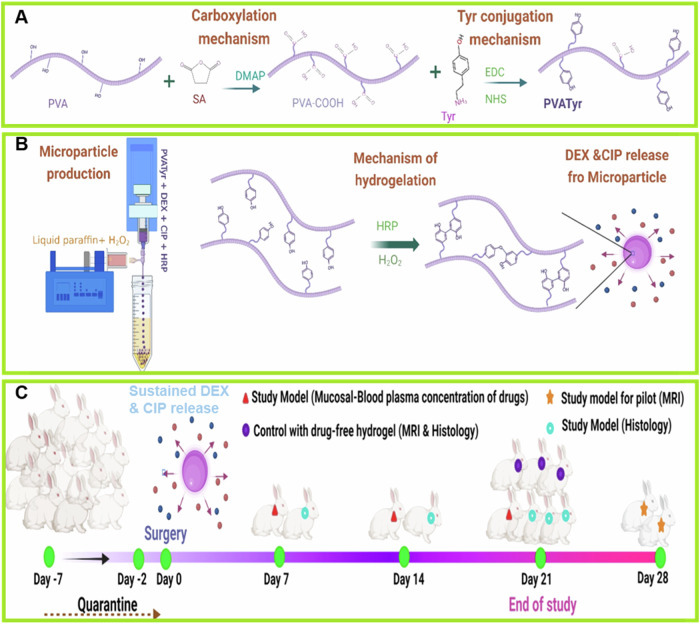
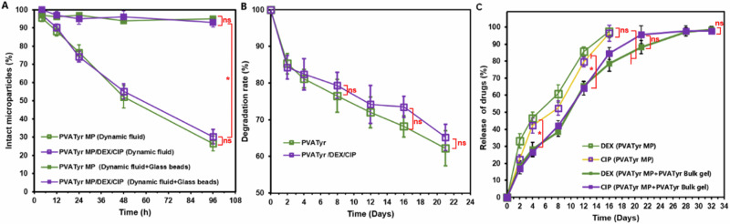
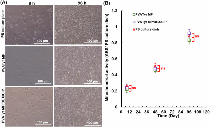
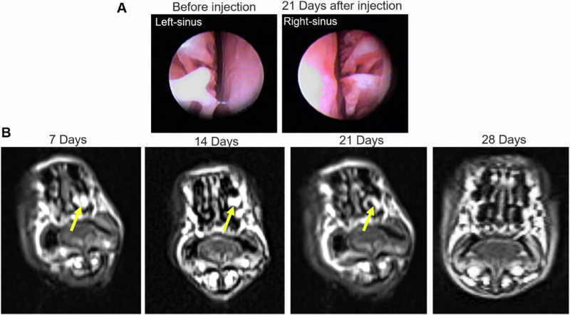
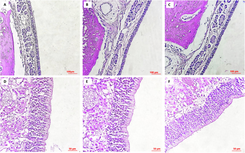
Topical delivery to paranasal sinuses through sustained-release stents is one of the new horizons in treating chronic rhinosinusitis (CRS). This study aims to introduce and evaluate sustained co-release of encapsulated ciprofloxacin (CIP) and dexamethasone (DEX) in polyvinyl alcohol-based carriers within the maxillary sinus of rabbit animals. DEX and CIP were loaded in a tyramine-substituted polyvinyl alcohol microparticle (PVATyr MP). The mechanical stability, degradability, and sustained-release patterns of both drugs as well as cellular cytocompatibility were assessed in vitro. The PVATyr MPs were then injected into the maxillary sinus of rabbits and they were monitored weekly for 21 days. Nasal endoscopy, MRI imaging, and tissue microscopy were used to follow the changes and compared them with the control condition. Also, the concentrations of drugs were evaluated in the maxillary sinus and blood samples over the study period. Produced PVA-based MPs possessed a relatively narrow particle size distribution (CV 7.7%) with proper physical stability until 30 days of incubation. The uniform-sized PVATyr MPs and their surrounding hydrogel showed sustained-release profiles for DEX and CIP for up to 32 days in vitro. The injected drugs-loaded hydrogel showed complete clearance from the maxillary sinus of rabbits within 28 days. The concentrations of DEX and CIP in mucosal remained within the therapeutic window when measured on days 7, 14, and 21, which were well above the plasma concentrations without any pathological changes in endoscopy, MRI imaging, and histological examinations. DEX/CIP loaded PVATyr MPs provided an effective, controlled, and safe sustained-drug delivery in both in vitro and in vivo analyses at therapeutic concentrations with minimal systemic absorption, suggesting a promising treatment approach for CRS.


Conflict of interest statement

The authors declare no competing interests.

Figures

Fig. 1
Fig. 1
Schematic diagram of study including; A Carboxylation of PVA and phenolation of PVA-COOH through carbodiimide condensation reaction, B Fabrication of PVATyr microparticles through co-flow droplet generation device and hydrogelation of PVATyr through horseradish peroxidase-mediated reaction, C Time table of the animal study on rabbits. PVA: Polyvinyl alcohol, SA: Succinic anhydride, Tyr: Tyramine, EDC/NHS: 1-ethyl-3-(3-dimethylaminopropyl) carbodiimide/N-hydroxysulfosuccinimide
Fig. 2
Fig. 2
A Schematic view of injection site, B Coronal section of injection site. (Arrow: nasolacrimal duct. Stars: maxillary sinus), CE Cross-sectional slice from nose with pseudostratified and olfactory epithelium
Fig. 3
Fig. 3
A 1H-NMR spectrums of intact PVA, synthesized PVA-COOH and PVATyr, B UV-spectrophotometry of synthesized PVATyr, CE Microphotograph, size distribution, and SEM of PVATyr microparticles loaded with dexamethasone and ciprofloxacin respectively
Fig. 4
Fig. 4
A Durability of microparticles in the exposure of dynamic fluid and glass bead stresses, B Degradation rate of hydrogels, C Sustained-release profile of DEX and CIP from PVATyr MP and PVATyr MP loaded in injectable PVATyr hydrogel. Data are means ± SD, n = 4–5, ns: P-value > 0.05, and *P-value < 0.05
Fig. 5
Fig. 5
Microphotographs (A) and mitochondrial activity measurement (B) of sub-cultured cells in presence of PVATyr MPs and DEX/CIP/PVATyr MPs in different time lapses (8 h and 96 h) which showing cellular morphology and proliferation. Data are means ± SD, n = 4, ns: P-value > 0.05
Fig. 6
Fig. 6
A Endoscopy images of rabbit sinus before and after 21 days of drugs-loaded MPs injection, B Transverse views of nasal cavity MRI which show the gel in the maxillary sinus in different time lapses after injection. The yellow arrows demonstrate the existed hydrogel in maxillary sinus of rabbit
Fig. 7
Fig. 7
Histology of pseudostratified (AC) and olfactory (DF) epithelium of nasal region. End of 1st week (A, D), 2nd week (B, E), and 3rd week (C, F) post-injection of gel into the maxillary sinus. There is no sign of significant submucosal edema, hypertrophy/hyperplasia of submucosa glands, and infiltration of inflammatory cells in both kind of epitheliums

Similar articles

Cited by

References

    1. Albu S. Chronic rhinosinusitis—an update on epidemiology, pathogenesis and management. J Clin Med. 2020;9:2285 - PMC - PubMed
    1. Tai J, Lee K, Kim TH. Current perspective on nasal delivery systems for chronic rhinosinusitis. Pharmaceutics. 2021;13:246. - PMC - PubMed
    1. Thompson HM, Lim DJ, Banks C, Grayson JW, Ayinala S, Cho DY, et al. Antibiotic eluting sinus stents. Laryngoscope Invest Otolaryngol. 2020;5:598–607. - PMC - PubMed
    1. Mah T-FC, O’Toole GA. Mechanisms of biofilm resistance to antimicrobial agents. Trends Microbiol. 2001;9:34–9. - PubMed
    1. Bhattacharyya N, Orlandi RR, Grebner J, Martinson M. Cost burden of chronic rhinosinusitis: a claims-based study. Otolaryngol Head Neck Surg. 2011;144:440–5. - PubMed

LinkOut - more resources