. 2024 Dec 3;22(12):1128-1142.
doi: 10.1158/1541-7786.MCR-23-0958.

Defining Splicing Factor Requirements for Androgen Receptor Variant Synthesis in Advanced Prostate Cancer

Affiliations

Defining Splicing Factor Requirements for Androgen Receptor Variant Synthesis in Advanced Prostate Cancer

Laura Walker et al. Mol Cancer Res. .

Abstract

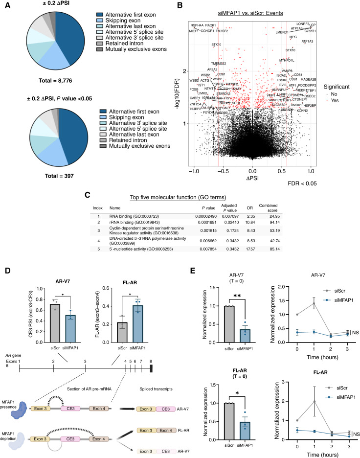
Resistance to androgen receptor (AR)-targeted therapies represents a major challenge in prostate cancer. A key mechanism of treatment resistance in patients who progress to castration-resistant prostate cancer (CRPC) is the generation of alternatively spliced AR variants (AR-V). Unlike full-length AR isoforms, AR-Vs are constitutively active and refractory to current receptor-targeting agents and hence drive tumor progression. Identifying regulators of AR-V synthesis may therefore provide new therapeutic opportunities in combination with conventional AR-targeting agents. Our understanding of AR transcript splicing, and the factors that control the synthesis of AR-Vs, remains limited. Although candidate-based approaches have identified a small number of AR-V splicing regulators, an unbiased analysis of splicing factors important for AR-V generation is required to fill an important knowledge gap and furnish the field with novel and tractable targets for prostate cancer treatment. To that end, we conducted a bespoke CRISPR screen to profile splicing factor requirements for AR-V synthesis. MFAP1 and CWC22 were shown to be required for the generation of AR-V mRNA transcripts, and their depletion resulted in reduced AR-V protein abundance and cell proliferation in several CRPC models. Global transcriptomic analysis of MFAP1-depleted cells revealed both AR-dependent and -independent transcriptional impacts, including genes associated with DNA damage response. As such, MFAP1 downregulation sensitized prostate cancer cells to ionizing radiation, suggesting that therapeutically targeting AR-V splicing could provide novel cellular vulnerabilities which can be exploited in CRPC. Implications: We have utilized a CRISPR screening approach to identify key regulators of pathogenic AR splicing in prostate cancer.


Conflict of interest statement

C. Crafter reports employment with AstraZeneca and ownership of AstraZeneca shares. D.J. O’Neill reports employment with AstraZeneca and ownership of AstraZeneca shares. No disclosures were reported by the other authors.

Figures

Figure 1.
Figure 1.
Cas9-mediated knockout of MFAP1 and CWC22 diminishes AR-V levels in CWR22Rv1-AR-EK-iCas9. A, Schematic of the CRISPR/Cas9 knockout screening protocol optimized for use in the CWR22Rv1-AR-EK-iCas9 cell line. B, CWR22Rv1-AR-EK-iCas9 cells were reverse-transfected with 25 nmol/L of either sgRNA targeting AR exon 1 (sgAR-1) or PLK1 (sgPLK1-Pool) compared with a nontargeting scrambled (sgScr) control, as per the optimized screening protocol for 96 hours. Cells were fixed and stained with Hoechst nuclear stain, anti–AR 441 (Dako) at a concentration of 1:1,000 followed by the secondary antibody Alexa Fluor 647 in modified blocking buffer (1% BSA/0.1% Triton X/PBS). GFP expression and Hoechst staining were imaged, and nuclear AR-V abundance in response to AR knockout using sgAR-1 was compared with the sgScr control. Statistical significance was determined using an unpaired t test (**, P < 0.01; ***, P < 0.001). C, CWR22Rv1-AR-EK-iCas9 cells were transfected as in B with 25 nmol/L sgRNAs targeting 211 splicing factors for 96 hours prior to AR-V immunofluorescence analysis. The resultant data are presented as a heatmap showing the top 50 ranked splicing factors impacting AR levels compared with sgScr control. D, The top 10 ranked splicing factors shown to diminish AR abundance are shown in graphical form. Data are representative of four independent experiments ± SEM. Statistical significance was determined by a one-way ANOVA with the Dunnett multiple comparison test (**, P < 0.01; ***, P < 0.001). E, Protein levels of AR-Vs, Cas9, CWC22, MFAP1, and α-tubulin were assessed using Western blot analysis in the CWR22Rv1-AR-EK-iCas9 cell line. Data are representative of three independent experiments.
Figure 2.
Figure 2.
MFAP1 and CWC22 depletion causes a reduction in expression of AR-FL and AR-V target genes across several CRPC cell line models. A, Protein levels of AR species (FL-AR and AR-Vs) and α-tubulin were assessed using Western blot analysis in the CWR22Rv1 cell line in response to siRNA-mediated splicing factor knockdown. Protein changes were compared with the scrambled (siScr) control for gene knockdown studies. CWR22Rv1-AR-EK (B), CWR22Rv1 (C), and VCaP (D) cell lines were treated with 25 nmol/L siRNA for 72 hours targeting MFAP1 (siMFAP1, pool of siMFAP1-1, and siMFAP1-4) and CWC22 (siCWC22, pool of siCWC22-2, and siCWC22-4) compared with a nontargeting scrambled (siScr) control. CWR22Rv1 and VCaP cells were cultured in steroid-depleted media to allow AR-Vs to drive growth. FL-AR, AR-V1, AR-V6, AR-V7, and AR-V9 transcripts were quantified by RT-qPCR. Top, Diagrammatic representation of AR gene exons 3, CEs, and exon 4, and how the distinct AR-Vs were detected. E, CWR22Rv1-AR-EK-iCas9 cell lines were treated with 1 μg/mL doxycycline and were reverse-transfected with 25 nmol/L sgRNA for 72 hours targeting CWC22 (sgCWC22-1, sgCWC22-2, sgCWC22-3, and sgCWC2-4) and MFAP1 (sgMFAP1-1, sgMFAP1-2, sgMFAP1-3, and sgMFAP1-Pool), along with a positive control targeting AR exon 1 (sgAR-1) to deplete AR-Vs, compared with a nontargeting scrambled (sgScr) control. AR-V1 and AR-V7 transcripts were quantified by RT-qPCR. Ct values were normalized to RPL13A and the scrambled control. Data represent three independent experiments ± SEM. Statistical significance was determined by a one-way ANOVA with the Dunnett multiple comparison test (*, P < 0.05; **, P < 0.01; ***, P < 0.001; ns, not significant). CWR22Rv1-AR-EK (F), CWR22Rv1 (G), and VCaP (H) cell lines were cultured as above and subjected to RT-qPCR to analyze AR target gene expression UBE2C, CCNA2, and TMPRSS2. Ct values were normalized to RPL13A and the scrambled control. Data represent three (B, C, F, G) and two (D and H) independent experiments ± SEM. Statistical significance was determined by a one-way ANOVA with the Dunnett multiple comparison test (*, P < 0.05; **, P < 0.01; ***, P < 0.001; ns, not significant).
Figure 3.
Figure 3.
MFAP1 and CWC22 depletion causes a selective reduction in prostate cancer cell growth. Prostate cancer cell lines CWR22Rv1 (A), CWR22Rv1-AR-EK (B), VCaP (C), CWR-R1-AD1 (D), CWR-R1-D567 (E), and PC3 (F) and prostate epithelial cells PNT1-A (G) and PNT2C-2 (H) were transfected with 25 nmol/L siRNA for 120 hours targeting MFAP1 (siMFAP1, pool of siMFAP1-1, and siMFAP1-4) or CWC22 (siCWC22, pool of siCWC22-2, and siCWC22-4), along with a positive control targeting AR exon 1 (A–C), compared with a nontargeting scrambled (siScr) control. Cell growth was assessed by Sulforhodamine B (SRB) assay after 120 hours compared with the scrambled control for each cell line. Data represent two (E) and three (A–D and F–H) independent experiments ± SEM. Statistical significance was determined by a one-way ANOVA with the Dunnett multiple comparison test (*, P < 0.05; **, P < 0.01; ***, P < 0.001; ns, not significant). I, CWR22Rv1-AR-EK-iCas9 cell lines were treated with 1 μg/mL doxycycline and transfected with 25 nmol/L sgRNA for 120 hours targeting CWC22 (sgCWC22-1, sgCWC22-2, sgCWC22-3, and sgCWC2-Pool) and MFAP1 (sgMFAP1-1, sgMFAP1-2, sgMFAP1-3, and sgMFAP1-Pool), along with a positive control targeting AR exon 1 (sgAR-1) to deplete AR-Vs, compared with a nontargeting scrambled (sgScr) control. Cell growth was assessed after 120 hours compared with the scrambled control. Data represent independent experiments ± SEM. Statistical significance was determined by a one-way ANOVA with the Dunnett multiple comparison test (**, P < 0.01; ***, P < 0.001). J, CWR22Rv1-AR-EK cells cultured under serum-containing medium conditions were transfected with 25 nmol/L siRNA to deplete MFAP1 (siMFAP1, pool of siMFAP1-1, and siMFAP1-4) and AR-Vs (siAREx1; CWR22Rv1-AR-EK cells only) compared with a nontargeting scrambled (siScr) control. Cells were incubated for 72 hours, and cell pellets were collected and subjected to propidium iodide–based flow cytometry. Data are representative of two independent experiments ± SEM. Statistical significance was determined by a two-way ANOVA with the Dunnett multiple comparison test (*, P <0.05; **, P <0.01; ns, not significant). K, CWR22Rv1 cells were transfected with 25 nmol/L siRNA for 120 hours targeting MFAP1 (siMFAP1, pool of siMFAP1-1, and siMFAP1-4), compared with a nontargeting scrambled (siScr) control, along with increasing concentrations of enzalutamide, compared with a DMSO control. Cell growth was assessed after 120 hours compared with the scrambled, DMSO treatment arm of the experiment. Data are representative of three independent experiments, in which average cell growth across the three independent experiments was plotted as a heatmap. L, Enzalutamide-resistant VCaP cells grown in 10 μmol/L enzalutamide were subjected to MFAP1 knockdown as in K for 120 hours prior to cell count analysis. Data represent three independent experiments, and statistical significance was determined using a paired t test (***, P < 0.001).
Figure 4.
Figure 4.
CWC22 and MFAP1 knockdown compromises splicing of constitutive exons and CEs without impacting AR pre-mRNA levels. A, Diagrammatic representation of the primers used to detect AR pre-mRNA and AR-V7 in the CWR22Rv1 cell line. CWR22Rv1 cells were cultured under steroid-depleted conditions and were reverse-transfected with 25 nmol/L siRNA targeted to CWC22 (siCWC22, pool of siCWC22-2, and siCWC22-4) or MFAP1 (siMFAP1, pool of siMFAP1-1, and siMFAP1-4), compared with a nontargeting scrambled (siScr) control, for 72 hours. Samples were harvested for RT-qPCR, and comparative protein lysates were generated to assess knockdown efficiency by Western blot analysis. Gene expression of AR species (AR pre-mRNA; AR-V7 mature transcript; B), CWC22 (C), and MFAP1 (D) were quantified by RT-qPCR. Ct values were normalized to RPL13A and the scrambled control. Data represent three independent experiments ± SEM. Statistical significance was determined by a one-way ANOVA with the Dunnett multiple comparison test (B) and an unpaired t test (C and D; *, P < 0.05; **, P < 0.01; ***, P < 0.001; ns, not significant). E, Diagrammatic representation of the minigene construct and primer-binding sites. Primers are designed to be specific to AR-V7 (binding exon 3 and CE3) or FL-AR (binding exon 3 and exon 4). PC3 cells were transfected with 1 μg of the minigene plasmid and 25 nmol/L siRNA targeting CWC22 (siCWC22, pool of siCWC22-2, and siCWC22-4) or MFAP1 (siMFAP1, pool of siMFAP1-1, and siMFAP1-4), compared with a nontargeting scrambled (siScr) control for 72 hours. A nontransfected (NT) experimental arm was also included. Cells were incubated for 72 hours and harvested for RT-qPCR and Western blot analyses. AR-V7 and FL-AR transcripts (F) and CWC22 (G) and MFAP1 (H) transcript and protein levels were quantified by RT-qPCR and Western blot analysis, respectively. Ct values were normalized to RPL13A and the scrambled control. Data represent three independent experiments ± SEM. Statistical significance was determined by a one-way ANOVA with the Dunnett multiple comparison test (F) and an unpaired t test (G and H; *, P < 0.05; ***, P < 0.001; ns, not significant).
Figure 5.
Figure 5.
MFAP1 depletion impacts AR-dependent and -independent signaling in prostate cancer. A, DEGs were assessed for MFAP1 (siMFAP1, pool if siMFAP1-1, and siMFAP1-4) and AR-V (siCE3) depletion compared with the nontargeting scrambled (siScr) control in the CWR22Rv1 cell line. Dots represent genes up- or downregulated, demonstrating a 1.5-fold change. Blue dots represent any genes that were up- or downregulated demonstrating a 1.5-fold change which have a P value < 0.05. N = number of SDEGs up- or downregulated. B, Downregulated SDEGs from MFAP1 and AR-V7 knockdown in CWR22Rv1 cells were compared with an AR-V transcriptome derived from CWR22Rv1-AR-EK cells [Kounatidou and colleagues (32)] to determine AR-dependent and -independent gene signatures of MFAP1. C, DEG lists from depletion of MFAP1 (siMFAP1, pool of siMFAP1-1, and siMFAP1-4) in CWR22Rv1 cells were compared with an AR hallmark gene list and custom lists of AR-V7–regulated genes taken from Hu and colleagues (47) and Cai and colleagues (48) using gene set enrichment analysis (GSEA). Nominal P value and NES (normalized enrichment score) for each treatment arm are shown. DEG lists from MFAP1 depletion in CWR22Rv1 cells were compared with (D) hallmark and (E) Kyoto Encyclopedia of Genes and Genomes (KEGG) gene lists using GSEA. Graphs show the top 15 (or less) positively and negatively enriched pathways with a P value < 0.05 and an FDR < 25%. F, Downregulated AR-independent, MFAP1-regulated genes (SDEGs) were profiled using Enrichr pathway analysis.
Figure 6.
Figure 6.
MFAP1 knockdown impacts global splicing patterns, including AR exon inclusion dynamics. A, Global splicing patterns of CWR22Rv1 cells depleted of MFAP1 (siMFAP1, pool of siMFAP1-1, and siMFAP1-4) were quantified by category (e.g., alternative first exon and skipping exon). Events that passed a ∆proportion spliced in (ΔPSI) value ± 0.2 were plotted (left); events that were considered significant (P value of <0.05) were plotted (right). B, Volcano plots were plotted of differential splicing events that passed cutoffs of ΔPSI ± 0.6 and FDR <0.05, annotated with their gene ID (red). C, 397 significantly altered splicing events (∆PSI and P value) which accounted for 227 unique genes were ran through Enrichr using molecular function gene ontology (GO) terms filters to identify involvement in cellular functions. D, RNA-seq data derived from CWR22Rv1 cells depleted of MFAP1 were analyzed for altered exon composition of distinct AR transcripts as calculated by investigating relative exon inclusion (PSI) for all junctions measured using SUPPA2. Bottom, Diagrammatic representation of the AR gene exon 3, CE3, and exon 4 and shift in splicing activity in response to MFAP1 depletion. E, CWR22Rv1 cells cultured under steroid-depleted conditions were transfected with 25 nmol/L siRNA targeted to MFAP1 (siMFAP1, pool of siMFAP1-1, and siMFAP1-4,) or scrambled (siScr) control for 72 hours prior to treatment with 5 μmol/L actinomycin D for 0–3 hours and FL-AR/AR-V7 transcript profiling by RT-qPCR. Ct values were normalized to RPL13A transcript and the scrambled control. Data represent three independent experiments ± SEM. Statistical significance was determined by an unpaired t test at time point 0 hour or at the 3 hours endpoint (*, P < 0.05; **, P < 0.01; ns, not significant).
Figure 7.
Figure 7.
MFAP1 regulates an AR-independent DDR gene signature in which knockdown sensitizes prostate cancer cells to IR. A, DEG lists from depletion of MFAP1 (siMFAP1, pool of siMFAP1-1, and siMFAP1-4) and AR-V7 (siCE3) in CWR22Rv1 cells were compared with KEGG gene lists from gene set enrichment analysis (GSEA) for nucleotide excision repair and homologous recombination. Log2 FC for each gene was plotted as a heatmap. B, CWR22Rv1-AR-EK cells were transfected with 25 nmol/L siRNA to deplete MFAP1 (siMFAP1, pool of siMFAP1-1, and siMFAP1-4), AR-Vs (siAREx1), or both MFAP1 and AR-V7 (siDual, pool of siMFAP1-1, siMFAP1-4, and siCE3) or scrambled (siScr) control for 72 hours prior to irradiation with 2 Gy IR and harvested for immunofluorescence analysis of γH2AX foci 1, 6 and 24 hours after IR. A minus IR (−IR) arm was also included to compare with endogenous DNA damage. γH2AX foci were quantified using ImageJ software, and data are presented as average foci/cell. Data are representative of three independent experiments ± SEM. C, CWR22Rv1 cells were transfected with 25 nmol/L siRNA to deplete MFAP1 (siMFAP1, pool of siMFAP1-1, and siMFAP1-4) or scrambled (siScr) control in the presence and absence of 10 μmol/L enzalutamide for 72 hours prior to irradiation with 2 Gy IR and harvested for immunofluorescence as in B. D, PC3 cells were transfected as in C and harvested for immunofluorescence analysis of γH2AX foci 1, 6, and 24 hours after IR as in B. E, CWR22Rv1-AR-EK cells were transfected with 25 nmol/L siRNA to deplete MFAP1 (siMFAP1, pool of siMFAP1-1, and siMFAP1-4), AR-Vs (siAREx1), or both and a scrambled (siScr) control for 72 hours prior to re-seeding at a density of 125, 250, or 500 cells/well on six-well plates, irradiated with 2 Gy IR (plus IR arm only) 8 hours later and left for 3 weeks to allow colonies to form. The resultant colonies were counted, and high-resolution scanned images of the plates were generated for representative images. Data are representative of three independent experiments ± SEM. Statistical significance was determined by a one-way ANOVA with the Dunnett multiple comparison test (*, P < 0.05; **, P < 0.01; ***, P < 0.001). F, PC3 cells were transfected as in E and irradiated with 2 Gy IR (plus IR arm only) 8 hours later and left for 3 weeks to allow colonies to form. The resultant colonies were counted, and high-resolution scanned images of the plates were generated for representative images. Data are representative of three independent experiments ± SEM. Statistical significance was determined by a one-way ANOVA with the Dunnett multiple comparison test (*, P < 0.05; **, P < 0.01; ***, P < 0.001).

Similar articles

Cited by

References

    1. Westaby D, Viscuse PV, Ravilla R, de la Maza MLDF, Hahn A, Sharp A, et al. . Beyond the androgen receptor: the sequence, the mutants, and new avengers in the treatment of castrate-resistant metastatic prostate cancer. Am Soc Clin Oncol Educ Book 2021;41:e190–202. - PubMed
    1. Nevedomskaya E, Baumgart SJ, Haendler B. Recent advances in prostate cancer treatment and drug discovery. Int J Mol Sci 2018;19:1359. - PMC - PubMed
    1. Ross RW, Xie W, Regan MM, Pomerantz M, Nakabayashi M, Daskivich TJ, et al. . Efficacy of androgen deprivation therapy (ADT) in patients with advanced prostate cancer: association between Gleason score, prostate-specific antigen level, and prior ADT exposure with duration of ADT effect. Cancer 2008;112:1247–53. - PubMed
    1. Sumanasuriya S, De Bono J. Treatment of advanced prostate cancer—a review of current therapies and future promise. Cold Spring Harbor Perspect Med 2018;8:a030635. - PMC - PubMed
    1. Attard G, Murphy L, Clarke NW, Cross W, Jones RJ, Parker CC, et al. . Abiraterone acetate and prednisolone with or without enzalutamide for high-risk non-metastatic prostate cancer: a meta-analysis of primary results from two randomised controlled phase 3 trials of the STAMPEDE platform protocol. Lancet 2022;399:447–60. - PMC - PubMed

Publication types

MeSH terms