. 2024 Sep 30;19(9):e0309533.
doi: 10.1371/journal.pone.0309533. eCollection 2024.

Cytotoxicity of bendamustine, alone and in combination with novel agents, toward adult T-cell leukemia cells

Affiliations

Cytotoxicity of bendamustine, alone and in combination with novel agents, toward adult T-cell leukemia cells

Naoki Osada et al. PLoS One. .

Abstract

Adult T-cell leukemia/lymphoma (ATL) develops from the infection of T cells with human T lymphotropic virus type 1 (HTLV-1). There are an estimated 5-20 million HTLV-1 carriers worldwide and the patients are frequently observed in subtropical Africa, the Caribbean, Middle East, South America, and South West Japan. The prognosis of ATL remains dismal due to rapid acquired resistance to treatment with cytotoxic chemotherapeutic agents. In particular, the development of novel therapies for relapsed or refractory (R/R) ATL is an unmet need. Previous clinical trials revealed that bendamustine (BDM) was effective as the first-line treatment for indolent lymphoma and R/R cases of diffuse large B-cell lymphoma. Its major advantage is that it has few side effects such as hair loss and peripheral neuropathy, and does not impair the quality of life. However, its efficacy has not been verified for ATL in pre-clinical or clinical studies. In this study, we have shown the cytotoxicity of BDM alone and in combination with novel agents including the histone deacetylase (HDAC) inhibitor tucidinostat, the enhancer of zeste homolog 1/2 (EZH1/2) dual inhibitor valemetostat, and the Bcl2 family inhibitor ABT-737. The combined in vitro effects of BDM and tucidinostat were reproduced in a murine model without any obvious hematological toxicity. Our present results suggest that the combination of tucidinostat and BDM could additively prolong the survival of patients with R/R progressive ATL. The efficacy and safety of this combination are thus worthy of investigation in clinical settings.


Conflict of interest statement

Yusuke Furukawa received research funding from SymBio Pharmaceuticals Ltd. This does not alter our adherence to PLOS ONE policies on sharing data and materials. Other authors have no conflict of interest.

Figures

Fig 1
Fig 1. In vitro cytotoxicity toward ATL cells of bendamustine alone and in combination with novel agents.
(A) We cultured the indicated cell lines with various concentrations of bendamustine (BDM) (0–100 μM) and cell viability was determined by the MTT (3-(4,5-dimethylthiazol-2-yl)-5-(3-carboxymethophenyl-2-(4-sulfophenyl)-2H-tetrazolium) reduction assay after 72 h to obtain dose-response curves. IC50 values are defined as the concentrations of drug that produce 50% inhibition of cell growth. (B) Upper panel; Su9, S1T, MT2 and ATN-1 cells were cultured with various concentrations of BDM for 72 h. Cell viability was determined by the MTT reduction assay and expressed as a percentage of the untreated control. The means ± S.D. (bars) of multiple independent experiments (n = 3) are shown. Asterisks indicate P < 0.05 vs untreated control. P values were determined by one-way analysis of variance (ANOVA) with a Student–Newman–Keuls multiple comparison test. Lower panel; Su9, S1T, MT2 and ATN-1 cells were cultured with various concentrations of BDM for 24 h. Whole cell lysates were simultaneously obtained and subjected to immunoblotting for cleaved caspase-3 and glyceraldehyde 3-phosphate dehydrogenase (GAPDH) proteins (loading control). (C) S1T, TLSu, MT2, and ATN-1 cells were treated with BDM in combination with tucidinostat, valemetostat, or ABT-737 for 72 h. Cell viability was determined by the MTT reduction assay to obtain dose-response curves for each combination. Combination index plots were generated by CompuSyn software. Combination indexes (CI) <0.8 and 0.8–1.2 indicate synergism and additivity between two drugs, respectively. Bars indicate the means of four ATL cell lines. (D) Upper panel: MT2 and ATN-1 cells were treated with vehicle alone (control), BDM (MT2: 40 μM, ATN-1: 80 μM), tucidinostat (MT2 and ATN-1: 150 nM), or a combination of both (combination) for 72 h. Cell viability was determined by the MTT reduction assay and expressed as a percentage of the untreated control. The means ± S.D. (bars) of multiple independent experiments (n = 3) are shown. P values were determined by one-way analysis of variance (ANOVA) with a Student–Newman–Keuls multiple comparison test. Lower panel: Whole cell lysates were simultaneously obtained and subjected to immunoblotting for cleaved caspase-3 and GAPDH (loading control) proteins.
Fig 2
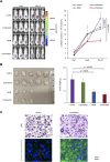
Fig 2. In vivo cytotoxicity toward ATL cells of bendamustine alone and in combination with tucidinostat.
(A) We inoculated 5 × 10⁵ luciferase expressing MT2 cells subcutaneously into the right thigh of male NOD/SCID mice and randomized them into four treatment groups when measurable tumors developed over 5 x 10⁵ photons/s (day 1). Each group was treated with the vehicle alone (0.9% NaCl, orally, 5 times a week) (n = 5), tucidinostat alone (20 mg/kg, orally, 5 times a week) (n = 5), BDM alone (30 mg/kg, intraperitoneally, 3 times a week) (n = 3), or the combination of tucidinostat and BDM (n = 5 mice in each group) (n = 3) for 2 weeks. Left panel: tumor-derived luciferase activity was measured ex vivo by the IVIS Imaging System after D-luciferin injection. Representative photographs of NOD/SCID mice on day 1 and day 15 are shown (original magnification: × 2). Right panel: quantitative data of in vivo bioluminescence imaging shown in the left panel. The signal intensity is shown as photon units (photons/s). P values were determined by one-way analysis of variance (ANOVA) with a Student–Newman–Keuls multiple comparison test. (B) Left panel: representative photographs of tumors resected on day 30 (original magnification: × 2). Right panel: the means ± S.D. (bars) of the weights of resected tumors shown in the left panel. P values were determined by one-way analysis of variance (ANOVA) with a Student–Newman–Keuls multiple comparison test. (C) Upper panel: Tumor sections were prepared from mice and stained with hematoxylin eosin (HE). Lower panel: Serial tumor sections were immunostained for cleaved caspase-3 (green), then counterstained with 4’,6-diamidino-2-phenylindole (DAPI, blue). Only merged images are shown. Data shown are representative of multiple independent experiments.

References

    1. Proietti FA, Carneiro-Proietti AB, Catalan-Soares BC, Murphy EL. Global epidemiology of HTLV-I infection and associated diseases. Oncogene. 2005;24:6058–68. doi: 10.1038/sj.onc.1208968 - DOI - PubMed
    1. Iwanaga M, Watanabe T, Yamaguchi K. Adult T-cell leukemia: a review of epidemiological evidence. Front Microbiol. 2012;3:322. doi: 10.3389/fmicb.2012.00322 - DOI - PMC - PubMed
    1. Katsuya H. Current and emerging therapeutic strategies in adult T-cell leukemia-lymphoma. Int J Hematol. 2023;117:512–522. doi: 10.1007/s12185-023-03572-4 - DOI - PubMed
    1. Kataoka K, Nagata Y, Kitanaka A, Shiraishi Y, Shimamura T, Yasunaga J, et al.. Integrated molecular analysis of adult T cell leukemia/lymphoma. Nat Genet. 2015;47:1304–1315. doi: 10.1038/ng.3415 - DOI - PubMed
    1. Hartmann M, Zimmer C. Investigation of cross-link formation in DNA by the alkylating cytostatic IMET 3106, 3393 and 3943. Biochim Biophys Acta. 1972;287:386–389. doi: 10.1016/0005-2787(72)90282-1 - DOI - PubMed

MeSH terms

Substances

LinkOut - more resources