. 2024 Sep 30;18(9):e0012553.
doi: 10.1371/journal.pntd.0012553. eCollection 2024 Sep.

HuR (ELAVL1) regulates the CCHFV minigenome and HAZV replication by associating with viral genomic RNA

Affiliations

HuR (ELAVL1) regulates the CCHFV minigenome and HAZV replication by associating with viral genomic RNA

Moe Ikegawa et al. PLoS Negl Trop Dis. .

Abstract

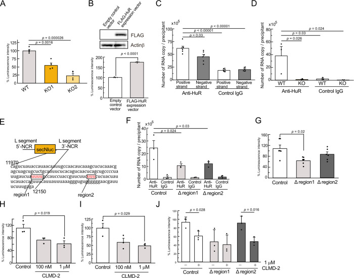
Crimean-Congo Hemorrhagic Fever virus (CCHFV) is a tick-borne pathogen that causes severe acute fever disease in humans and requires a biosafety level 4 laboratory for handling. Hazara virus (HAZV), belonging to the same virus genus as CCHFV, does not exhibit pathogenesis in humans. To investigate host RNA-binding proteins (RBPs) that regulate CCHFV replication, we generated a series of mutant RAW264.7 cells by CRISPR/Cas9 system and these cells were infected with HAZV. The viral titers in the supernatant of these cells was investigated, and HuR (ELAVL1) was identified. HuR KO RAW264.7 cells reduced HAZV replication. HuR is an RBP that enhances mRNA stability by binding to adenyl-uridine (AU)-rich regions in their 3' non-coding region (NCR). HuR regulates innate immune response by binding to host mRNAs of signaling molecules. The expression of cytokine genes such as Ifnb, Il6, and Tnf was reduced in HuR KO cells after HAZV infection. Although HuR supports the innate immune response during HAZV infection, we found that innate immune activation by HAZV infection did not affect its replication. We then investigated whether HuR regulates HAZV genome RNA stability. HAZV RNA genome was precipitated with an anti-HuR antibody, and HAZV genome RNA stability was lowered in HuR KO cells. We found that HuR associated with HAZV RNA and stabilized it to enhance HAZV replication. Furthermore, HuR-deficiency reduced CCHFV minigenome replication. CCHFV is a negative-strand RNA virus and positive-strand RNA is produced during replication. HuR was associated with positive-strand RNA rather than negative-strand RNA, and AU-rich region in 3'-NCR of S segment was responsible for immunoprecipitation with anti-HuR antibody and minigenome replication. Additionally, HuR inhibitor treatment reduced CCHFV minigenome replication. Our results indicate that HuR aids replication of the CCHFV minigenome by associating with the AU-rich region in the 3'-NCR.


Conflict of interest statement

The authors have declared that no competing interests exist.

Figures

Fig 1
Fig 1. Identification of HuR as a regulator for HAZV replication in RAW264.7 cells.
A, RAW264.7 cells and MEFs were infected with 0.1 MOI HAZV and virus titers in the supernatant were measured. B, WT and mutant RAW264.7 cells were infected with 0.1 MOI HAZV and the virus titers in the supernatants were measured at 48 h post-infection. Virus titers of the mutant cells were plotted, and cell lines that showed higher or lower titers than WT were highlighted. C, Cell lysates were extracted and immunoblotted using anti-HuR and anti-Actinβ antibodies. D, WT and HuR KO RAW264.7 cells were infected with 0.1 MOI of HAZV, TBEV or JEV, and the time courses of virus titer in the supernatants were measured at the indicated time points. E, The copy number of HAZV S segment inside cells was measured by real-time PCR at 48 h after 0.1 MOI HAZV infection. One-way ANOVA with Tukey’s multiple comparison test (C,D); *p < 0.05.
Fig 2
Fig 2. Innate immune response after HAZV infection in HuR KO cells.
A,B, WT and HuR KO RAW264.7 cells were infected with 1 MOI HAZV or UV-treated HAZV, and cytokine gene expression was measured at 9 h after infection by real-time PCR (A). Gene expression was calculated by fold increase compared to non-infected control WT cells. Copy number of S segment inside cells was calculated by real-time PCR (B). C, IL-6 production in the supernatant was measured by ELISA after 1 MOI HAZV infection. D, WT and HuR KO RAW264.7 cells were infected with 1 MOI HAZV. Cell lysates were extracted and immunoblotted with the indicated antibodies. E, HuR KO HEK293 cells were generated by genome editing and the deficiency was confirmed by western blotting. F,G, WT and HuR KO HEK293 cells were transfected with a reporter plasmid driven by IFN-β promoter (F) or ISRE promoter (G) with internal control promoter plasmid, and these cells were stimulated with 1 MOI HAZV or poly(I:C). Luciferase activity was measured 24 h after stimulation. The ratio of intensity of firefly luciferase (IFN-β/ISRE promoter) to intensity of renilla luciferase (internal control promoter) was plotted. One-way ANOVA with Tukey’s multiple comparison test (A,C,F,G).
Fig 3
Fig 3. Innate immune activation by HAZV infection is not involved in its replication.
A, WT, Rig-I/Mda5 KO, IPS1 KO and Tbk1/Ikk-i KO MEFs were infected with 1 MOI HAZV and the expression of cytokine genes was measured by real-time PCR at 24 h post infection. B,C, WT and KO MEFs with or without pre-poly(I:C) for 6 h were infected with 0.1 MOI HAZV, and virus titers in the supernatant (B) or copy number of S segment inside cells (C) were measured at 48 h post-infection. D,E, WT and HuR KO RAW264.7 cells with or without pre-poly(I:C) for 6 h were infected with 0.1 MOI HAZV, and virus titers in the supernatant (D) or copy number of S segment inside cells (E) were measured at 48 h post-infection. Ono-way ANOVA with Tukey’s multiple comparison test (A, C).
Fig 4
Fig 4. HuR stabilizes HAZV RNA.
A, WT and HuR KO RAW264.7 cells were infected with 0.1 MOI HAZV, and RNA inside cells was isolated. RNA was transcribed using random primers, and the copy number of RNA inside cells was measured by real-time PCR. B, WT and HuR KO RAW264.7 cells were infected with 0.1 MOI HAZV, and RNA inside cells was isolated at 48 h post infection. RNA was transcribed using specific primers for positive or negative strands of the S, M, and L segments, and the copy number of RNA inside cells was measured by real-time PCR. C, WT and HuR KO HEK293 cells were transfected with HAZV L and N protein expression plasmids and reporter RNA, and the luciferase activity of secNluc in the supernatant was measured. D, HAZV minigenome replication in HEK293 cells was measured in the presence of control or FLAG-HuR expression vectors. FLAG-HuR expression was confirmed by western blotting with anti-FLAG antibody (left) and the luciferase activity of secNluc in the supernatant was measured (right). E, WT and HuR KO RAW264.7 cells were infected with 0.1 MOI HAZV, and cell lysates at 48 h post-infection were blotted with the indicated antibodies. F, HAZV RNA in the cell lysates of WT and HuR KO RAW264.7 cells at 48 h post infection with HAZV was precipitated with anti-HuR or control IgG antibody. RNA was transcribed using random primers and copy numbers in the precipitants were measured by real-time PCR. G, WT and HuR KO RAW264.7 cells were infected with 0.1 MOI HAZV and cells at 48 h post infection were treated with actinomycin D for the indicated time. Cell lysates were blotted with the indicated antibodies. H, RAW264.7 cells at 48 h post-infection with HAZV were treated with or without actinomycin D, and the S segment of RNA inside cells at the indicated time points was measured by real-time PCR. I, WT and HuR KO RAW264.7 cells at 48 h post-infection were treated with actinomycin D, and S, M, and L segments of RNA inside cells at the indicated time points were measured by real-time PCR. Unpaired two-tailed t-test (A,B,D); n.s., not significant. Ono-way ANOVA with Tukey’s multiple comparison test (C, F).
Fig 5
Fig 5. HuR associates with L segment of CCHFV RNA to support minigenome replication.
A, WT and HuR KO HEK293 cells were transfected with CCHFV L and N protein expression plasmids and reporter RNA, and the luciferase activity of secNluc in the supernatant was measured. B, CCHFV minigenome replication in HEK293 cells was measured in the presence of control or FLAG-HuR expression vectors. FLAG-HuR expression was confirmed by western blotting with anti-FLAG antibody (upper panel) and the luciferase activity of secNluc in the supernatant was measured (lower panel). C, Positive and negative strands of the CCHFV reporter RNA were synthesized and transfected into HEK293 cells for 3 h. RNA in the cell lysate was precipitated using anti-HuR or control IgG antibody. The reporter RNA in the precipitants was transcribed using random primers and the copy number of reporter RNA was measured by real-time PCR. D, RNA in the cell lysate from WT and HuR KO HEK293 cells at 3 h post-transfection with the positive strand of the CCHFV reporter RNA was precipitated with anti-HuR or control IgG antibody. The reporter RNA in the precipitants was transcribed using random primers and the copy number of reporter RNA was measured by real-time PCR. E, Schematic diagram of positive-strand reporter RNA. AU-rich regions [12029–12032 (region1) and 12088–12090 (region2)] were highlighted in the 3′-NCR sequence. F, Positive strand of control, 12029–12032 (Δregion1) and 12088–12090 (Δregion2) reporter RNA were transfected into HEK293 cells, and RNA in the cell lysate was precipitated with anti-HuR or control IgG antibody. G, HEK293 cells were transfected with CCHFV L and N protein expression plasmids with reporter RNA for control, Δregion1 and Δregion2, and the luciferase activity of secNluc in the supernatant was measured. H, I, Minigenome replication of CCHFV (H) and HAZV (I) in HEK293 cells was measured after treatment with CLMD-2. J, Minigenome replication of control, Δregion1 and Δregion2 was measured after treatment with CLMD-2. Ono-way ANOVA with Tukey’s multiple comparison test (A,B, C, D, F, G,H,I). Unpaired two-tailed t-test (B,J).

Similar articles

Cited by

References

    1. Bente DA, Forrester NL, Watts DM, McAuley AJ, Whitehouse CA, Bray M. Crimean-Congo hemorrhagic fever: history, epidemiology, pathogenesis, clinical syndrome and genetic diversity. Antiviral Res. 2013;100(1):159–89. doi: 10.1016/j.antiviral.2013.07.006 - DOI - PubMed
    1. Abudurexiti A, Adkins S, Alioto D, Alkhovsky SV, Avsic-Zupanc T, Ballinger MJ, et al.. Taxonomy of the order Bunyavirales: update 2019. Arch Virol. 2019;164(7):1949–65. doi: 10.1007/s00705-019-04253-6 - DOI - PMC - PubMed
    1. Hawman DW, Feldmann H. Crimean-Congo haemorrhagic fever virus. Nat Rev Microbiol. 2023;21(7):463–77. doi: 10.1038/s41579-023-00871-9 - DOI - PMC - PubMed
    1. Spengler JR, Bente DA, Bray M, Burt F, Hewson R, Korukluoglu G, et al.. Second International Conference on Crimean-Congo Hemorrhagic Fever. Antiviral Res. 2018;150:137–47. doi: 10.1016/j.antiviral.2017.11.019 - DOI - PMC - PubMed
    1. Ergonul O, Celikbas A, Baykam N, Eren S, Dokuzoguz B. Analysis of risk-factors among patients with Crimean-Congo haemorrhagic fever virus infection: severity criteria revisited. Clin Microbiol Infect. 2006;12(6):551–4. doi: 10.1111/j.1469-0691.2006.01445.x - DOI - PubMed