. 2024 Sep 30;22(9):e3002813.
doi: 10.1371/journal.pbio.3002813. eCollection 2024 Sep.

Mycobacteria that cause tuberculosis have retained ancestrally acquired genes for the biosynthesis of chemically diverse terpene nucleosides

Affiliations

Mycobacteria that cause tuberculosis have retained ancestrally acquired genes for the biosynthesis of chemically diverse terpene nucleosides

Jacob A Mayfield et al. PLoS Biol. .

Abstract

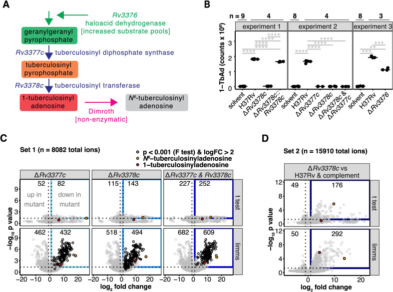
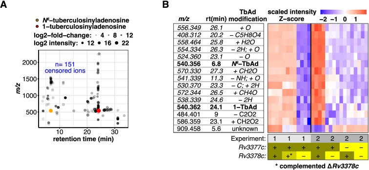
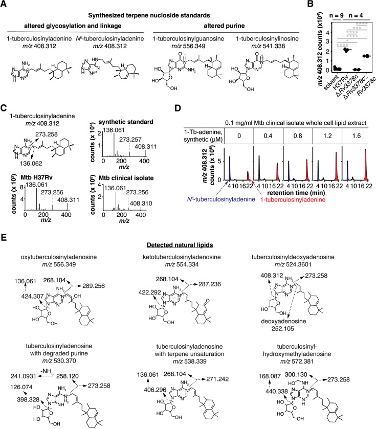
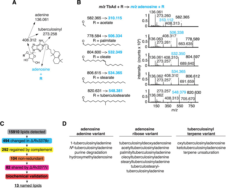
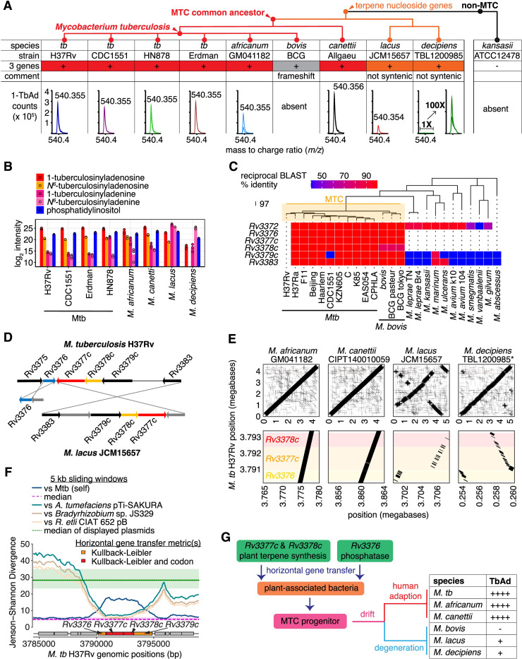
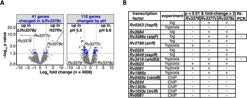
Mycobacterium tuberculosis (Mtb) releases the unusual terpene nucleoside 1-tuberculosinyladenosine (1-TbAd) to block lysosomal function and promote survival in human macrophages. Using conventional approaches, we found that genes Rv3377c and Rv3378c, but not Rv3376, were necessary for 1-TbAd biosynthesis. Here, we introduce linear models for mass spectrometry (limms) software as a next-generation lipidomics tool to study the essential functions of lipid biosynthetic enzymes on a whole-cell basis. Using limms, whole-cell lipid profiles deepened the phenotypic landscape of comparative mass spectrometry experiments and identified a large family of approximately 100 terpene nucleoside metabolites downstream of Rv3378c. We validated the identity of previously unknown adenine-, adenosine-, and lipid-modified tuberculosinol-containing molecules using synthetic chemistry and collisional mass spectrometry, including comprehensive profiling of bacterial lipids that fragment to adenine. We tracked terpene nucleoside genotypes and lipid phenotypes among Mycobacterium tuberculosis complex (MTC) species that did or did not evolve to productively infect either human or nonhuman mammals. Although 1-TbAd biosynthesis genes were thought to be restricted to the MTC, we identified the locus in unexpected species outside the MTC. Sequence analysis of the locus showed nucleotide usage characteristic of plasmids from plant-associated bacteria, clarifying the origin and timing of horizontal gene transfer to a pre-MTC progenitor. The data demonstrated correlation between high level terpene nucleoside biosynthesis and mycobacterial competence for human infection, and 2 mechanisms of 1-TbAd biosynthesis loss. Overall, the selective gain and evolutionary retention of tuberculosinyl metabolites in modern species that cause human TB suggest a role in human TB disease, and the newly discovered molecules represent candidate disease-specific biomarkers.


Conflict of interest statement

The authors have declared that no competing interests exist.

Figures

Fig 1
Fig 1. Engineered deletions of 1-TbAd biosynthesis genes reveal gene functions and greatly expand the lipid signature.
(A) Schematic shows the 1-TbAd biosynthetic pathway. (B) Area-under-the-curve of extracted ion chromatograms tested 1-TbAd production by the parental Mtb strain (H37Rv) and single or two-gene knockouts as well as the Rv3378c deletion complemented with Rv3378c. A Benjamini–Hochberg adjusted p value is indicated only for significant pairwise t tests (*: p < 0.05, **: p < 0.01, ***: p < 0.001, ****: p < 0.0001). The peak area in the retention time window corresponding to Mtb H37Rv 1-TbAd [M+H]+ was measured in intervening solvent blank samples to indicate the measurement threshold. (C) Comparative metabolomics analysis showed genetic control of differentially abundant molecules. Positive mode mass spectrometry data were analyzed by comparing deletions in Rv3377c, Rv3378c, or a double deletion of both genes to the H37Rv parental strain. Differential abundance determined using t tests or a linear model fit using limms was compared. The number of significant events (p < 0.05 after adjustment using the Benjamini–Hochberg method) that also changed more than 2-fold were indicated (blue rectangle). The most abundant 1- and N6-tuberculosinyladenosine peaks are flagged (red and orange circles, respectively). Using limms, events with similar patterns of change in all 3 comparisons were determined by F-test. Events with >4-fold decrease in all 3 mutants and p < 0.001 (Benjamini–Hochberg adjusted p value of the F-test) are shown in black. (D) An independent metabolomic comparison of the Rv3378c deletion to H37Rv parent strain and the Rv3378c deletion complemented with Rv3378c was analyzed for differentially abundant positive mode events. Significantly changed events were determined using t tests or limms. The numbers of changed events (Benjamini–Hochberg adjusted p < 0.05 and 2-fold or greater change), the gene-dependent events (blue rectangle), and the most abundant 1- and N6-tuberculosinyladenosine peaks are indicated as in Fig 2A. The data in Fig 1B–1D can be found in S1 Data.
Fig 2
Fig 2. Rv3377-3378c-dependent lipids share biochemical properties consistent with a larger family of terpene nucleosides.
(A) The 254 significantly changed events in the blue box in panel Fig 1D were filtered to remove 151 recognizable isotopes and alternate adducts, yielding 104 unique molecules. These Rv3378c-dependent events restored by complementation clustered in mass and time with either 1- and N6-TbAd. (B) Heatmap of 15 Rv3378c-dependent events that had mass (within 10 ppm) and retention time (within 3 min) analogs across the 2 independent experimental data sets shown in Fig 1C and 1D. Rows show the ion intensity scaled separately for each experiment. Chemical modifications to TbAd consistent with the observed mass are indicated. Rows in italics were subsequently validated. Raw data for Fig 2B and 2C can be found in S1 Data.
Fig 3
Fig 3. Unknown Rv3377-3378c-dependent lipids were identified as new terpene nucleoside family members.
(A) Structures of synthetic molecules used to analyze natural compounds. (B) Intensities of ion chromatograms corresponding to m/z 408.312, the most abundant non-TbAd lipid found in the differential abundance analysis. Significant pairwise t tests after Benjamini–Hochberg adjustment are indicated (*: p < 0.05, **: p < 0.01, ***: p < 0.001, ****: p < 0.0001). Raw data for these measurements were provided in S1 Data. (C) CID-MS of m/z 408.312 showed fragmentation patterns diagnostic of 1-tuberculosinyladenine. The chemical structure with fragmentation shows calculated masses while spectra show observed masses. (D) Mtb clinical isolate M0014870-1 total lipid extracts were spiked with synthetic 1-tuberculosinyladenine, which showed co-elution with natural 1-tuberculosinyladenine and established its chemical identity and absolute yield in Mtb. (E) Annotated fragments from CID-MS established structures of 6 previously unknown terpene nucleosides where the calculated masses are shown. Collision localized modifications to the ribose, adenosine, or terpene but linkage within the moiety was inferred based on known analogous compounds.
Fig 4
Fig 4. Fragmentation and identification of adenosine-containing lipids revealed terpene nucleoside family members.
(A) Annotated fragments and calculated masses of lipid-linked 1-TbAd derivatives detected by CID-MS. The R group was assigned as ribose-linked based on ribose-fatty acyl fragments with the 2-linkage favored based on its chemical reactivity and known structures; however, linkage position could not be assigned directly from MS. (B) CID-MS showed diverse fragmentation patterns containing adenosine and consistent with parent terpene nucleosides containing lipid-conjugated ribose. Observed masses are shown. (C) Schematic shows the unsupervised lipidomic discovery process for the new TbAd-like lipids. (D) Thirteen lipids were identified as Rv3378c dependent and validated through CID-MS and synthetic chemistry.
Fig 5
Fig 5. The timing and origin of horizontal transfer of biosynthetic genes for terpene nucleosides.
(A) Mass spectrometry tested 1-TbAd production and abundance in MTC species that contained the three-gene locus. Positive mode extracted ion chromatograms for 1-TbAd show lipid counts near m/z 540.354 (observed mass shown) at a retention time of approximately 23 min in samples at 1 mg/ml total lipid. The lack of 1-TbAd in M. bovis and M. kansasii was documented previously [5,12]. A cladogram of arbitrary branch lengths reflected plausible organization [33]. (B) Peak intensity of a predominant membrane lipid, phosphatidylinositol, along with 1- and N6-TbAd, and 1- and N6-tuberculosinyladenine were measured among a panel of 8 MTC strains and species. (C) Reciprocal BLAST hit scores versus H37Rv are shown as a heatmap for the terpene nucleoside biosynthetic genes Rv3376, Rv3377c, and Rv3378c along with flanking genes. The neighbor-joining species dendrogram was based on whole-genome presence/absence of orthogroups. (D) Locus organization in M. lacus is rearranged relative to Mtb H37Rv. (E) Synteny of Mycobacterium species is shown for the whole genome and Rv3376-8c locus as dot plots compared to the reference Mtb H37Rv genome. The M. decipiens TBL1200985 genome is not fully assembled; hence, genome positions were inferred by scaffolding using the Mtb H37Rv genome. (F) Jenson–Shannon divergence profiles of Mtb H37Rv comparing to Mtb H37Rv genome itself or to DNA sequences from other bacteria using a 5 kb sliding window with 100 bp step. The gene schematic is colored according to Kullback–Leibler divergence. (G) Schematic of gene acquisition shows divergence and function. The data for Fig 5B and 5C can be found in S1 Data.
Fig 6
Fig 6. Terpene nucleoside gene regulation.
(A) Gene expression measured by RNAseq was compared in the Rv3378c mutant and the parental strain at pH 6.6 or 5.5. A nested contrast of the Rv3378c mutant versus the parental strain at each pH indicated the genes with differential abundance with respect to the Rv3378c mutant (left), while a nested contrast of pH 5.5 versus 6.6 shared by both strains showed pH responsive genes (right). Differentially abundant genes (blue; Benjamini–Hochberg adjusted p value <0.05) and the TbAd biosynthesis pathway genes are indicated. The underlying transcriptomics data can be found in S1 Data. (B) Transcription factor overexpression strains that caused significant alterations in Rv3376, Rv3377c, or Rv3378c transcripts in log-phase growth or during the induction and release of hypoxia are shown. Transcription factors physically associated with Rv3376, Rv3377c, or Rv3378c, measured by ChIP-seq, were included.

Similar articles

Cited by

References

    1. Ruhl CR, Pasko BL, Khan HS, Kindt LM, Stamm CE, Franco LH, et al.. Mycobacterium tuberculosis Sulfolipid-1 Activates Nociceptive Neurons and Induces Cough. Cell. 2020;181(2):293–305 e11. doi: 10.1016/j.cell.2020.02.026 - DOI - PMC - PubMed
    1. Ishikawa E, Ishikawa T, Morita YS, Toyonaga K, Yamada H, Takeuchi O, et al.. Direct recognition of the mycobacterial glycolipid, trehalose dimycolate, by C-type lectin Mincle. J Exp Med. 2009;206(13):2879–88. doi: 10.1084/jem.20091750 - DOI - PMC - PubMed
    1. Wang Q, Boshoff HIM, Harrison JR, Ray PC, Green SR, Wyatt PG, et al.. PE/PPE proteins mediate nutrient transport across the outer membrane of Mycobacterium tuberculosis. Science. 2020;367(6482):1147–51. doi: 10.1126/science.aav5912 - DOI - PMC - PubMed
    1. Layre E, Sweet L, Hong S, Madigan CA, Desjardins D, Young DC, et al.. A comparative lipidomics platform for chemotaxonomic analysis of Mycobacterium tuberculosis. Chem Biol. 2011;18(12):1537–49. doi: 10.1016/j.chembiol.2011.10.013 - DOI - PMC - PubMed
    1. Layre E, Lee HJ, Young DC, Martinot AJ, Buter J, Minnaard AJ, et al.. Molecular profiling of Mycobacterium tuberculosis identifies tuberculosinyl nucleoside products of the virulence-associated enzyme Rv3378c. Proc Natl Acad Sci U S A. 2014;111(8):2978–83. doi: 10.1073/pnas.1315883111 - DOI - PMC - PubMed