. 2024 Oct 14;64(19):7709-7724.
doi: 10.1021/acs.jcim.4c01392. Epub 2024 Sep 30.

Capturing Autoinhibited PDK1 Reveals the Linker's Regulatory Role, Informing Innovative Inhibitor Design

Affiliations

Capturing Autoinhibited PDK1 Reveals the Linker's Regulatory Role, Informing Innovative Inhibitor Design

Liang Xu et al. J Chem Inf Model. .

Abstract

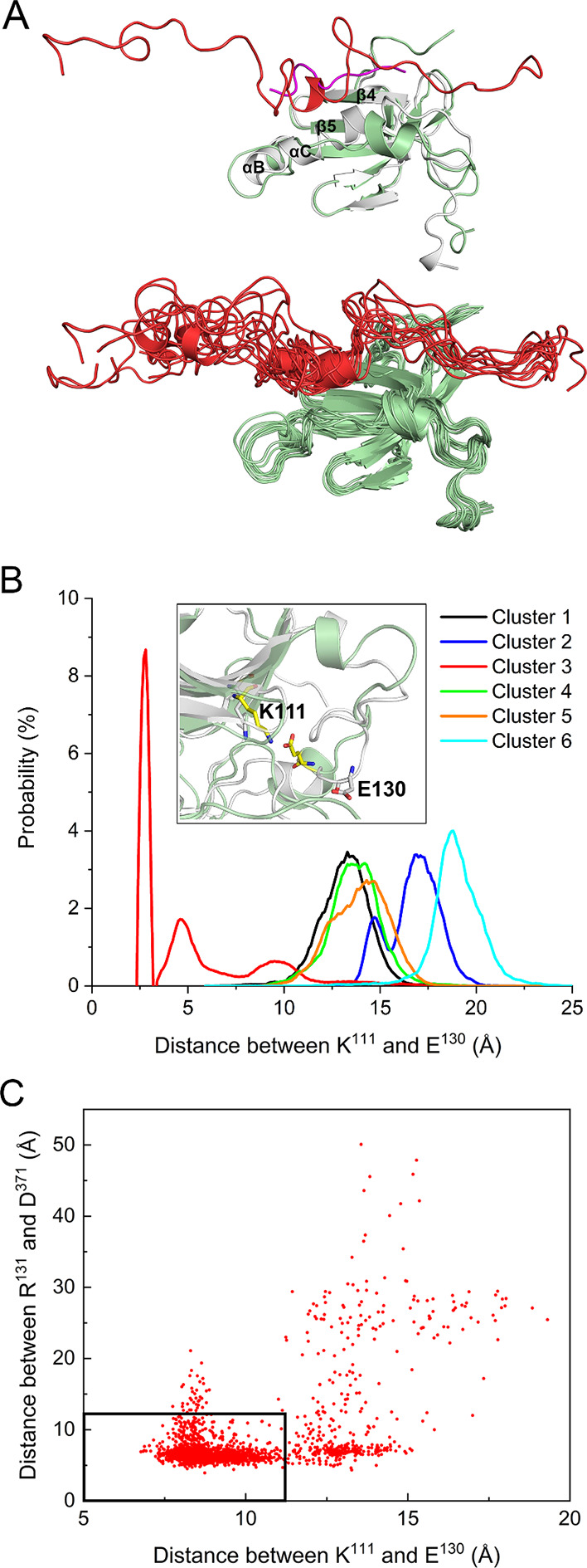
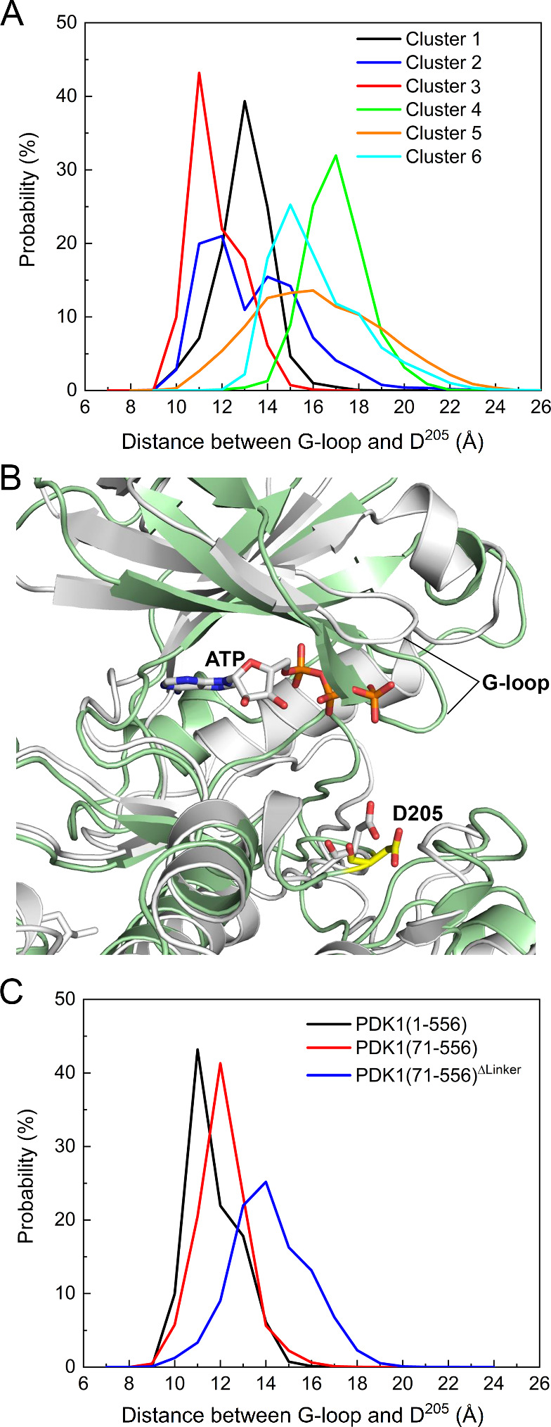
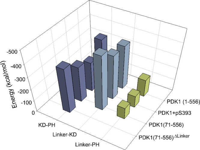
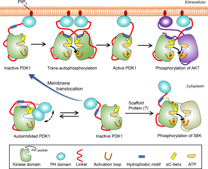
PDK1 is crucial for PI3K/AKT/mTOR and Ras/MAPK cancer signaling. It phosphorylates AKT in a PIP3-dependent but S6K, SGK, and RSK kinases in a PIP3-independent manner. Unlike its substrates, its autoinhibited monomeric state has been unclear, likely due to its low population time, and phosphorylation in the absence of PIP3 has been puzzling too. Here, guided by experimental data, we constructed models and performed all-atom molecular dynamics simulations. In the autoinhibited PDK1 conformation that resembles autoinhibited AKT, binding of the linker between the kinase and PH domains to the PIF-binding pocket promotes the formation of the Glu130-Lys111 salt bridge and weakens the association of the kinase domain with the PH domain, shifting the population from the autoinhibited state to states accessible to the membrane and its kinase substrates. The interaction of the substrates' hydrophobic motif and the PDK1 PIF-binding pocket facilitates the release of the autoinhibition even in the absence of PIP3. Phosphorylation of the serine-rich motif within the linker further attenuates the association of the PH domain with the kinase domain. These suggest that while the monomeric autoinhibited state is relatively stable, it can readily shift to its active, catalysis-prone state to phosphorylate its diverse substrates. Our findings reveal the PDK1 activation mechanism and discover the regulatory role of PDK1's linker, which lead to two innovative linker-based inhibitor strategies: (i) locking the autoinhibited PDK1 through optimization of the interactions of AKT inhibitors with the PH domain of PDK1 and (ii) analogs (small molecules or peptidomimetics) that mimic the linker interactions with the PIF-binding pocket.


Figures

1
1
Sequence and crystal structure of PDK1. (A) Sequence of the full-length PDK1. The sequences corresponding to the N-terminal region, kinase domain, linker, PH domain, and C-terminal region are underlined. The nonpolar, basic, acidic, and polar residues are colored black, blue, red, and green, respectively. Two motifs, 375NYD377 and the equivalent hydrophobic motif 383FGCM386 are in the linker region. (B) The crystal structure of the PDK1 kinase domain in the DFG-out conformation (left panel). The PIF-binding pocket is on the kinase surface, formed by αB-helix, αC-helix, β4-strand, and β5-strand. The crystal structure of the isolated PDK1 PH domain (right panel). The N-lobe and C-lobe of the kinase domain are colored in blue and cyan, respectively. The αB-helix, αC-helix, β4-strand, and β5-strand are colored in yellow.
2
2
Clustering of autoinhibited PDK1 conformations. Overlay of all simulated configurations with respect to the kinase domain in a cartoon representation, resulting in clustered locations of the PH domain in a thread representation with different colors (left panel). The best representative conformations from the top 6 ensemble clusters with different populations (right panels). The kinase domain is colored light green, the PH domain in different clusters is colored differently, and the linker region is colored cyan. The population of each cluster is shown in parentheses. Cluster 3 corresponds to the AKT-like autoinhibited conformation.
3
3
Key charged residues on the surface of the kinase and PH domains involved in the interdomain interactions of PDK1. Residue–residue contact probabilities for residues on the surface of (A) the kinase domain and (B) the PH domain, obtained by statistical analysis of all simulations. Charged residues with a probability >0.5% are labeled. Mapping of those charged residues with a probability >1.0% on the surface of the kinase domain and the PH domain is also shown. A contact occurs when the Cβ atom of a residue (Cα for Gly) in the kinase domain is within 10 Å of the Cβ atom of a residue (Cα for Gly) in the PH domain. The electrostatic interactions between the kinase and PH domains over the large surface area lead to different autoinhibited conformations.
4
4
Electrostatic interactions contribute to the stability of the AKT-like autoinhibited PDK1 (cluster 3). Contact maps between the kinase and PH domains (A), between the kinase domain and the linker (B), and between the PH domain and the linker (C) calculated for the conformations in the AKT-like autoinhibited PDK1 (cluster 3). Snapshots representing the key residue pairs involved in the interdomain interactions are also shown. The contacting residue pair with a probability >50% is labeled. The underlined residues (Lys76, Arg131, Thr148, and Gln150) correspond to the phosphate-binding site of the kinase domain of PDK1 (PDB ID: 1H1W). S389–S393 denotes the serine-rich motif 389SSSSS393.
5
5
Salt bridge formation in the AKT-like autoinhibited PDK1 (cluster 3). (A) Superimposition of the PIF-binding pocket with the crystal structure of PDK1 kinase domain in complex with PIFtide (purple, PDB ID: 4RRV). The bound linker in the autoinhibited PDK1 is shown in red. The dynamic conformations of the linker in the PIF-binding pocket are also shown. (B) The probability of the Glu130-Lys111 salt bridge formation in different PDK1 clusters. This salt bridge is only formed in cluster 3, and the inset shows an illustration of the formation of this salt bridge. For comparison, the salt bridge in the active kinase domain is also shown (white cartoon, PDB ID: 4RRV). (C) Correlation between the binding of the linker to the PIF-pocket and the formation of the Glu130-Lys111 salt bridge. The center of mass distance between Arg131 and Asp371 is used to characterize the binding of the linker to the PIF-binding pocket. The dense data points in the black rectangle (12 Å × 12 Å) suggest that the decrease in the distance between Arg131 and Asp371 corresponds to the decrease in the distance between Glu130 and Lys111 (formation of the salt bridge).
6
6
The AKT-like autoinhibited PDK1 shows a closed conformation of the ATP binding site. (A) Distribution of the distance between the center of mass of the glycine-rich loop (G-loop, residues 90–94) and Asp205 for full-length PDK1 in different clusters. Cluster 3 displays the shortest distance between the G-loop and Asp205, resulting in a closed conformation of the active site. (B) Comparison of the positions of the G-loop and Asp205 in the autoinhibited PDK1 (cluster 3) and in the crystal structure after superimposition of the conformation of the autoinhibited PDK1 (green) with the crystal structure of the active kinase domain (white, PDB ID: 4RRV). The conformation of cluster 3 is not accessible to ATP. (C) Distribution of the distance between the G-loop and Asp205 for truncated PDK1. PDK1(71–556) denotes the N-terminal truncated PDK1, and PDK1(71–556)ΔLinker denotes the deletion of both the N-terminal tail and linker. The conformation of the full-length PDK1 at 1 μs from the M1 simulation is used as the starting conformation for both PDK1(71–556) and PDK1(71–556)ΔLinker. All PDK1 refers to the AKT-like autoinhibited conformation (cluster 3).
7
7
The presence of the linker or phosphorylation of S 393 weakens the interactions between the kinase and the PH domains. Interdomain interaction energies calculated for the full-length PDK1 with S393 phosphorylation, N-terminal truncated PDK1, and N-terminal truncated PDK1 without the linker. For comparison, the interaction energy of the full-length PDK1 is included. All PDK1 refer to the AKT-like conformation (cluster 3). PDK1(1–556)+pS393 denotes the full-length PDK1 with S393 phosphorylation. PDK1(71–556) denotes the N-terminal truncated PDK1, and PDK1(71–556)ΔLinker denotes the N-terminal truncated PDK1 without the linker.
8
8
Proposed mechanism for the activation of PDK1, highlighting the regulatory role of the linker in PDK1 activation. PDK1 can exit in equilibrium between autoinhibited conformations and other inactive states with free PH domain in the cytoplasm. The autoinhibited states are more populated, with the AKT-like state displaying the high-affinity binding between the kinase and PH domains. Binding of the linker to the PIF-pocket promotes the formation of the Glu130-Lys111 salt bridge and changes the orientation of the αC-helix (from αC-out to αC-in), a hallmark of the active kinase domain. In the presence of PIP3, the population of the autoinhibited PDK1 shifts to the conformation with high affinity for PIP3. PDK1 is then recruited to the membrane and becomes active via trans-autophosphorylation. Once activated, PDK1 phosphorylates the activation loop of AKT. Binding of the hydrophobic motif in the C-terminal region of AKT to the PIF-binding pocket of PDK1 may facilitate phosphorylation. In cytoplasm, the inactive PDK1 may interact with scaffold proteins, become active and phosphorylate other substrate, independent of PIP3, which needs further investigation.
9
9
Proposed strategies for the design of PDK1 inhibitors. (A) Inhibitors in the crystal structures of autoinhibited AKT. (B) Superimposition of autoinhibited AKT conformations. The kinase and PH domains of the AKT (PDB ID: 3O96) are shown and colored in orange and light blue, respectively. Residues interacting with inhibitors are shown in surface and colored based on their types (nonpolar: white; polar: green; basic: blue; and acidic: red). (C) Sequence alignment of those interacting residues shown in (B) between AKT and PDK1. For clarity, the nonpolar residues are colored in black. (D) The binding site of those AKT inhibitors in the autoinhibited PDK1. The kinase and PH domains of PDK1 are separately superimposed to the kinase and PH domains of the autoinhibited AKT. The interacting residues based on the sequence alignment in (C) are shown and labeled. The kinase and PH domains of PDK1 are also colored in orange and light blue, respectively. (E) Interaction of the PIFtide 13MFRDFDYIA21 with the PIF-binding pocket (PDB ID: 4RRV). The binding of two inhibitors based on the PIFtide are also shown (PDB IDs: 4RQK and 4RQV). The PIF-binding pocket is shown in surface, and the PIFtide is shown in yellow tube. Residues in the PIFtide are shown as sticks, with Met13 and Ala21 labeled. Residues that define the binding site of the inhibitors are labeled and colored according to their types. The structures of the inhibitors are shown in Table S2. (F) Interaction of the linker segment (368DDEDCYGNYDNLLSQF383) with the PIF-binding pocket. The linker is shown as yellow tube. The binding site of the two inhibitors shown in (E) is obtained by superimposition of the crystal structures (PDB IDs: 4RQK and 4RQV) with the conformation of PDK1. The first inhibitor binding site, as well as the second potential binding site, are indicated as dashed lines.

Similar articles

Cited by

References

    1. Levina A., Fleming K. D., Burke J. E., Leonard T. A.. Activation of the essential kinase PDK1 by phosphoinositide-driven trans-autophosphorylation. Nat. Commun. 2022;13:1874. doi: 10.1038/s41467-022-29368-4. - DOI - PMC - PubMed
    1. Cordon-Barris L., Pascual-Guiral S., Yang S., Gimenez-Llort L., Lope-Piedrafita S., Niemeyer C., Claro E., Lizcano J. M., Bayascas J. R.. Mutation of the 3-Phosphoinositide-Dependent Protein Kinase 1 (PDK1) Substrate-Docking Site in the Developing Brain Causes Microcephaly with Abnormal Brain Morphogenesis Independently of Akt, Leading to Impaired Cognition and Disruptive Behaviors. Mol. Cell. Biol. 2016;36:2967–2982. doi: 10.1128/MCB.00230-16. - DOI - PMC - PubMed
    1. Niba E. T., Nagaya H., Kanno T., Tsuchiya A., Gotoh A., Tabata C., Kuribayashi K., Nakano T., Nishizaki T.. Crosstalk between PI3 kinase/PDK1/Akt/Rac1 and Ras/Raf/MEK/ERK pathways downstream PDGF receptor. Cell Physiol. Biochem. 2013;31:905–913. doi: 10.1159/000350108. - DOI - PubMed
    1. Molinaro C., Martoriati A., Lescuyer A., Fliniaux I., Tulasne D., Cailliau K.. 3-phosphoinositide-dependent protein kinase 1 (PDK1) mediates crosstalk between Src and Akt pathways in MET receptor signaling. FEBS Lett. 2021;595:2655–2664. doi: 10.1002/1873-3468.14195. - DOI - PubMed
    1. Xia H., Dai X., Yu H., Zhou S., Fan Z., Wei G., Tang Q., Gong Q., Bi F.. EGFR-PI3K-PDK1 pathway regulates YAP signaling in hepatocellular carcinoma: the mechanism and its implications in targeted therapy. Cell Death Dis. 2018;9:269. doi: 10.1038/s41419-018-0302-x. - DOI - PMC - PubMed

MeSH terms

Substances

LinkOut - more resources