. 2024 Oct 9;24(40):12529-12535.
doi: 10.1021/acs.nanolett.4c03472. Epub 2024 Sep 30.

Photothermal Properties of Solid-Supported Gold Nanorods

Affiliations

Photothermal Properties of Solid-Supported Gold Nanorods

Maja Uusitalo et al. Nano Lett. .

Abstract

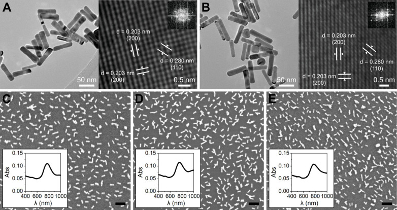
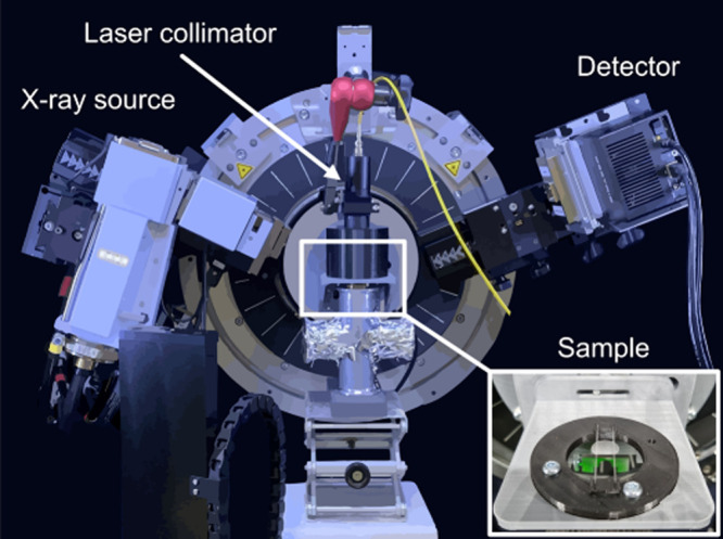
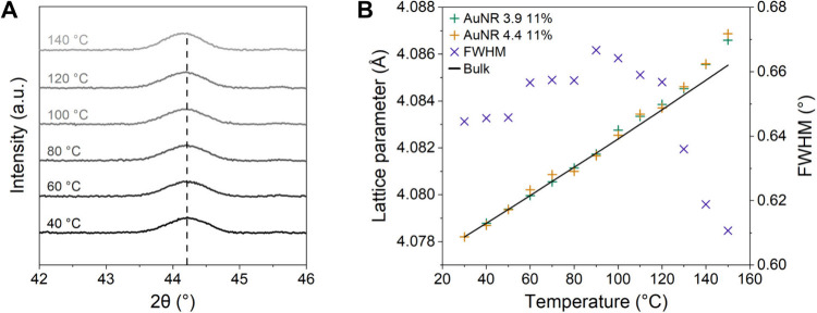
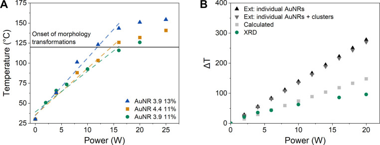
Gold nanoparticles possess unique photothermal properties and have gained considerable interest in biomedical research, particularly for photothermal therapy (PTT). This study focuses on evaluating the photothermal properties of gold nanorods (AuNRs) supported on glass substrates upon excitation with near-infrared (NIR) light. Two aspect ratios of AuNRs were electrostatically immobilized onto glass with controlled coverage. In situ X-ray diffraction (XRD) was performed to evaluate the photothermal behavior and morphological changes of the supported AuNRs during NIR laser irradiation. The XRD data sets were corroborated with scanning electron microscopy and Vis-NIR spectroscopy characterization. XRD revealed a linear temperature increase with laser power, aligning with theoretical predictions, and a slope dependent on the AuNR coverage, until the onset of morphology transformations around 120 °C. This study provides valuable insights into the photothermal properties of supported AuNRs, crucial for their application in PTT.

Keywords: X-ray diffraction; gold nanorods; near-infrared; thermal expansion; thermoplasmonics.


Conflict of interest statement

The authors declare no competing financial interest.

Figures

Figure 1
Figure 1
TEM characterization of (A) AuNR 3.9, and (B) AuNR 4.4. SEM micrographs and Vis-NIR absorption spectra of the AuNRs supported on glass for (C) AuNR 3.9 with 10.7 ± 2.0% surface coverage (AuNR 3.9 11%), (D) AuNR 3.9 with 13.4 ± 1.4% surface coverage (AuNR 3.9 13%), and (E) AuNR 4.4 with 11.0 ± 1.4% surface coverage (AuNR 4.4 11%). Scale bars in (C), (D) and (E) are 200 nm.
Figure 2
Figure 2
Schematic illustration of the setup used for the in situ XRD NIR laser experiments. The samples were placed on a support stage consisting of two glass capillaries (inserted picture), ensuring that the NIR beam did not heat any other elements than the sample. The NIR laser system’s collimator was placed 10 cm away from the sample at a 90° angle, illuminating the entire sample surface and ensuring that the flux was comparable between experiments.
Figure 3
Figure 3
(A) The evolution of the (200) peak during the furnace heating for AuNR 4.4 11%. (B) Lattice parameter of the gold cubic structure for AuNR 3.9 11% and AuNR 4.4 11%, together with FWHM for AuNR 4.4 11%, plotted against temperature. The solid line represents the thermal expansion of bulk gold (eq 1). We attribute the sharp decrease in FWHM above 120 °C to changes in the morphology of the AuNRs.
Figure 4
Figure 4
(A) Temperature as a function of laser power determined from comparing the measured lattice parameters with the thermal expansion of gold, for samples AuNR 3.9 11%, AuNR 4.4 11% and AuNR 3.9 13%. Dashed lines represent linear trends up to 120 °C. (B) Theoretically predicted (eq 2) and experimentally determined temperature increase as a function of laser power for AuNR 3.9 11%. The absorption cross sections used in the theoretical predictions were determined by calculations based on the AuNR dimensions (“Calculated”), or based on extinction spectroscopy measurements assuming either only individual AuNRs (“Ext: individual AuNRs”) or both individual and clustered AuNRs (“Ext: individual AuNRs + clusters”) contribute to the extinction at 808 nm.
Figure 5
Figure 5
Postexperiment Vis-NIR spectroscopy and SEM characterization of AuNR 3.9 11%, showing the morphology transformations and the resulting absorption peak shifts induced by furnace heating or NIR laser irradiation. Scale bars in the SEM micrographs are 100 nm.

References

    1. Huang X.; Jain P. K.; El-Sayed I. H.; El-Sayed M. A. Plasmonic Photothermal Therapy (PPTT) Using Gold Nanoparticles. Lasers Med. Sci. 2008, 23 (3), 217–228. 10.1007/s10103-007-0470-x. - DOI - PubMed
    1. Baffou G.; Cichos F.; Quidant R. Applications and Challenges of Thermoplasmonics. Nat. Mater. 2020, 19 (9), 946–958. 10.1038/s41563-020-0740-6. - DOI - PubMed
    1. Weissleder R. A Clearer Vision for in Vivo Imaging. Nat. Biotechnol. 2001, 19, 316–317. 10.1038/86684. - DOI - PubMed
    1. Vines J. B.; Yoon J. H.; Ryu N. E.; Lim D. J.; Park H. Gold Nanoparticles for Photothermal Cancer Therapy. Front Chem. 2019, 7, 167.10.3389/fchem.2019.00167. - DOI - PMC - PubMed
    1. Zharov V. P.; Mercer K. E.; Galitovskaya E. N.; Smeltzer M. S. Photothermal Nanotherapeutics and Nanodiagnostics for Selective Killing of Bacteria Targeted with Gold Nanoparticles. Biophys. J. 2006, 90 (2), 619–627. 10.1529/biophysj.105.061895. - DOI - PMC - PubMed

LinkOut - more resources